BY11 - MODULE 1: Cells as the Basis of Life
1.1 - CELL STRUCTURE
INQUIRY QUESTION: WHAT DISTINGUISHES ONE CELL FROM ANOTHER?
COMPONENTS OF CELLS
CELL ORGANELLES (PARTS 1 & 2)
What is a cell?
A cell is the smallest structural and functional unit of a living organism.
THE THREE PRINCIPLES OF CELL THEORY
Cells are the building blocks for life. It is the basic unit of living organisms.
All living things are composed of one or more cells.
All cells come from pre-existing cells.
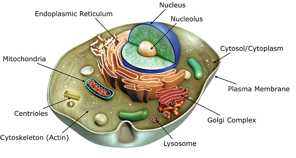
PARTS OF A CELL
CELL MEMBRANE
CYTOPLASM
ORGANELLES
Structure and function of main organelles
NUCLEUS - Contains the genetic information (DNA chromosomes) of the cell. This is needed for growth, repair, and proper functioning. It tells the rest of the cell what to do. It is surrounded in a nuclear membrane containing gaps called nuclear pores.
NUCLEOLUS - A small organelle found within the nucleus. It is made of proteins and ribonucleic acid (a.k.a. RNA). It is where ribosomes are made.
RIBOSOMES - Blobs made of ribosomal RNA and protein. They are NOT MEMBRANE BOUND. Ribosomes create proteins, and they are found floating around in cytoplasm, but are generally attached to the endoplasmic reticulum.
ENDOPLASMIC RETICULUM - Connected to the nucleus in some places. It is a network of flattened, interconnected membranes. Creates a tunnel through which substances can move. There are two types of endoplasmic reticulum: rough and smooth. Rough ER has many ribosomes attached to it, while smooth ER has no ribosomes. In the rough ER, proteins are made by ribosomes, which are then processed and modified. The smooth ER makes lipids (fats).
GOLGI BODY - Also known as the Golgi apparatus. Made out of flat membrane sacs stacked on top of each other. The membrane sacs are not interconnected. It processes and packages substances made by the cell. After processing, it pinches off around the substance to form a bubble with the substance inside, known as a vesicle. The vesicle then transports the substance to where it is needed, which can be inside or outside the cell.
LYSOSOMES - A vesicle made by the Golgi body. They are membrane-bound sacs that contain digestive enzymes that are needed to break down other substances such as cellular waste and foreign particles.
Structure and function of organelles involved in energy transformation
CHLOROPLASTS - (PLANT CELL ONLY) A disc-shaped organelle that has a double membrane, their own DNA, and chlorophyll (a green pigment). They capture light energy from the sun and use it to perform photosynthesis, that creates glucose (a source of energy).
MITOCHONDRIA - (PLANT AND ANIMAL) Bean-shaped organelle that has a double membrane and their own DNA. Performs chemical respiration by combining oxygen and glucose to create ATP (energy) that the cell can use. The powerhouse of the cell. The number of mitochondria in a cell depends on how much energy that specific type of cell needs. (e.g., muscle cells need lots of mitochondria because they need a lot of energy to work!)
Structure and function of organelles involved in cell structure and storage
CELL MEMBRANE - Provides protection for a cell and separates the interior of a cell from external environment. Also known as a plasma membrane. It is semi-permeable, meaning it regulates the movement of substances entering and exiting the cell.
CELL WALL - Found in plant cells, fungal cells, and some prokaryotic cells. What it is made of depends on the type of cell. It is an external structure surrounding the cell membrane. It gives the cell structural strength and protection.
CYTOPLASM - Gelatinous fluid that fills up the inside of a cell, giving it shape. All organelles are suspended in it.
CYTOSKELETON - A network of microtubules, microfilaments, and intermediate filaments. A “protein spiderweb” throughout the cell, holding all the organelles in place.
CENTRIOLES - Small, cylindrical structures made of microtubules wound up together. Involved in cell division; they pull chromosomes apart.
PILI AND FLAGELLA - Hair-like appendages made of microtubules, enclosed by an extension of the cell membrane. Used to help the cell move around. The flagella is a longer version of pili.
VACUOLE - A membrane-bound vesicle containing fluid. The fluid is made up of water and other dissolved substances e.g., sugars and salts. Its main job is to store substances for when the cell wants to use it. They are found in most cells, but size and quantity varies. Animal cells have small, temporary vacuoles, while plant cells have one very large, permanent vacuole. In a plant cell, vacuoles give the cell structural support (this is known as turgor pressure)
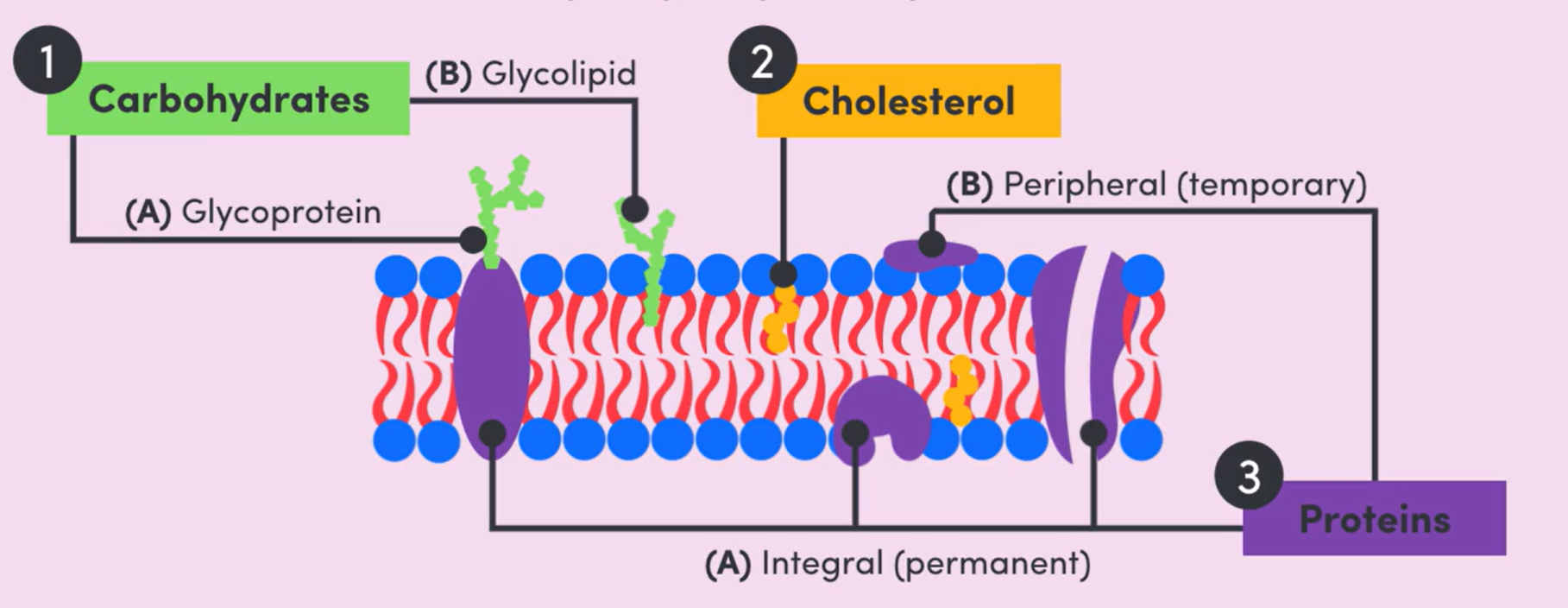
 THE FLUID MOSAIC MODEL
The fluid mosaic model is the currently accepted model for the cell membrane. It is used to explain the function and structure of all cell membranes.
Structure of the fluid mosaic model
Cell membranes are phospholipid bilayers. In other words, they consist of 2 layers of phospholipid molecules.
A phospholipid molecule consists of a phosphate head and two tails made of lipids (fats).
The lipid tails are hydrophobic, meaning they are repelled by water.
The phosphate head is hydrophilic, meaning it is attracted to water.
The hydrophobic tails face the inside, away from water, while the hydrophilic head faces the outside, towards the water.
Within the phospholipid bilayer, there are carbohydrates, cholesterol molecules, and proteins.
CARBOHYDRATES: attached to the surface of the cell membrane. If they are attached to a protein, they are called glycoproteins, and if they are attached to a lipid, they are called glycolipids. They are NEVER attached to the phosphates.
CHOLESTOROL MOLECULES: Squished in between the phospholipids
PROTEINS: Proteins that are a permanent part of the cell membrane are known as integral proteins. Proteins that are a temporary part of the cell membrane are known as peripheral proteins. INTEGRAL = LODGED DEEP, PERIPHERAL = ATTACHED LOOSELY ON THE OUTSIDE
Function of the fluid mosaic model
SEPARATE, REGULATE, COMMUNICATE
SEPARATE: The cell membrane separates the contents of the cell from the external environment (intracellular fluid = cytoplasm inside the cell, extracellular fluid = watery environment outside the cell). Applies to BOTH prokaryotic and eukaryotic cells! However, in eukaryotes, membranes are used to create compartments within the cell (organelles).
REGULATE: The cell membrane is able to control which substances go through it; which means that it is responsible for regulating the contents of the cell. It is semi-permeable, meaning it will allow certain molecules to pass through it via passive or active transport. It is able to get the chemicals it needs, while getting rid of what it doesn’t.
COMMUNICATE: Involved in cell recognition and communication with other cells. The components responsible for this are glycoproteins, glycolipids, and integral proteins.
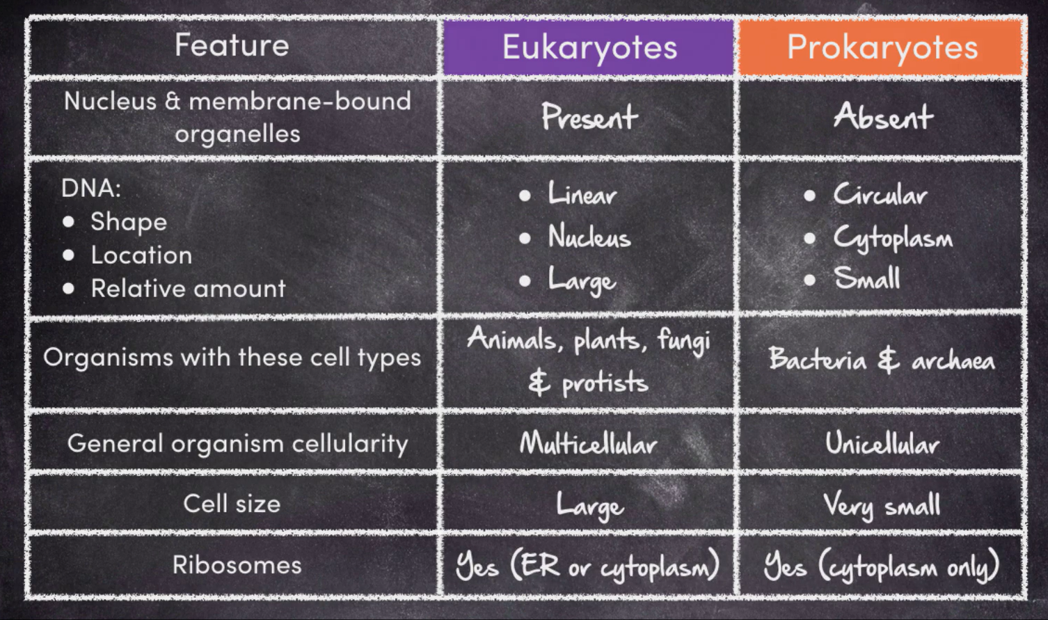
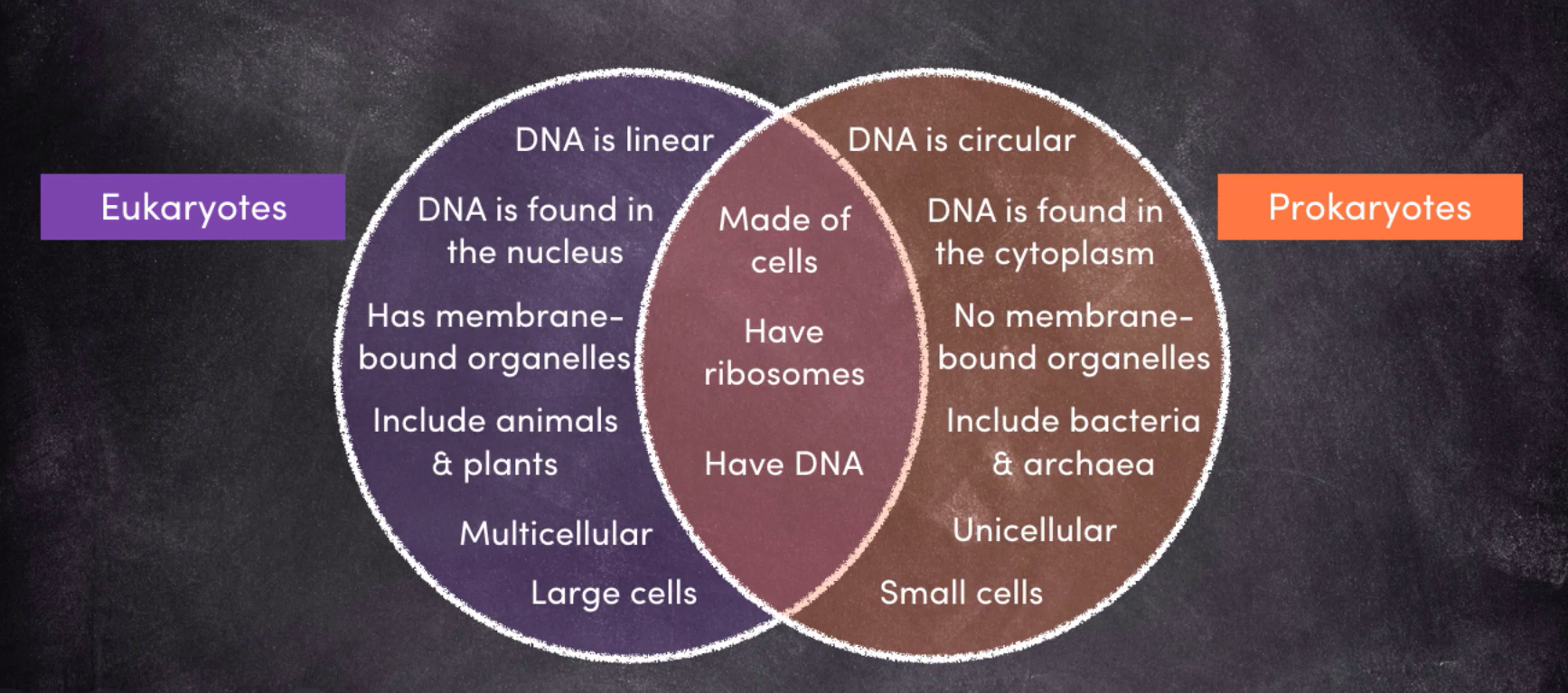
PROKARYOTES AND EUKARYOTES
Eukaryotes
A cell is eukaryotic if it has a nucleus and membrane-bound organelles.
Eukaryotic DNA is found inside the nucleus, and is straight (does not reconnect with itself).
Large amount of DNA
“Eukaryotes” are organisms that are made of eukaryotic cells. This includes animals, plants, fungi, and protists.
Eukaryotes are usually multicellular, but can be unicellular (e.g., protists).
Prokaryotes
A cell is prokaryotic if it does NOT have a nucleus or membrane-bound organelles.
They are also very small in comparison to eukaryotic cells.
Ribosomes are not technically an organelle, and they are present in prokaryotic cells and float around in the cytoplasm.
They have a cell wall, but it is made out of a different substance called peptidoglycan.
The DNA is not enclosed in a nucleus, so it floats around in the cytoplasm. It is small and circular
Prokaryotes are only ever unicellular.
They have flagella and pili to help them move around.
Recognising different cells
PROKARYOTES: Bacteria, archaea
EUKARYOTES: Animal cells, plant cells, fungi, protists
Prokaryotes vs. Eukaryotes
 DRAWING CELLS
Materials
Unlined paper, pencil, eraser, ruler
DO NOT DRAW IN PEN! The pencil makes it so that it is easy to fix mistakes!
Positioning + size
Centre the drawing in the middle of the page so that you have room to label it
The diagram should take up around ½ of a page so that it is clear
Simple, clear lines, no shading!
Accuracy + labels
When drawing from a reference, only draw a few of the cells, not all of them. You don’t need to draw all of them to get your point across, so just do a few to save time. Only draw what you see!
LABEL THE DRAWING! Use a ruler to draw the label lines, and make sure the DON’T CROSS!
Do NOT add arrowheads.
Title + scale
The title must include the name of what has been drawn, as well as the magnification of the image. e.g., “A human cheek cell under 400x magnification”
Underline the title!
A biological drawing needs either magnification or a scale bar
Magnification: How close up the specimen is. This is calculated by multiplying the objective lens by the ocular (eyepiece) lens. In a light microscope, the ocular lens is usually 10x.
Scale bar: A capped, horizontal line that represents the ratio between the drawn size of the object and the actual size. It is put at the bottom corner of the drawing.
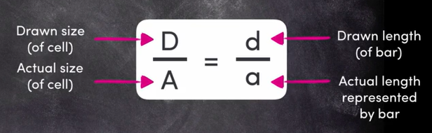
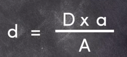 REMEMBER: 1mm = 1000µm
VIEWING OF CELLS
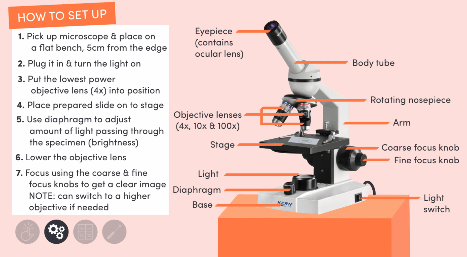
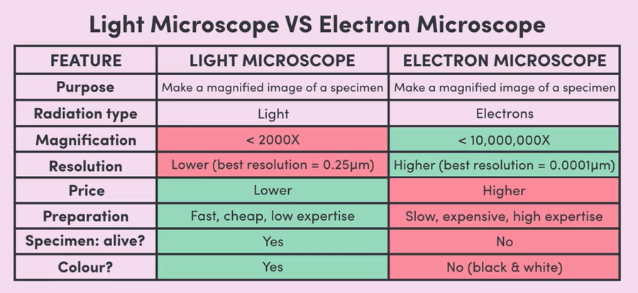
THE LIGHT MICROSCOPE
Uses visible light and 2 lenses to make a specimen look bigger.
The light shines up through the specimen, then travels through the objective and ocular lenses
The light is refracted as it moves through the lenses, creating a magnified image.
STAINING: Adding dye to a specimen that stains certain cell components. The increase in colour contrast makes it so you can see the different structures in the specimen easier. Used in the preparation of all specimens (whole organisms, cell smears, thin slices)
 Calculating magnification
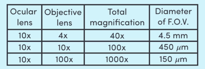 MAGNIFICATION = magnified size / actual size
The confocal microscope
One of the most advanced light microscopes
Works by passing a highly focused laser through the specimen
End product: high quality image of a tiny part of the specimen
A computer then enhances these images and stitches them together to create a three dimensional image of the specimen
THE ELECTRON MICROSCOPE
The internal chamber of the electron microscope is under vacuum conditions, meaning there is nothing inside, not even air!
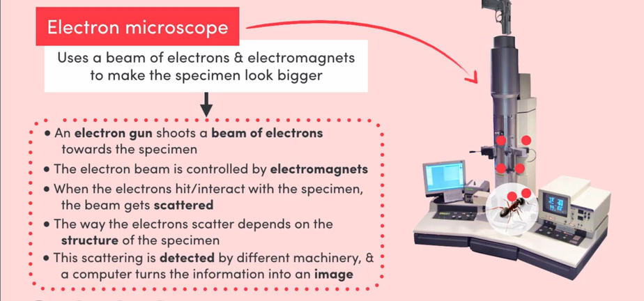 Transmission electron microscope
Specimen is prepared by being treated with chemicals, giving it increased structural strength… so that the electron beam doesn’t just blow it up lmao
Water is removed from the specimen using alcohol. This is because water evaporates in vacuum conditions, so if water isn’t removed from the specimen beforehand, it will be destroyed.
The specimen is then embedded in a resin and cut into very thin slices. The slices make it so the inside can be seen.
Scanning electron microscope
Specimen is treated with chemicals
Water is removed from the specimen
The specimen is coated in a thin layer of gold
TRANSMISSION VS. SCANNING ELECTRON MICROSCOPES: Transmission microscopes shoot one broad beam at the specimen, while scanning electron microscopes shoot one super fine beam focused to a specific point, that is then systematically scanned across the whole specimen. The electron beam bounces straight off of the surface of the specimen, meaning you see all of the outside, but not the inside.
Light microscope vs. electron microscope
1.2 - CELL FUNCTION
INQUIRY QUESTION: HOW DO CELLS COORDINATE ACTIVITIES WITHIN THEIR INTERNAL ENVIRONMENT AND THE EXTERNAL ENVIRONMENT?
TRANSPORT OF MATERIALS
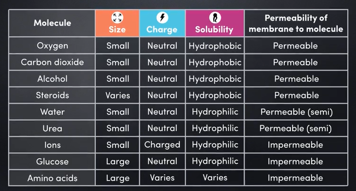
CHARACTERISTICS OF MATERIALS
Characteristics of materials determine whether or not they will pass through the semi-permeable membrane
Size
The cell membrane is permeable to small molecules (e.g., urea and water)
The cell membrane is impermeable to large molecules (e.g., glucose and amino acids)
Small molecules can fit in between the phospholipids and slip through, whereas the larger ones cannot.
Charge
This refers to whether the molecule has a positive, negative, or neutral charge.
The cell membrane is permeable to uncharged (neutral) molecules (e.g., oxygen and carbon dioxide)
The cell membrane is impermeable to charged molecules, known as ions (e.g., potassium and sodium)
If a molecule isn’t called an ion, it’s neutral!
Solubility
Refers to how quickly and easily materials in the cell membrane can dissolve/move across the membrane.
Only applies to materials that are small and/or neutral.
Solubility has no effect on impermeable materials because they are already blocked from entering!
“LIKE DISSOLVES LIKE” - A hydrophilic substance only dissolves in a hydrophilic substance, and a hydrophobic substance can only dissolve in a hydrophobic substance
Water and urea are hydrophilic, meaning they don’t dissolve across the cell membrane as quickly as hydrophobic molecules like alcohol and steroids.
Hydrophilic substances will find it more difficult to get past the hydrophobic lipid barrier.
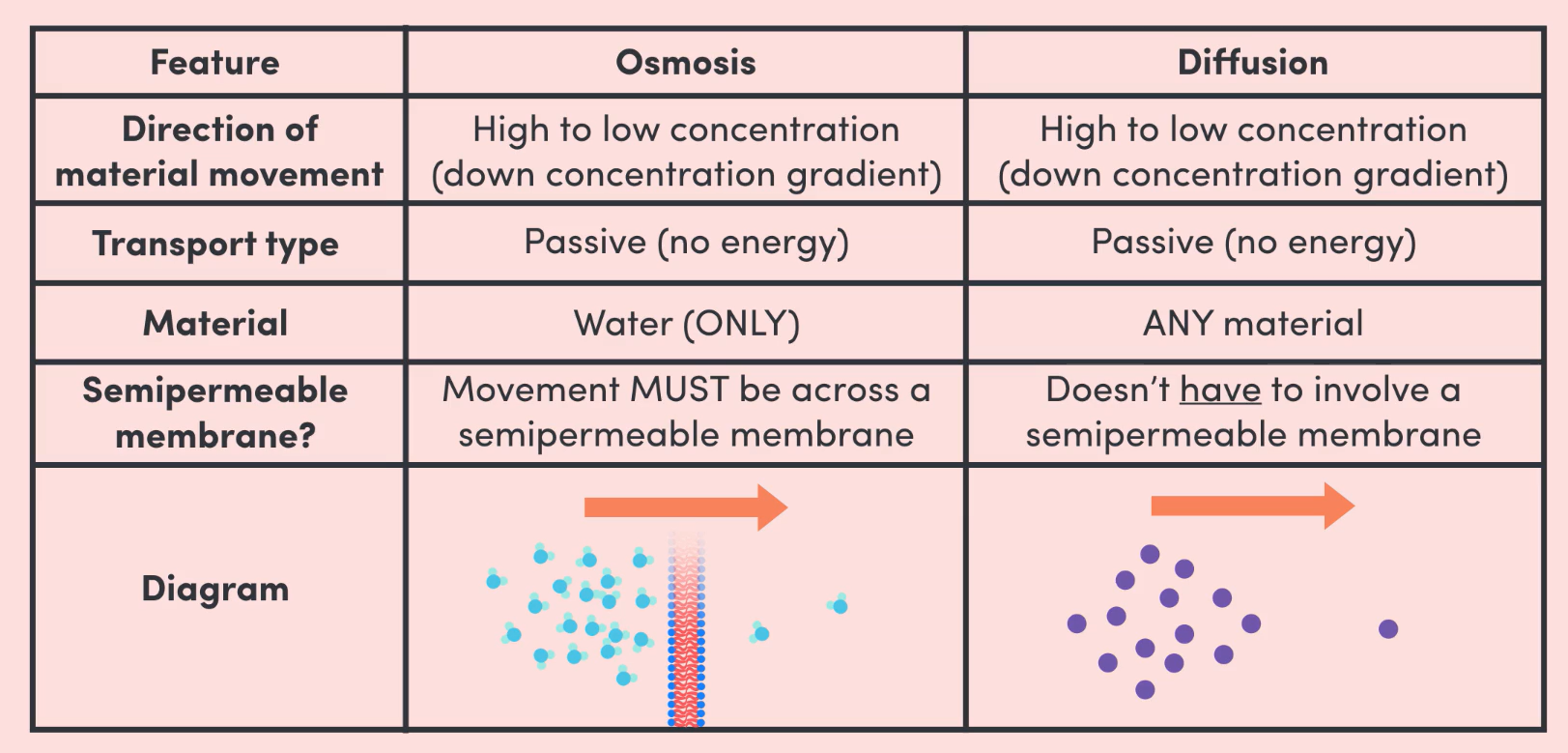
DIFFUSION
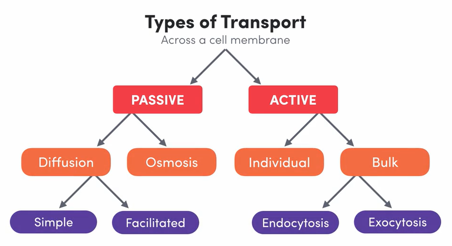
Diffusion is a type of passive transport.
What is passive transport?
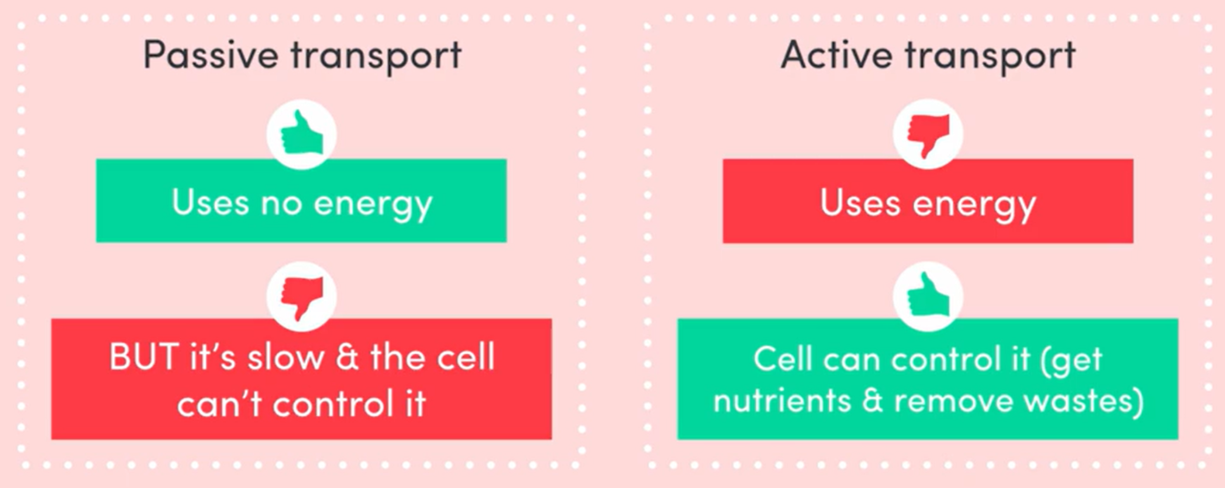
Passive transport is the movement of materials across the cell membrane WITHOUT the expenditure of energy.
This means that certain substances will move across the cell membrane without the cell needing to do anything.
It happens spontaneously!
All passive transport happens due to differences in concentration.
A concentration gradient is just a situation where the concentration gradient is higher in one area and lower in the other.
It is a gradual change in concentration from one region to another
FUNDAMENTAL RULE: The universe always wants a balance.
Materials will move spontaneously from an area of high concentration to low concentration to achieve balance (equilibrium). This is known as moving down the concentration gradient.
TWO TYPES OF PASSIVE TRANSPORT: Diffusion and Osmosis
What is diffusion?
The movement of ANY molecule from high to low concentration (down the concentration gradient).
Diffusion with a semi-permeable membrane means that both concentration gradients and permeability must be considered.
Simple diffusion
The diffusion of substances directly through the phospholipid bilayer.
If the cell membrane is permeable to the material, it will diffuse through the membrane (high to low conc.)
Diffusion is generally used to transport materials that are small and neutral (water, carbon dioxide, urea, alcohol, lipids, steroids, etc.).
EXAMPLE: Oxygen diffusing across from capillaries which have high levels of concentration (due to lungs), to body cells which have low levels of concentration (due to respiration)
Facilitated diffusion
If the cell membrane is impermeable to the material, it will not diffuse through the membrane, no matter the strength of the gradient.
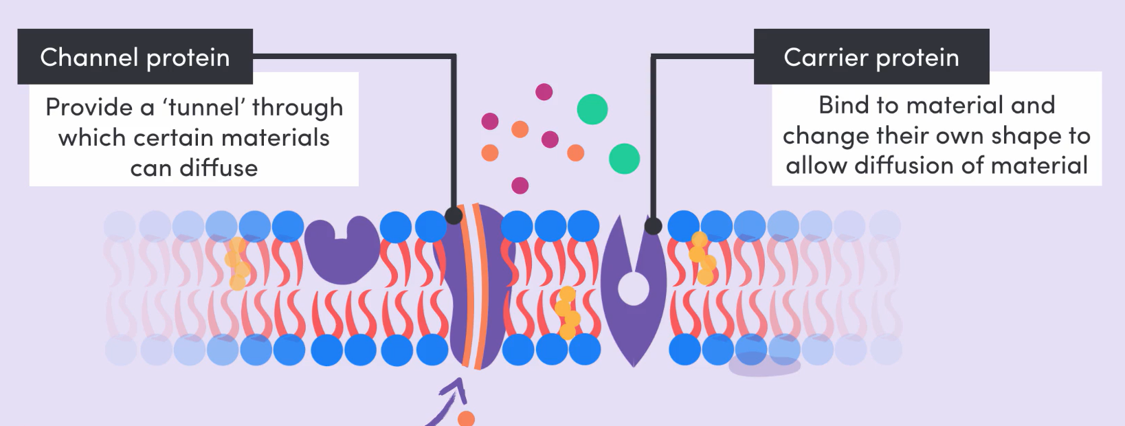
Facilitated diffusion is the diffusion of substances through the cell membrane via channel and carrier proteins found throughout.
CHANNEL PROTEINS: Proteins with tunnels all the way through where certain materials can diffuse. Each tunnel has a unique diameter and acid lining, so that only particular molecules can fit through them.
CARRIER PROTEINS: Bind to the material and change their own shape to allow diffusion across the membrane. Once the material has passed through, they change back to their original shape.
OSMOSIS
What is osmosis?
Osmosis refers to the movement of water from an area of high water concentration to an area of low water concentration across a semi-permeable membrane.
Generally, the “concentration” of a solute refers to the amount of solute, NOT water. In the case of osmosis, concentration refers to the amount of water, NOT solute.
EXAMPLE: 10% glucose solution has a HIGHER water concentration in comparison to a 30% glucose solution. TLDR whichever solution has LESS solute and MORE water has a higher WATER CONCENTRATION, but a lower SOLUTE CONCENTRATION. When looking at osmosis, WATER CONCENTRATION is the focus.
In osmosis, water particles (but NOT the solute particles!!) will diffuse across the semi-permeable membrane from high water concentration to low water concentration
REMEMBER: the practical experiment with glucose solution and dialysis tubing!!!!!!
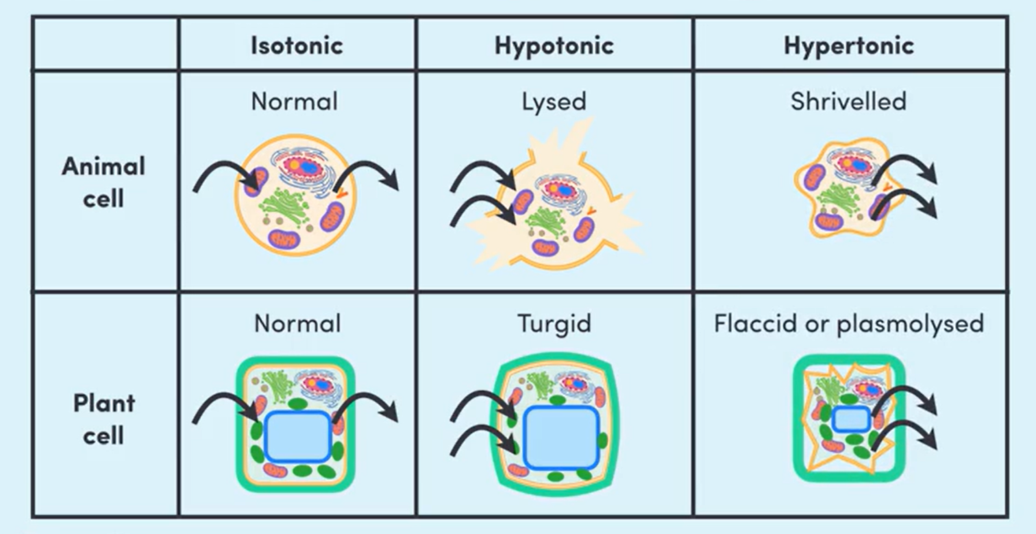
Isotonic, hypotonic, and hypertonic solutions
Terms used to describe the concentration differences between 2 solutions.
In an exam, this content would be useful when asked a question about what would happen if a cell was placed into a solution. This applies because the inside of a cell is filled with cytoplasm, and is separated by a cell membrane.
Isotonic
An isotonic solution is a solution that has equal solute concentrations.
A cell in an isotonic solution is 😄 happy because everything is balanced and at equal concentrations!
The rate at which water leaves the cell and the rate at which water is entering the cell is the same. The concentrations are balanced.
The cells are “normal”.
Hypotonic
A hypotonic solution is a solution with a lower solute concentration and a higher water concentration.
EXAMPLE: Fresh water.
If you place a cell into a hypotonic solution, water will move INTO the cell from the solution to achieve balance.
An animal cell will continue to absorb water until it pops! This popped cell is referred to as “lysed”
A plant cell will swell, but the cell wall will prevent it from popping! The plant cell is “turgid”.
REMEMBER: The French Orbeez guy. “hypOtonic”, o for Orbeez girl!! Orbeez take in water and swell up.
Hypertonic
A hypertonic solution is a solution with a higher solute concentration and a lower water concentration.
EXAMPLE: Salty ocean water.
If you place a cell into a hypertonic solution, water will move OUT of the cell and into the solution to achieve balance.
An animal cell is referred to as “shrivelled”, and a plant cell is “flaccid”. When a plant cell is completely dry, it is “plasmolysed”.
Osmosis vs. Diffusion
 Tip: Use dot points and tables in exams to save time and clearly demonstrate knowledge!
ACTIVE TRANSPORT
Active transport is the movement of materials across the cell membrane that require expenditure of energy.
The energy is called adenosine triphosphate, or ATP for short.
The cell will spend ATP to move materials where it wants.
This is done through specific carrier proteins. Energy is used to bind specific materials to the carrier protein, and open and close it.
Active transport is used when materials move from low to high concentration. This is known as moving against the concentration gradient.
It is the opposite of passive transport!
EXAMPLE: Uptake of glucose from the digestive system. Epithelial cells will use active transport to move glucose into them so that it can be transported to other cells around the body. It is going against the concentration gradient, so energy is needed to move it across.
Bulk transport (endocytosis and exocytosis)
Passive and active transport are suited to move most small, individual molecules across the cell membrane.
When a material cannot be moved in this way (either because it is too big or there is too much of it), endocytosis and exocytosis come into play (mr fu reference)
Bulk transport requires the formation of vesicles (pinched off cell membrane). They are both, technically, forms of active transport.
Endocytosis
Used to transport materials into the cell (endo = inside)
Cell membrane engulfs external substance and pinches off, forming a vesicle inside the cell.
THREE TYPES OF ENDOCYTOSIS: Pinocytosis, phagocytosis, and receptor-mediated endocytosis.
PINOCYTOSIS: Membrane engulfing a liquid
PHAGOCYTOSIS: Membrane engulfing a solid material. The food vesicle formed is known as a phagosome. To digest the contents of a phagosome, it will bind to a lysosome and use its digestive enzymes to break it down.
RECEPTOR-MEDIATED: Protein receptors detect specific molecules in extracellular fluid, triggering endocytosis. The receptors bind to the molecules and this triggers the membrane to engulf them.
Exocytosis
Used to transport materials out of the cell (ex = outside)
Gets rid of waste materials, the opposite of endocytosis
Vesicle (created by the Golgi body), approaches cell membrane. When they come into contact, the phospholipids are re-arranged and the membranes fuse.
The vesicle contents are then released out of the cell, and the vesicle membrane is now a permanent part of the cell membrane! It plays an important role in cell growth because of this 😃
THE RATE OF EXCHANGE
Concentration gradients
When there is an area of high concentration and an area of low concentration, a concentration gradient exists.
It is a gradual change in concentration from one region to another.
PASSIVE: along the gradient (high to low)
ACTIVE: against the gradient (low to high)
Like a hill - rolling down the hill with no effort vs. being pushed up the hill
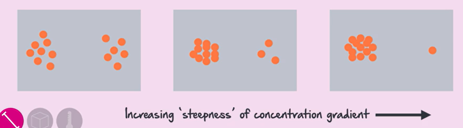
The STEEPER the gradient, the FASTER the material will move towards an area of low concentration passively, and the SLOWER the material will move towards the area of high concentration actively.
The cell CANNOT change the rate of passive transport! However, in active transport, the cell can use more or less energy (ATP) to change the rate.
STEEPER GRADIENT: bigger difference between high and low areas of concentration
Surface area to volume ratio
SURFACE AREA: Total area of all of the outside surfaces of an object. This is measured in squared units (e.g., mm², cm², m², etc.). The most common unit is cm².
VOLUME: Amount of space an object takes up. This is measured in cubed units (e.g., mm³, cm³, m³, etc.). The most common unit is cm³.
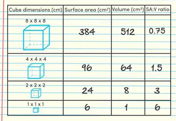
Surface area to volume ratio is calculated by dividing a cube’s surface area by its volume. This can be written either as a number or left in its simplest ratio (usually the ratio to one).
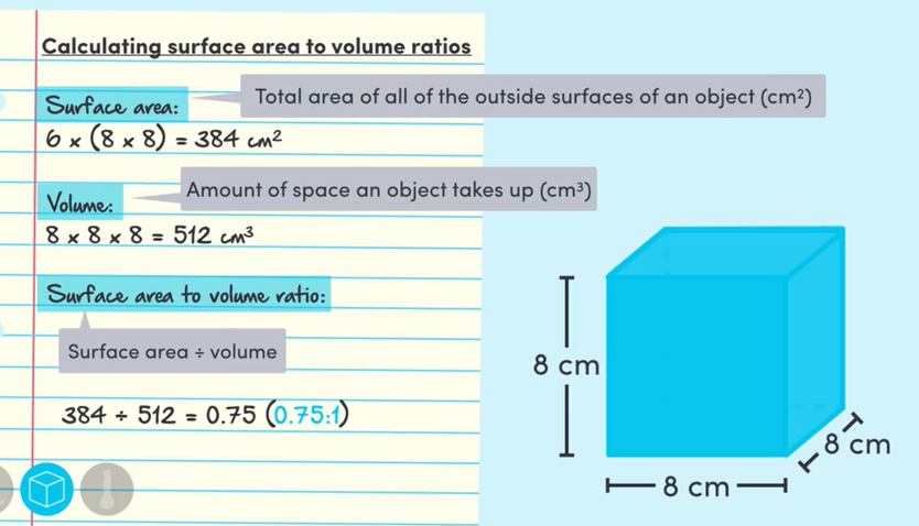 Calculating SA:V ratio
As the size of an object decreases, its SA:V ratio increases.
Cells and SA:V ratios
For cells, a higher SA:V ratio is ideal. The higher the ratio, the more efficiently materials can be exchanged across its surface (i.e., the cell membrane), particularly via diffusion and osmosis.
This is important for cells, which get nutrients and remove waste through their cell membrane.
This is also why cells are so small! The bigger the cell, the more nutrients it needs, but the smaller its SA:V ratio. If a cell is too big, it will not be able to get the nutrients it needs or get rid of waste fast enough to survive.
Though a cell can’t change its volume, it can increase its surface area through TWO ways: flattening itself, or extending its cell membrane.
FLATTENING: The flatter the shape, the larger the ratio. This strategy is used by lots of different cells, such as red blood cells and lung epithelial cells to increase rate of transport. However, not all cells can afford to be flat! The organelles within the cells prevent them from doing so.
EXTENDING: Because of this, the most common strategy used by cells is to extend the cell membrane. The cell membrane folds in half, creating little tentacles and thus creating a very large surface area in comparison to their volume. Organelles are kept down the bottom, with the extensions used purely for exchange.
Extending is particularly used in cells that require the absorption of lots of nutrients in a short period of time. e.g., microvilli in stomach lining absorbing nutrients from a meal, and root hair cells in plants.
Particle size and temperature
This only effects the rate of passive transport (diffusion + osmosis)
PARTICLE SIZE: the smaller the particle, the faster the rate of diffusion. This is because they can move through the phospholipid bilayer easier.
TEMPERATURE: Increasing temperature increases the speed at which molecules move. Faster moving particles move across the membrane quicker.
NUTRIENTS AND WASTES
CELL REQUIREMENTS
How organisms get nutrients
Cells need ATP. They are the only form of energy that cells can directly use.
Cells do not obtain ATP from food. They use chemical energy from the breakdown of food to make ATP.
AUTOTROPH: An organism that can produce its own food (e.g., plants, algae). They harness external energy to turn inorganic compounds (e.g., air, soil, sunlight) into food.
HETEROTROPH: An organism that can’t make its own food (e.g., animals, fungi, most bacteria, protists). They get nutrients and energy by consuming other organisms.
The nutrients cells need
There are two categories: organic and inorganic compounds
ORGANIC: Compounds that contain carbon and hydrogen. They are called organic because they are made only by living organisms. These include carbohydrates, lipids, proteins, nucleic acids, and vitamins.
INORGANIC: Compounds which don’t contain carbon bonded to hydrogen. These include water, oxygen, carbon dioxide, and minerals.
Organic compounds
CARBOHYDRATES: Chains which consist of carbon, hydrogen, and oxygen. They provide cells with an easily accessible energy source. In plants, certain carbohydrates such as cellulose (found in plant cell wall) also play a structural role.
LIPIDS: Chains of fatty acids and glycerol which consist of carbon, hydrogen and oxygen. Densely packed, allowing for more efficient, long-term energy storage. Integral part of cell membranes (lipid tails)!
PROTEINS: Chains of amino acids which consist of carbon, hydrogen, oxygen, and nitrogen. Nitrogen is the main thing that makes proteins different! They are important for structure (are in cell membrane and tissues). They also assist in growth, repair, and maintenance, as well as formation of enzymes.
NUCLEIC ACIDS: Chains of nucleotides which consist of carbon, hydrogen, oxygen, nitrogen, and phosphorus. Depending on the type of nucleotide, these are either DNA or RNA. DNA carries genetic info, and RNA helps the cells make proteins.
VITAMINS: A “mixed bag” of all other organic compounds needed for cell survival. IMPORTANT VITAMINS: A, B, C, D, E, and K. Involved with enzymes.
Inorganic compounds
WATER: H₂O molecule, consists of 2 hydrogen atoms bonded to an oxygen atom. Makes up majority of an organism (usually around 70-90%!!). Takes part in reactions, as either a reactant or the reaction takes place in it,
OXYGEN: Consists of two oxygen atoms bonded together (O₂). Needed for cellular respiration.
CARBON DIOXIDE: CO₂ molecule, consists of 2 oxygen atoms bonded to a carbon atom. Main source of carbon atoms for all organic molecules. Required by plants to create sugars via the process of photosynthesis.
MINERALS: A “mixed bag” of all other inorganic compounds needed for cell survival. IMPORTANT EXAMPLE: iron (component of haemoglobin in red blood cells).
WASTE REMOVAL
Main types of cellular waste
CARDON DIOXIDE: When carbs or lipids are broken down during cellular respiration, CO₂ is produced. It needs to be gotten rid of as it makes cells and their surroundings very acidic. Reacts with water and makes an acid that can destroy cellular structures, especially enzymes.
NITROGENOUS: Waste products that contain nitrogen (e.g., ammonia, uric acid, urea). Formed in the breakdown of unwanted proteins and nucleic acids. It needs to be gotten rid of as it makes cells and their surroundings very basic. Destroys cellular structures like enzymes.
WATER: Water becomes an issue if the organism drinks a lot of water or lives in a freshwater environment and water moves in via osmosis. Excess water not only takes up space, but can cause a cell to pop (lysed!) and die.
Waste removal
CELLULAR LEVEL: Occurs in both unicellular and multicellular organisms. The movement of wastes from the cytoplasm into the extracellular environment. Waste can be removed by passive or active transport. Wastes commonly removed via passive transport include CO₂, alcohol, and ammonia (simple diffusion), as well as urea, glucose, ions (facilitated diffusion), and waster (osmosis). Wastes commonly removed via active transport include nitrogenous waste. Ions can be removed either passively or actively, depending on how much there is, and how badly the cell needs it gone.
BODY SYSTEM LEVEL: Occurs in multicellular organisms. In plants, CO₂ and H₂O are excreted as gases through leaves. Other wastes can be released into soil, but most is moved into older leaves or to outer bark, which will eventually leave the plant and take the waste with it.
In animals, the circulatory system transports waste from cells to the organs of the excretory system (lungs, liver, kidneys).
Lungs - carbon dioxide and water diffused across respiratory membranes, excreted as gas when exhaling.
Liver - prepares wastes for removal e.g, turns ammonia into urea which is less toxic.
Kidneys - filter blood and return useful substances back to the body, while excreting unwanted wastes as urine.
PHOTOSYNTHESIS AND RESPIRATION
PHOTOSYNTHESIS
lalalala
RESPIRATION
lalalala
ENZYMES
ENZYMES
haha
FACTORS AFFECTING ENZYME RATE
haha