A&P Exam 2
Know the following: The functions of osseous (bone) tissue, 7 parts of a long bone, 5 parts of an osteon, 4 bone cells
6 different functions of osseous tissues
Support
Protection
Movement
Mineral homeostasis (store minerals: calcium & phosphorus // releases minerals into the blood to maintain critical mineral balance (homeostasis)
Energy storage (yellow bone marrow, involved with fat, provides energy)
Blood cell production (hemopoiesis in the red bone marrow)
7 parts of a long bone
Diaphysis - shaft area
Epiphysis - ends of the long bones
Metaphysis - junction of the diaphysis
Articular cartilage - hyaline cartilage covers the ends of the long bone
Periosteum - dense regular C.T. surrounds the entire bone except the articular cartilage is located
Medullary cavity - marrow, hollowed area inside the diaphysis
Endosteum - thin layer of potential bone producing cells that lines the marrow area(medullary cavity)
4 bone cells
Osteoprogenitor or Osteogenic cells - can become when needed; develop into osteoblast
Osteoblast - cells produce bone
Osteocyte - old, mature cell that maintains the bone
Osteoclast - bone resorption, breaks down the bone
Intramembranous ossification vs. intracartilaginous ossification
Intramembranous - Less common ossification
Happens in the flat bones like the skull and scapula bone.
Producing a bone on or within a membrane.
Intracarilaginous aka. endochondral - Most common ossification
Converting a cartilage into a bone
Requires blood supply. Cartilage = Avascular // Bone = Vascular
Factors influencing bone development and growth
Nutrients, Minerals: calcium, phosphorus, magnesium
Vitamins: D(absorbs calcium), C(important for producing C.T., bone is C.T), B12(essential for producing red blood cells)
Hormones: human growth hormones(GH), thyroid hormones(T3 & T4), parathyroid hormone, insulin-like growth factors, sex hormones (progesterone and estrogen & testosterone)
4 zone of the epiphyseal growth plate
Starting at the diaphysis, working towards the epiphysis (middle -> end areas) (Alphabetical order)
1. Zone of Calcified cartilage
2. Zone of Hypertrophic cartilage
3. Zone of Proliferating cartilage
4. Zone of Resting cartilage
Definitions: Osteogenic sarcoma, osteoporosis, osteoarthritis
Osteogenic sarcoma: bone cancer, usually associated with teenage boys. It can be treated if caught early but might need amputation if not.
Osteoporosis: demineralization of bones, occurring in women over age 50. As they go through menopause and their estrogen levels drop, the calcium levels drops and cartilage weakens.
Osteoarthritis: wear and tear type of arthritis where the hyalin cartilage which makes up the reticular cartilage at the end of the long bones begins to wear away because cartilage is avascular and can’t replenish easily.
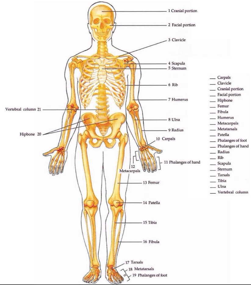
Know all the axial skeletal bones including their locations, grouping (such as cranial, facial, vertebral, etc), and only the following prominent features: sella turcica (hypophyseal fossa), parts of the sternum, true ribs vs. false ribs (vertebrosternal, vertebrochondral, floating), external auditory meatus (canal), crista galli, foramen magnum
8 cranial = frontal, parietal, temporal, occipital, sphenoid, ethmoid
14 facial = nasal, lacrimal, zygomatic, maxillary, palatine, inferior nasal conchae, vomer, mandible.
Hyoid bone - non-articulating (free-floating) bone in the neck
26 vertebral -
7 cervical: found in neck
12 thoracic: attached to ribs
5 lumbar: lower back area
1 sacrum (5 fused): pelvic cavity area
1 coccyx (4 fused): pelvic cavity area
12 pairs of ribs, connected by different types of cartilage connections
Sternum: flat bone, breastbone containing
Manubrium: suprasternal notch, 2 clavicular notches
Body: main part
Xiphoid: process
Sella turcica: located in the body of the sphenoid bone, forms hypophyseal fossa which supports and protects the pituitary gland.
True ribs vs false ribs:
Vertebrosternal: first 7 pairs of ribs are true ribs because attach directly to sternum with costal cartilage.
Vertebrochondral: ribs 8-10 are false ribs because connect to vertebrae and costal cartilage of rib 7.
Floating ribs: ribs 11-12 are false ribs and don't have any anterior attachment
External auditory meatus (canal): part of temporal bone in ear
Crista galli: part of ethmoid bone that sticks up and is an attachment for meninges
Foramen magnum: “large opening” through occipital bone for spinal cord to travel down
Know the 4 main sutures and the bones that form them. The fontanels and their locations
Coronal suture: located between frontal and parietal bone
Sagittal suture: located between parietal bones
Squamous suture: located between parietal and temporal bones on both sides
Lambdoid suture: located in back of head, looks like upside down triangle between occipital and parietal bones.
Fontanelles: soft parts on head when intramembranous ossification hasn’t completed. Allows for compression of head during labor. 6 of them:
Anterior: between parietal and frontal bones
Posterior: between parietal and occipital bones
Anterolateral (2): at junction of frontal, parietal, temporal, and sphenoid bones
Posterolateral (2): at junction of occipital, parietal and temporal bones
Know the 5 classifications of bones (long, short, flat, irregular, and sesamoid) plus sutural and an example.
Long: length is greater than its width
Eg: humorous, radius, ulna, meda carpal, phalanges, femur, shin, fibula, metatarsals
Short: length and width are approximately equal like
Eg: carpal (wrist) bones: scaphoid, lunate, triquetral, pisiform, trapezius, trapezoid, capitate, and hamate
Eg: ankle: Talus, calcaneus, navicular (scaphoid), medial, intermediate, lateral cuneiforms, cuboid
Flat: bone that has 2 parallel plates of compact bone with spongy bone in between
Eg: sternum, ribs, parietal bones, frontal bones
Irregular: not long, short, or flat
Eg: vertebrae, hip, facial bones
Sesamoid: specializes bones found in areas with there’s a lot of pressure an friction
Eg: patellae (knee caps), some have it in base of thumb or big toe
Sutral: little bones that develop in suture lines of skull
Know the main parts of typical vertebrae.
Body
Vertebral arches (2): made up of pendicles and lamina on left and right sides, they meet and come together at vertebral foramen where spinal cord is
7 processes: 2 transverse processes, 1 spinous process, 2 superior articulating processes, 2 inferior articulating processes
Structure and function of the intervertebral disc (IVD)
Structure: fibrocartilage, outer annulus fibrosis and inner nucleus pulposus which is egg-yolk material inside. Its like a jelly donut: firm on outside and soft on inside.
Function: cushion, shock absorber between vertebral bodies
Know the parts of atypical vertebrae (atlas and axis)
C1 (atlas): lacks body, lacks spinous process, has 2 lateral masses associated with it, anterior and posterior arch
C2 (axis): typical vertebrae with everything a regular one has but with a projection called “dens” that atlas pivots around
Know all the appendicular skeletal bones including location, grouping (upper or lower limb) and only the following prominent features: acetabulum, olecranon fossa, olecranon, glenoid fossa (cavity), greater trochanter, trochlear notch, medial and lateral malleoli, head of the radius
Appendicular(upper & lower limb) skeletal bones
Upper limb: clavicle, scapula, humerus, radius(lateral bone), ulna(medial bone), carpal, metacarpal, 14 phalanges bones(fingers),
Lower limb: hip bone, femur bone(thigh bone), patella(knee cap), tibia, fibula(lateral bone), 7 ankle bones: tarsal, talus, calcaneus(heel bone), navicular, 1st, 2nd, 3rd cuneiform, metatarsal, 14 phangage bones(toes).
KNOW THE GENERAL LOCATIONS OF ABOVE.
EX) Arm → Humerus
Forearm → Radius & Ulna
Leg → Tibia & Fibula
Thigh → Femur
Scaphoid & lunate → part of the carpal bone
Calcaneus → heel bone
Which of the following is not the part of the lower limb?
Acetabulum: lateral side of the hip bone where the head of the femur fits in
Olecranon fossa: distal humerus posterior surface, when we extend the forearm, the olecranon fits in together.
Olecranon: proximal end on the posterior surface of the ulna, tip of the elbow
Glenoid fossa(cavity): part of the scapula, where the head of the humerus fits in. Little shallow cup. socket.
Greater trochanter: proximal end of the femur. The muscles attached to it help the movement of the thigh.
Trochlear notch: associated with the ulna, proximal, fits to the trochlear at the distal end of the humerus where the hinge joint is.
Medial & lateral malleoli: Medial malleoli → distal tibia, Lateral malleoli → distal fibula. Malleoli → think of the ankle region, ankle joint, between the tibia, fibula, and talus all fit together.
Head of the radius: proximal end of the radius, it articulates with the capitulum of the humerus, the movement of the arm pronation & supination happen there
Know the 3 structural and 3 functional classifications of joints, including examples (including the characteristics of synovial joints)
3 structural classifications
Fibrous joint: lacks a joint cavity, bones are held together by fibrous connective tissues.
Examples of fibrous joints
Sutures of the skull
Gomphosis: where the tooth fits into the jaw bone
Syndesmosis: located on the distal end of the tibia and fibula
Cartilaginous joint: 2 types
Synchondrosis: no joint cavity. Bones meeting cartilage.
growth plate. bone → growth plate(cartilage) → bone
Ex) Rib → costal cartilage → sternum → costal cartilage
Symphysis: bones are joined by fibrocartilage, a cushion in between.
Ex) Pubic symphysis, intervertebral disc joints(cushion between the vertebrae)
Synovial joint: ①diarthrosis(freely moveable) // ②has joint cavity associated // ③ends of the bone is covered with articular cartilage // ④surrounded by the articular capsules // ⑤outer layer is fibrous, strong & dens irregular CT // ⑥inner layer is made up of synovial membrane which secretes synovial fluid(lubricates & nourishes the joints) // ⑦has ligaments(bind bone to bone) // ⑧some synovial joints have bursa (pl. bursae) which reduces frictions // ⑨meniscus(articular discs)
Functional classification
Synarthrosis: immobile joints
Amphiarthrosis: joints that allow for slight movement
Diarthrosis: Freely moveable, the most mobile joints, also known as synovial joints
What is the function of a ligament
Dense regular CT, binds bone to bone
Know the structure and function of a bursa (pl. bursae)
nickel to a quarter-sized small fluid sac, contains synovial fluid, reduces friction between the tendon & bone, ligament & bone, muscle & bone
Bursa injury - Bursitis
Know the difference types of movement (flexion, extension, adduction, abduction, circumduction, inversion, eversion) + examples of where they occur.
Flexion: decrease in angle between bone
Ex: forearm going closer to shoulder, fingers, neck when you bring chin to chest, arm at shoulder
Extension: increase in angle
Ex: forearm back into position, fingers back into place, toes, knees, hip
Adduction: adding something - moving body part to midline
Abduction: something being abducted - moving away from midline
Ex: shoulder, hip, fingers if middle finger is midline
Circumduction: movement of proximal bone in circle while proximal end remains stable
Ex: shoulder and hip
Inversion: movement of foot so sole of foot faces toward midline - special movement
Eversion: movement of foot so sole of foot faces away from midline - special movement
Ex: only happen at ankle area
Defintions: sprain, strain, spina bifida, cleft palate, scoliosis
Sprain: forcible wrenching or twisting of a joint that stretches or tears its ligaments but does not dislocate the bones. It occurs when the ligaments are stretched beyond their capacity (pg 290)
Strain: stretched or partially torn muscle or muscle and tendon. It often occurs when a muscle contracts suddenly and powerfully (pg 290).
Spina bifida: congenital (present from birth) defect of the vertebral column in which laminae of L5 or S1 fail to develop normally and unite at the midline (pg 229).
Cleft palate: the palatine processes of the maxillary bone fail to unite during weeks 10 to 12 of embryonic development. The condition may also involve incomplete fusion of the horizontal plates of the palatine bones. It often occurs together with a cleft lip (pg 208).
Scoliosis: the most common of the abnormal curves, is a lateral bending of the vertebral column, usually in the thoracic region. It may result from congenitally (present at birth) malformed vertebrae, chronic sciatica (pain in the lower back and lower limb), paralysis of muscles on one side of the vertebral column, poor posture, or one leg being shorter than the other (pg 229).
Know the functions of muscle tissue
Movement: all types - moving arms, blood through vessels, air through lungs, baby out of the womb
Postural movement: need muscles along back to stand upright
Storage of movement within organs: maintaining of organ volume, like muscles in urinary bladder can hold matter, stomach can hold food
Thermogenesis: creating heat, muscle contraction can produce most of body heat like when you work out, when you sleep your muscles don’t move so get colder
Know the 3 types of muscle tissue and their location
Skeletal: striated (striped), voluntary and controlled by mind, one end at least must be connected to skeleton
Cardiac: striated, involuntary, heart tissue
Smooth: non-striated, involuntary, lines respiratory and digestive and cardiovascular and urogenital system
Know the following parts of a muscle cell: sarcolemma, sarcoplasm, sarcoplasmic reticulum, sarcomere, thin and thick filaments, troponin-tropomyosin filaments.
Sarcolemma: muscle cell membrane (same as plasma membrane)
Phospholipid bilayer with proteins and cholesterol and glycoproteins
Sarcoplasm: cytoplasm in muscle cell
Sarcoplasmic reticulum: endoplasmic reticulum of muscle cell
Involved in calcium storage
Sarcomere: small compartment in muscle cell separated by z-discs
Thin and thick filaments: proteins that make up muscle cells.
Thin filaments are made up of actin
Thick filaments are made of myosin
Body-builders will eat protein which is digested and made into amino acids which muscle cells turn into actin and myosin
Troponin-tropomyosin filaments: protein in muscle that block binding sites between actin and myosin
Know the 3 CT coverings of a muscle
From smaller units to bigger units
Endomysium: CT that covers each muscle cell
Perimysium: CT that surrounds fascicle - bundle of muscle cell
Epimysium: CT that surrounds entire muscle
What is the neuromuscular junction?
The neuromuscular junction is functional contact between neuron and muscle. When we move our finger, our brain sends a neurotransmitter acetylcholine down the spinal cord to the nerve to the muscles. They don’t actually meet, the neurotransmitter goes through space.
4 sources of energy for muscle contraction. By-product of oxygen debt
ATP (adenine triphosphate): energy storage and release molecule. Within muscles of body, we store 8 seconds worth of ATP
CP (phosphocreatine or creatine phosphate): tapped into after ATP is worn out
Sugar: glucose or glycogen - storage of glucose
Fat: used once sugar is used up
Lactic acid is the by-product of oxygen debt. We need oxygen to get energy from all of these sources so if we don’t have, lactic acid stops the process and we fatigue.