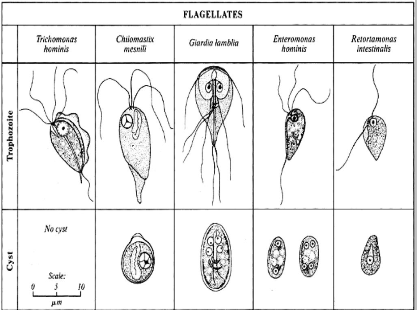
Flagellates-mastigophora
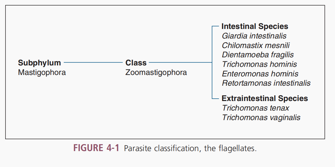
Phylum protozoa, Subphylum Mastigophora
2 groups
Intestinal flagellates
Atrial flagellates
Movement of flagellates
presence of whiplike structures known as flagella in trophozoite form
Life Cycle
Otrophozoite
similar to amebic trophozoites but with no known cyst stages; trophozoites are more resistant
appear to survive in the outside environment
Cysts
Encystation and Excystation
Location
small intestine
cecum
colon
duodenum (Giardia intestinalis)
Giardia intestinalis
Cercomonas intestinalis
Giardia duodenalis
1681
observed by Anthony van Leeuwenhoek
1859
Vilem Lambl observed in stool of children with diarrhea
Gr. Giard Czechslovakian scientist
1915
Stiles coined the term Giardia lamblia
multiplies by binary fission
DS
stool
MORPHOLOGY
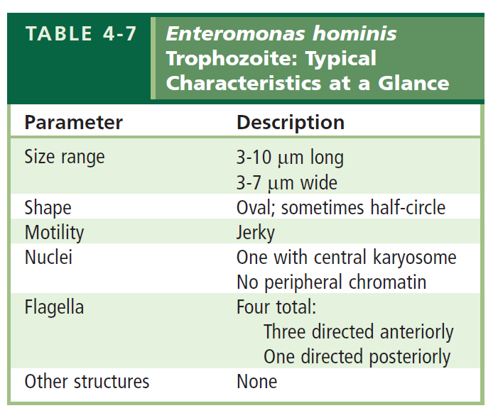
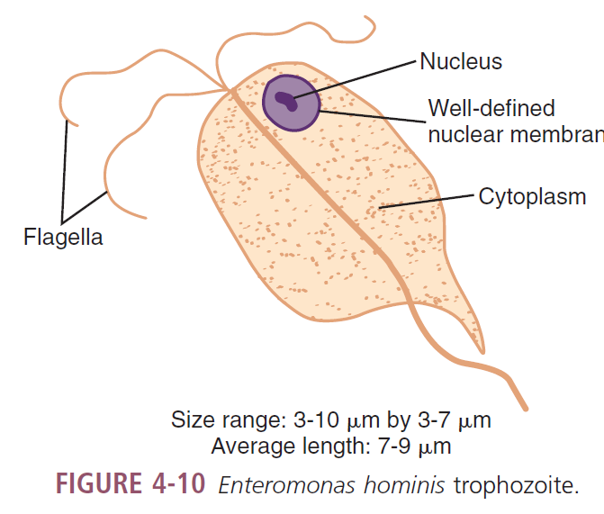
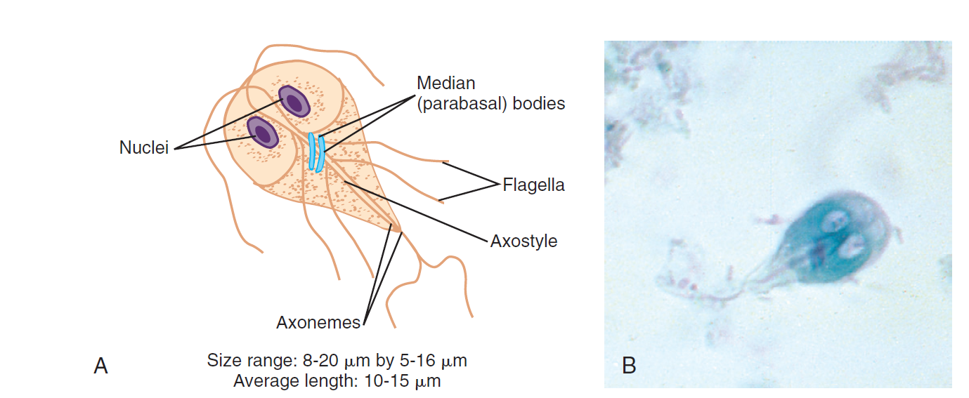
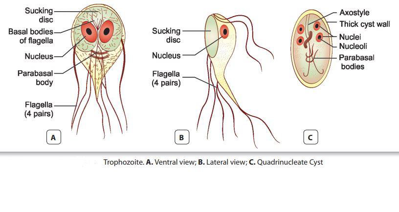
TROPHOZOITE
8-20 microns length; 5-16 microns width (10-15 microns long)
pear or teardrop-shaped
broad anterior end tapers off at the posterior end
Motility
falling leaf
bilaterally symmetrical
Nuclei
two ovoid to spherical
Karyosome
large, centrally located
best detected on permanently stained specimen
2 Axonemes
supports trophozoites as an axostyle— inner portions of the flagella
Median bodies
2 slightly curved rodlike structures
sit on the axonemes posterior to the nuclei
Flagella
difficult to detect
4 pairs
1 pair anterior end
1 pair posterior end
2 pairs laterally; axonemes extending
Sucking disc
50%-70% of the ventral surface
serves as nourishment point of entry
attaching to the intestinal villi of the infected human
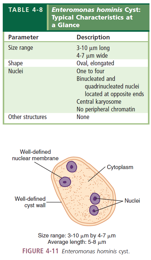
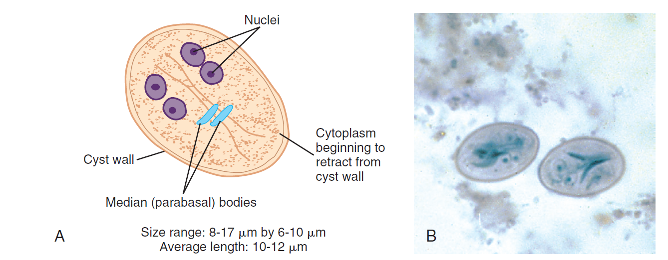
CYST
8-17microns long; 6-10 microns wide (10-12 microns)
cyst wall
colorless and smooth
prominent and distinct from the interior of the organism
Ovoid
Nuclei
Immature cyst
two
Mature cyst
four
Karyosomes
Central
Cytoplasm
retracted from cell wall
Median bodies/parabasal
immature
two
mature
four
Interior flagellar structures
twice as many in mature cyst compared with immature
 Life Cycle
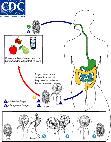
ingestion
infected cyst enter stomach
digestive juices (gastric acid)
stimulate cysts to excyst in duodenum
trophozoites in duodenum
becomes established and multiply every 8hrs.
longitudinal binary fission
feed
attaching sucking discs to the muscosa of the duodenum
may also infect common bile duct and gallbladder
Changes
result in unacceptable environment for multiplication stimulate encystation
trophozoites migrate to the large bowel
cysts
enter the outside environment via feces
remain viable for 3 months in water
Giardiasis
infection in small intestine
spreads through contact with infected people
Signs and Symptoms
Diarrhea
Gas
Greasy stools that tend to float
Stomach or abdominal cramps
Upset stomach/nausea
Dehydration (loss of fluids)
Treatment
accdng. to CDC
metronidazole
tinidazole
nitazoxanide
accdng. to FDA
metronidazole (not approved; carcinogenic in rats and mice)
tinidazole (approved; carcinogenic in rats and mice)
nitazoxanide
Lab Diagnosis
examine multiple samples
stool
duodenal contents by aspiration
ETEROTEST (string test)
upper small intestine (jejunum)
Concentration methods
Giardia western immunoblotting
Trichrome stain
Antigen detection tests by enzyme immunoassay
Wet mount
Direct fluorescent antibody (DFA)
Rapid immunochromatographic cartridge assays
Prevention and Control
practice good hygiene
avoid water that may be contaminated
avoid eating food that may be contaminated
prevent contact and contamination with feces during sex
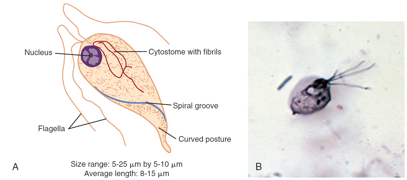
Chilomastix mesnili
found more frequently in warm climates
occur commensally in the intestine of various vertebrate
MORPHOLOGY
TROPHOZOITES
8-15 microns
pear-shaped
Motility
stiff
rotary
directional
Nuclei
one
Karyosome
small central or eccentric
Flagella
four
3 extending from anterior end
1 posteriorly from cytostome region
prominent cytosome
1/3 to ½ body length
spiral groove
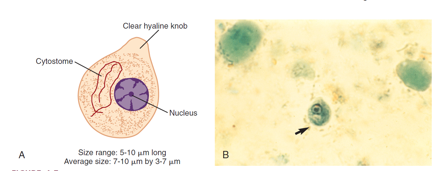
CYST
7-10 microns long; 2-7 microns width; 5-10 microns length
Nucleus
one
karyosome
large central
well-defined cytostome
w/ accompanying fibrils
found one side of the nucleus
Epidemiology
cosmopolitan in its distribution
warm climates
greatest risk reduction:
personal hygiene & poor sanitary
transmission
hand to mouth
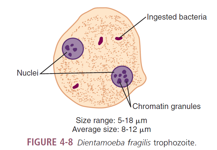
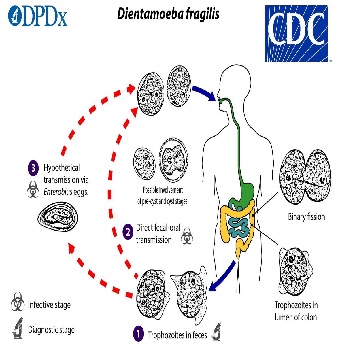
Diantamoeba fragilis
MORPHOLOGY
TROPHOZOITE
5-18 microns
irregularly round
Motility
progressive
broad hyaline pseudopodia
Nuclei
two
each consisting clumps of four to eight chromatin granules
Cytoplasm
bacteria-filled vacuoles common
Epidemiology
true MOT remains unknown
distributed in cosmopolitan areas
rarely collected and difficult to identify
IS
Trophozoite
MOT
via eggs of E. vermicularis or A. lumbricoides
fecal-oral
HABITAT
mucosal crypts of large intestine of man
symptomatic
mucus diarrhea
flatulence
fatigue
low-grade eosinophilia
pruritus
Lab Diagnosis
examination of fresh saline smear of stool
staining with iron hematoxylin is preferable
Conventional and real-time polymerase chain reaction (RT-PCR) methods
Treatment
iodoquinol
tetracycline (let)
paromomycin (humatin)
Prevention and Control
maintain personal and public sanitary conditions
avoidance of unprotected homosexual practices
Trichomonas hominis
non-pathogenic although it has been associated with diarrheic stools
most commonly found flagellate next to G. lamblia and D. fragilis
MORPHOLOGY
TROPHOZOITE
7-20 microns long; 5-18 microns wide
pear-shaped
Motility
nervous, jerky
Nuclei
one
karyosome
small central
Fllagella
three to five anterior
one posterior extending from the posterior end of the undulating membrane
axostyle
extends beyond the posterior end of the body
full body length undulating membrane
conical cytosome cleft (shepherd’s crook)
anterior region ventrally located opposite the undulating membrane
Lab Diagnosis
stool examination
flagellates
move very quickly in a jerky non-directional manner
hard to stain
axostyle and undulating membrane are diagnostic
axostyle
can be seen on a stained preparation
Life Cycle
trophozoites are shed in feces
fecal-oral route
resides in large intestine
MOT
ingestion of trophozoites
contaminated milk as one of the infectious causes
patients suffering from achlorydria act as shield of T. hominis trophozoites upon entry
fecal-oral
Trichomonas tenax
HABITAT
oral cavity
IS
use of contaminated dishes/utensils
IH
man
MORPHOLOGY
TROPHOZOITE
5-14 microns long
oval, pear-shaped
Nuclei
one
ovoid nucleus
vesicular region filled with chromatin granules
Flagella
five
all anteriorly
four anteriorly
one to posterior
undulating membrane extending two thirds of body length with accompanying costa
thick axostyle curves around nucleus
extends beyond body length
small anterior cytosome
opposite undulating membrane

Lab Diagnosis
examination of tonsillar crypts and pyorrheal pockets (mouth scrapings)
tartar bet. teeth and gingival margin of the gums
primary areas of the mouth that may harbor this
samples ay be cultured
Life Cycle
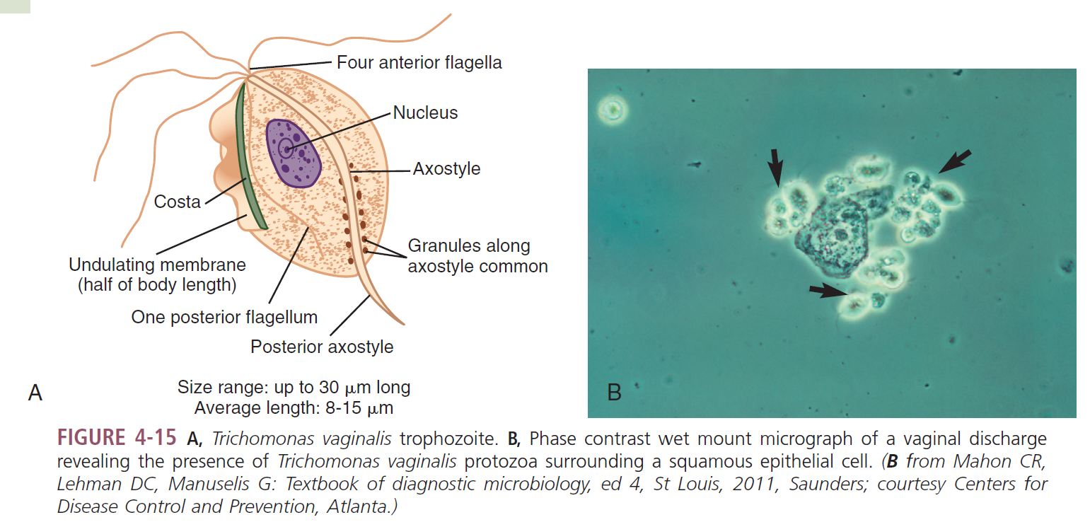
Trichomonas vaginalis
MORPHOLOGY
TROPHOZOITE
up to 30 microns long
ovoid, round or pear-shaped
Nuclei
one
ovoid
nondescript
Flagella
all originating anteriorly
three to five anteriorly
one to posterior
undulating membrane extending half of body length
prominent axostyle
often curves around nucleus
granules may be seen along
Lab Diagnosis
DNA-based assay
Affirm VPII (BD Diagnostics, Sparks, MD)
sensitivity and specificity is greater than standard methods
InPouch TV (Bio Med Diagnostics, White Cry, OR) culture system
vaginal swabs from women
urethral swabs from men and semen sediment
requires incubation time up to 3 days
Life Cycle
mucosal surface of the vagina
growing trophozoites
multiply by longitudinal binary fission
feed on local bacteria and leukocytes
thrive on alkine or slightly acidic pH
prostate gland region and epithelium of the urethra
unknown details
Epidemiology
worldwide
primary MOT
sexual intercourse
may migrate from mother’s birth canal and infect unborn child
rare transmission
via contaminated toilet articles or underclothing
sharing of douche supplies and communal bathing
trophozoites are known to survive
in urine, wet sponges, and on damp towels
several hours
in water
up to 40 minutes
Asymptomatic
most frequently occur in men
Persistent Urethritis
symptomatic men
enlarged tender prostate, dysuria, nocturia, epididymitis
patients often release a thin white urethral discharge
contains trophozoites
Persistent Vaginitis
foul-smelling
greenish-yellow liquid vaginal discharge
after incubation period of 4-28 days
burning, itching, ad chafing
Red punctate lesions
urethral involvement, dysuria, and increased frequency of urination
cystitis
less commonly observed but may occur
Infant infections
recovered from infants suffering from
respiratory infection
conjunctivitis
Treatment
metronidazole
treatment of all sexual partners is recommended
Prevention and Control
avoidance of unprotected sex
prompt diagnosis and treatment of asymptomatic men is essential
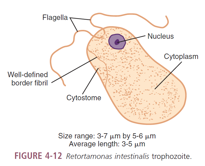
Retortamonas intestinalis
MORPHOLOGY
TROPHOZOITES
3-7 microns long; 5-6 microns wide
ovoid
Motility
jerky
Nuclei
one
karyosome
small central
ring of chromatin granules may be on the nuclear membrane
Fllagella
two; anterior
cytosome
extending halfway down body length
w/ well defined fibril border
opposite the nucleus in the anterior end
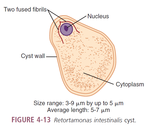
CYST
3-9 microns long; up to 5 microns wide
lemon-shaped, pear-shaped
Nucleus
one
located anterior canal central region
central karyosome
may be surrounded by a delicate ring of chromatin granules
two fused fibrils
resembling a bird’s beak in the anterior nuclear region
only visible in stained preps
Lab Diagnosis
best sample
stool
stained prep
difficulty of identification bc of small size
cyst and trophozoite in stool
Epidemiology
warm and temperate climates
MOT
ingestion of infected cyst
conditions to contract
poor sanitation and hygiene