WEEK 8 Lec- 2.1 Respiratory tract local immunity
Lecture Notes: Respiratory Tract Anatomy, Local Immunity, and Viral Infection
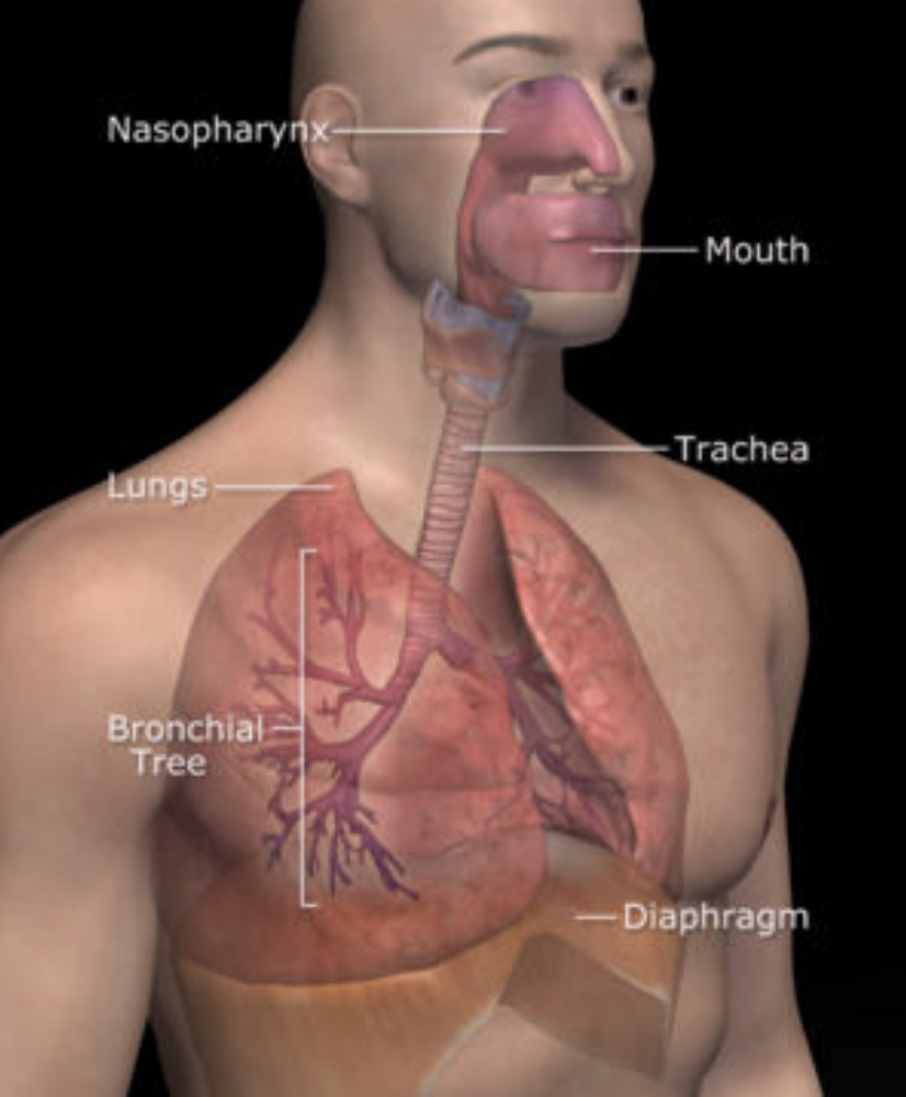
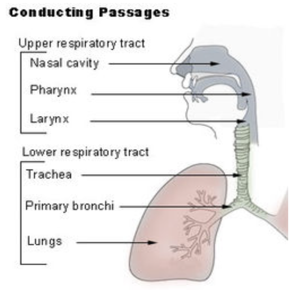
Page 2: Respiratory Tract Anatomy
Segments of the Respiratory Tract:
Upper Respiratory Tract:
Components: Nose, nasal passages, paranasal sinuses, throat (pharynx)
Respiratory Airways:
Components: Voice box (larynx), trachea, bronchi, bronchioles
Lungs:
Components: Respiratory bronchioles, alveolar ducts, alveolar sacs, alveoli
Page 3: Viral Respiratory Tract Infections
Commonality of Infections:
Upper respiratory tract infections are the most prevalent viral infections globally.
Typically result in mild irritation.
Lower Respiratory Tract Infections:
More severe and challenging to treat.
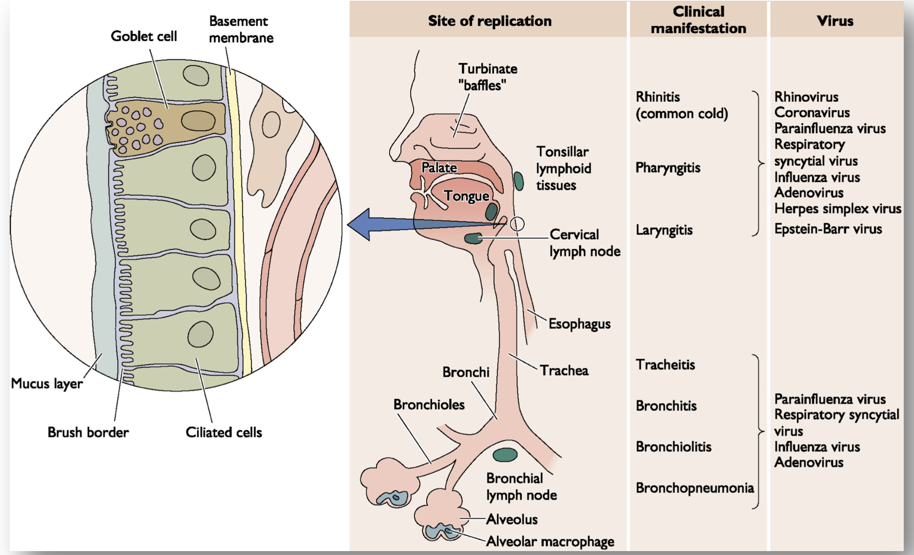
Page 4: Clinical Manifestations of Viral Infections
Infection Sites and Associated Viruses:
Goblet Cell Membrane:
Viruses: Rhinovirus (common cold), Coronavirus, Parainfluenza virus
Condition: Rhinitis
Tonsillar Lymphoid Tissues:
Viruses: Influenza virus, Adenovirus, Herpes simplex virus
Condition: Pharyngitis
Bronchi and Trachea:
Viruses: Parainfluenza virus
Condition: Tracheitis
Bronchioles:
Viruses: Respiratory syncytial virus, Influenza virus, Adenovirus
Condition: Bronchitis, Bronchiolitis
Alveolus:
Involvement of alveolar macrophages leading to Bronchopneumonia.
what differs between upper and lower respiratory infection is THE TYPE OF SECRETION
smaller aerosols and smaller droplets can make it all the way to lower areas of the lungs
however LARGE droplets most likely to be caught in nasal passage hence cause infection in URTI
WHAT ARE SOME EXAMPLES OF LARGE ANS SMALL DROPLETS IRL
In real life, examples of large and small droplets include:
Large Droplets: These can range in size from approximately 5 to 10 micrometers and larger. They are often produced from activities like coughing or sneezing. Common examples include:
Droplets expelled during a hard cough or sneeze.
Droplets from a talking person, particularly if they are shouting or projecting their voice.
Spit or saliva when a person speaks loudly or laughs.
Small Droplets: These are typically less than 5 micrometers in diameter. They can be created by aerosol-generating procedures or more subtle activities, and they can travel further into the respiratory tract. Examples include:
Aerosols produced from breathing or talking.
Fine mist from spray bottles (like disinfectants or perfumes).
Droplets generated by humidifiers or nebulizers when dispersing medication.
Understanding the differences between large and small droplets is crucial, especially regarding the transmission of respiratory infections.
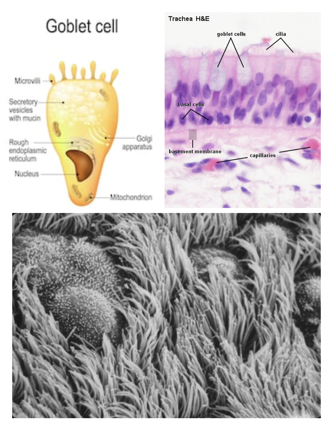
Page 5: Our respiratory tract – a tough barrier
Defense Mechanisms in the Respiratory Tract
Goblet Cells:
Function: Secrete mucin, which forms mucus when dissolved in water.
Mucus Role:
Mechanism: "Washes" away invading pathogens.
Ciliated Cells:
Function: Trap invading pathogens, enhancing respiratory defense.
ciliated cells “trap” invading pathogens
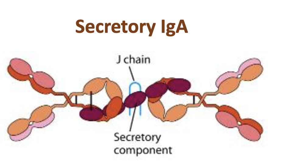
Page 6: Secretory IgA in Mucosal Immunity
Role of IgA:
First line of defense at epithelial surfaces.(mucosal immunity)
Daily production: Greater than any other immunoglobulin (5-15g/day).
IgA (Immunoglobulin A)
Type: Antibody
Function: Plays a crucial role in the immune system by protecting mucosal surfaces (like the gut, respiratory tract, and urogenital tract) from infections.
Location: Found in high concentrations in mucosal areas, as well as in saliva, tears, and breast milk.
Importance: Helps prevent pathogens from adhering to and penetrating epithelial cells, thus serving as a first line of defense against infections.
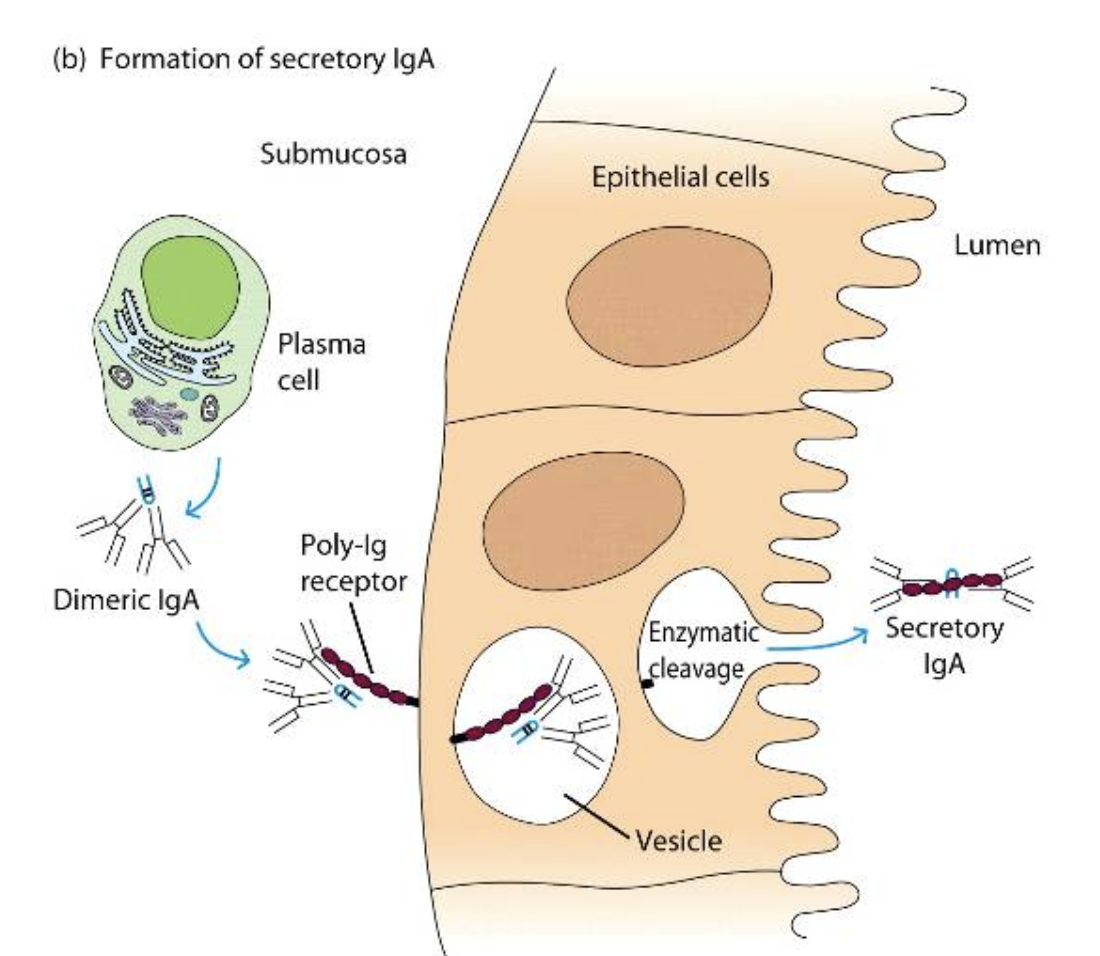
Page 7: Mechanism of Secretory IgA
Non-specific Hinderance:
IgA attaches to the mucous layer,covering epithelial cells creating a barrier against viral entry.
Virus Neutralization and Aggregation:
Promotes clearance through opsonization and complement activation.
Page 8: Learning Objectives
Understand the respiratory tract environment.
Explore local immune mechanisms in the respiratory tract.
Understanding the Respiratory Tract Environment
The respiratory tract is a complex system divided into several segments:
Upper Respiratory Tract:
Components: Nose, nasal passages, paranasal sinuses, throat (pharynx)
Respiratory Airways:
Components: Voice box (larynx), trachea, bronchi, bronchioles
Lungs:
Components: Respiratory bronchioles, alveolar ducts, alveolar sacs, alveoli
Defense Mechanisms in the Respiratory Tract:
Goblet Cells:
Function: Secrete mucin forming mucus when dissolved in water. Mucus "washes" away invading pathogens.
Ciliated Cells:
Function: Trap invading pathogens, enhancing respiratory defense.
Local Immune Mechanisms:
Mucociliary Escalator: Cilia and mucus trap and expel pathogens.
Alveolar Macrophages: Engulf and destroy inhaled pathogens.
Importance of Droplets:
Large Droplets (5-10 micrometers):
Typically caught in the nasal passage, causing upper respiratory infections. E.g., droplets from coughing or sneezing.
Small Droplets (<5 micrometers):
Can penetrate deeper into the lungs, potentially leading to lower respiratory infections. E.g., aerosolized particles from breathing or talking.
The respiratory tract's unique environment is vital for maintaining health and protecting against respiratory infections.
Local Immune Mechanisms in Body Niches
Skin:
Barrier Function: Physical barrier and antimicrobial peptides.
Langerhans Cells: Dendritic cells that capture and present antigens.
Respiratory Tract:
Mucociliary Escalator: Cilia and mucus trap and expel pathogens.
Alveolar Macrophages: Engulf and destroy inhaled pathogens.
Gastrointestinal Tract:
Acidic Environment: Stomach acid kills many pathogens.
Gut Microbiota: Competes with pathogens and produces antimicrobial substances.
Urogenital Tract:
Mucosal Immunity: Secretory IgA and antimicrobial peptides.
Normal Flora: Prevents colonization by pathogens.
Blood:
Circulating Immune Cells: Neutrophils, monocytes, and lymphocytes respond to infections.
Complement System: Enhances opsonization and lysis of pathogens.
Each niche has specialized mechanisms tailored to its unique environment and potential threats.
This note summarizes the key points from the lecture on respiratory tract anatomy, local immunity, and viral infections, providing a structured overview of the