Chapter 7: The Nervous System: Neurons and Synapses
Nervous System (NS)
Is divided into:
Central nervous system (CNS)
= brain & spinal cord
Peripheral nervous system (PNS)
= cranial & spinal nerves
Consists of 2 kinds of cells:
Neurons & supporting cells (= glial cells)
Neurons are functional units of NS.
Supporting cells maintain homeostasis.
Are 5X more common than neurons.
Neurons
Gather & transmit information by:
Responding to stimuli.
Sending electrochemical impulses.
Releasing chemical messages.
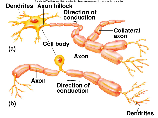
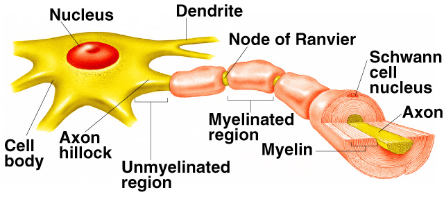
Have a cell body, dendrites, & axon.
Cell body contains nucleus.
Cell body makes macromolecules.
Groups of cell bodies in CNS are called nuclei; in PNS are called ganglia.
Dendrites receive information, convey it to cell body.
Axons conduct impulses away from cell body.
Axon length necessitates special transport systems:
Axoplasmic flow moves soluble compounds toward nerve endings.
Via rhythmic contractions of axon.
Axonal transport moves large & insoluble compounds bidirectionally.
Along microtubules; very fast.
Viruses & toxins enter CNS this way.
Functional Classification of Neurons
Sensory/Afferent neurons conduct impulses into CNS.
Motor/Efferent neurons carry impulses out of CNS.
Association/Interneurons integrate NS activity.
Located entirely inside CNS.
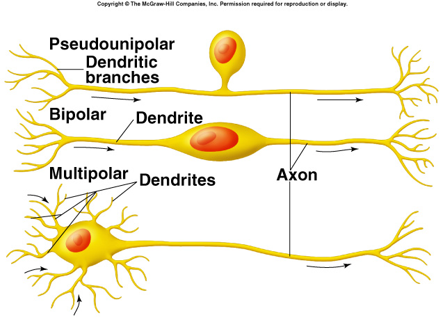
Structural Classification of Neurons
Pseudounipolar:
Cell body sits along side of single process.
e.g. sensory neurons
Bipolar:
Dendrite and axons arise from opposite ends of cell body.
e.g. retinal neurons/
Multipolar:
Have many dendrites and one axon.
e.g. motor neurons.
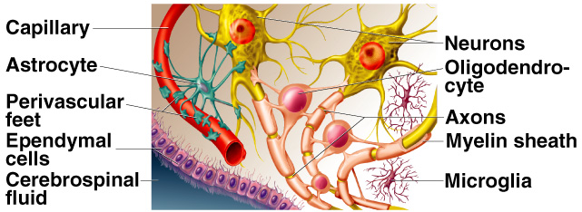
Supporting/Glial Cells
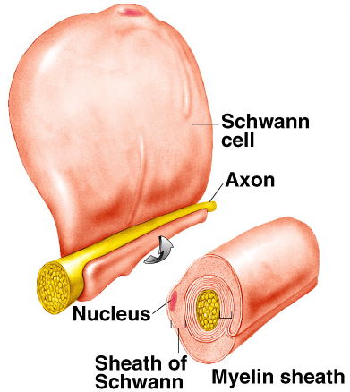
PNS has Schwann and satellite cells.
Schwann cells myelinate PNS axons.
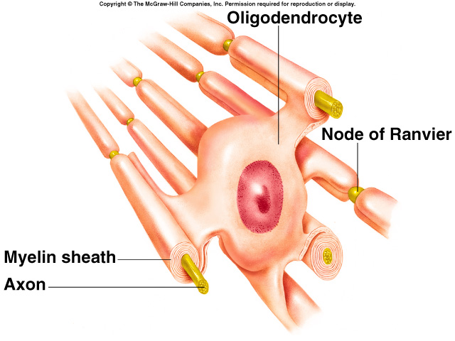
CNS has oligodendrocytes, microglia, astrocytes, and ependymal cells.
Each oligodendrocyte myelinates several CNS axons.
Ependymal cells are neural stem cells.
Other glial cells are involved in NS maintenance.
Myelination
In PNS each Schwann cell myelinates 1mm of 1 axon by wrapping round and round axon.
Electrically insulated axon.
Uninsulated gap between adjacent Schwann cells is called node of Ranvier.
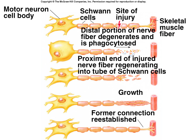
Nerve Regeneration
Occurs much more readily in PNS than CNS.
Oligodendrocytes produce proteins that inhibit regrowth.
Neurotrophins.
Promote fetal nerve growth.
Required for survival of many adult neurons.
Important in regeneration.
When axon in PNS is severed:
Distal part of axon degenerates.
Schwann cells survive; form regeneration tube.
Tube releases chemicals that attract growing axon.
Tube guides regrowing axon to synaptic site.
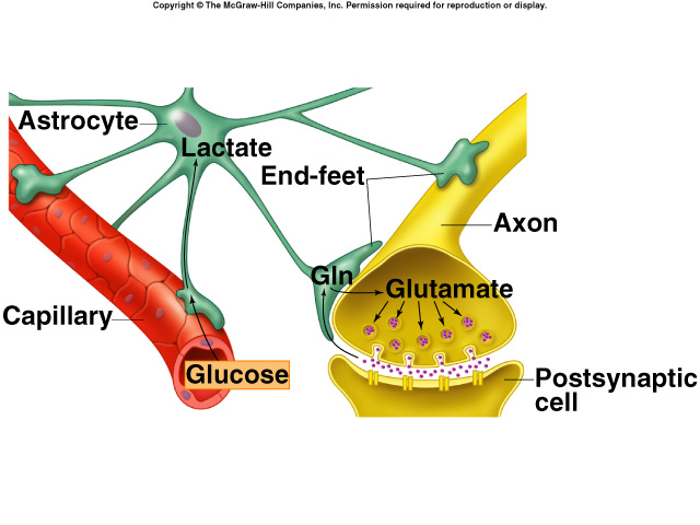
Astrocytes
Most common glial cell.
Involved in:
Inducing capillaries to form blood-brain barrier.
Buffering K+ levels.
recycling neurotransmitters.
Regulating adult neurogenesis.
Maintain interstitial fluid.
Blood-Brain Barrier
Allows only certain compounds to enter brain.
Formed by capillary specializations in brain.
Capillaries are not as leaky as those in body.
Do not have gaps between adjacent cells.
Closed by tight junctions.
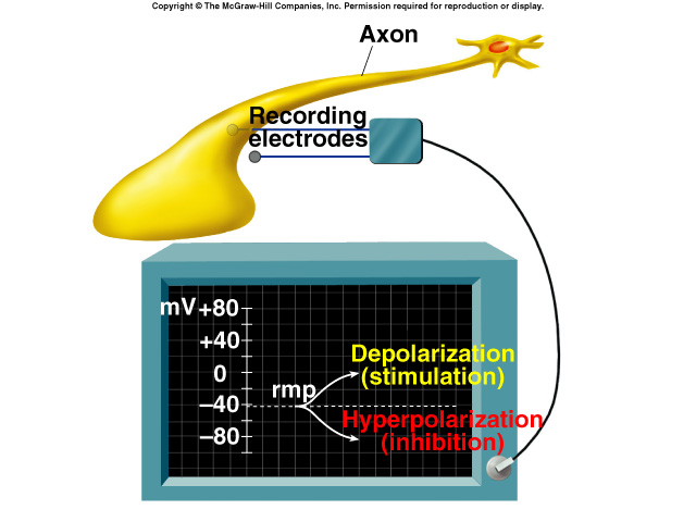
Resting Membrane Potential (RMP)
At rest, all cells have a negative internal charge and unequal distribution of ions:
Results from:
Large anions being trapped inside cell.
Na+/K+ pump and limited permeability keep Na+ high outside cell.
K+ is very permeable and is high inside cell.
Attracted by negative charges inside.
Excitability
Excitable cells can discharge their RMP quickly.
By rapid changes in permeability to ions.
Neurons and muscles do this to generate and conduct impulses.
Membrane Potential (MP) Changes
Measured by placing 1 electrode inside cell and 1 outside.
Depolarization occurs when MP becomes more positive.
Hyperpolarization: MP becomes more negative than RMP.
Repolarization: MP returns to RMP.
Membrane Ion Channels
MP changes occur by ion flow through membrane channels.
Some channels are normally open; some closed.
Closed channels have molecular gates that can be opened.
Voltage-gated (VG) channels are opened by depolarization.
1 type of K+ channel is always open; other type is VG and is closed in resting cell.
Na+ channels are VG; closed in resting cells.
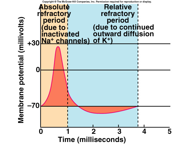
The Action Potential (AP)
Is a wave of MP change that sweeps along the axon from soma to synapse.
Wave is formed by rapid depolarization of the membrane by Na+ influx; followed by rapid repolarization by K+ efflux.
Depolarization causes more channels to open (positive feedback loop)
Mechanism of Action Potential
Depolarization and repolarization occur via diffusion.
Do not require active transport.
After an Ap, Na+/K+ pump extrudes Na+, recovers K+.
APs Are All-or-None
When MP reaches threshold, an AP is irreversibly fired.
Because positive feedback opens more and more Na+ channels.
Shortly after opening, Na+ channels close and become inactivated until repolarization.
Refractory Periods
Absolute refractory period:
Membrane cannot produce another AP because Na+ channels are inactivated.
Relative refractory period occurs when VG K+ channels are open, making it harder to depolarize to threshold.
Cable Properties
Refers to ability of axon to conduct current.
Axon cable properties are poor because:
Cytoplasm has high resistance.
Though resistance decreases as axon diameter increases.