Biology Notes - A1.2 Nucleic Acids
Genetic Material of Life
DNA is the genetic material of living organisms
Deoxyribonucleic acid (DNA) carries the genetic code in all living organisms.
This is the reason why the genetic code is said to be universal, it applies to all forms of life.
DNA is mainly found in the nucleus, where it forms chromosomes
It is also found in chloroplasts and mitochondria of eukaryotic cells
Ribonucleic acid (RNA) is another type of nucleic acid which is the main component of ribosomes, which play an important role in protein synthesis
Some RNA is also found in the nucleus and cytoplasm
Certain viruses (such as SARS-CoV-2) contain RNA as their genetic material instead of DNA
These viruses cause a variety of different diseases, such as COVID-19, Ebola, MERS, Mumps, and influenza
Viruses are not considered to be living organisms, since they are unable to replicate by themselves
They are dependent on other living cells for replication and survival
Viruses also lack a cellular structure, which is another reason they are not considered to be living
Nucleotide Components
Components of a nucleotide
Both DNA and RNA are polymers that are made up of many repeating units called nucleotides
Each nucleotide is formed from:
A pentose sugar (a sugar with 5 carbon atoms)
A nitrogen-containing organic base (with either 1 or 2 rings of atoms)
A phosphate group (this is acidic and negatively charged)
The base and phosphate group are both covalently bonded to the sugar
The nitrogenous bases in DNA are:
Adenine (A)
Guanine (G)
Cytosine (C)
Thymine (T)
share the same nitrogenous bases as DNA, except thymine, which is replaced by uracil (U) in RNA
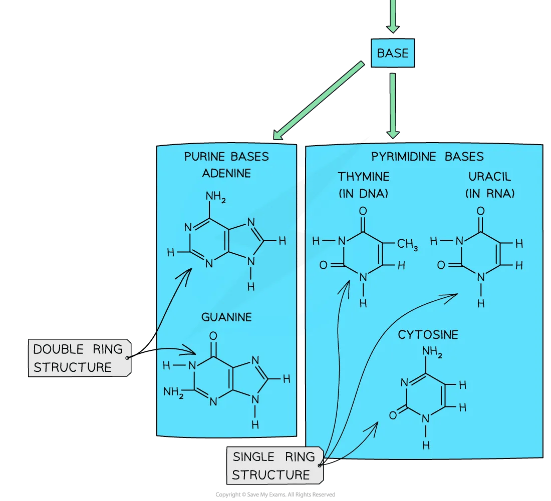
The nitrogenous bases can be grouped as either purine or pyrimidine bases:
Adenine and guanine are purine bases
Cytosine, thymine (in DNA) and uracil (in RNA) are pyrimidine bases
Nucleotide structure diagram

The basic structure of a nucleotide
Drawing simple diagrams of the structure of single nucleotides of DNA and RNA
Simple shapes can be used to draw the main building blocks of nucleotides and the DNA double helix
Advanced drawing skills are not required!
Pentagons can represent pentose sugars
Circles can represent phosphates
Often shown as a circle with the letter P inside: ℗
Rectangles can represent bases
Covalent bonds can be shown with solid lines
Hydrogen bonds can be shown with dashed lines
Or with complementary shapes that fit together (see diagrams)
Components of a nucleotide diagram

Simple shapes can be used to represent parts of nucleotide molecules
Linking Nucleotides
Forming the sugar-phosphate backbone
Nucleotides join together in chains to form DNA or RNA strands
The phosphate group of one nucleotide forms a covalent bond to the pentose sugar of the next one
This carries on to form a large polymer
These polymers form nucleic acids, which are also known as polynucleotides
The phosphate group of one nucleotide is linked to the pentose sugar of the next one by condensation reactions
This means a molecule of water is released during the formation of each covalent bond
This forms a 'sugar-phosphate backbone' with a base linked to each sugar
The polymer of nucleotides is known as a strand
DNA is double-stranded, and RNA is usually single-stranded
There are just 4 separate bases that can be joined in any combination/sequence
Because the sugar and phosphate are the same in every nucleotide
Linking nucleotides together diagram

Two nucleotides are shown bonded together covalently within a strand
RNA Structure
RNA structure
Unlike DNA, RNA molecules are relatively short, with lengths of between a hundred to a few thousand nucleotides
It usually forms a single-stranded polynucleotide with ribose as the pentose sugar in each nucleotide
RNA nucleotides contain the following nitrogenous bases:
Adenine
Guanine
Cytosine
Uracil (instead of thymine in DNA)
The carbon atoms in nucleotides are numbered from the right in a clockwise direction
This makes it easier to identify the bonds in the sugar-phosphate backbone of polynucleotides
It also indicates the orientation of the polynucleotide
RNA nucleotide diagram

The structure of an RNA nucleotide
Different types of RNA are found in the cells of living organisms:
messenger RNA (mRNA), which is formed in the nucleus and transported to the ribosomes in the cytoplasm
transfer RNA (tRNA), which is responsible for transporting amino acids to ribosomes during protein synthesis
ribosomal RNA (rRNA), which forms part of ribosomes
Adjacent RNA nucleotides are linked together by condensation reactions, during which a molecule of water is released.d
This forms a phosphodiester bond between the pentose sugar of one nucleotide and the phosphate group of the next nucleotide
The formation of an RNA polymer diagram

Linking RNA nucleotides together by condensation reactions will result in the formation of phosphodiester bonds.
DNA Structure
DNA structure
DNA is a double helix made of two antiparallel strands of nucleotides linked by hydrogen bonding between complementary base pairs
The nucleic acid DNA is a polynucleotide – it is made up of many nucleotides bonded together in a long chain.
DNA nucleotide diagram

A DNA nucleotide
DNA molecules are made up of two polynucleotide strands lying side by side, running in opposite directions – the strands are said to be antiparallel
Each DNA polynucleotide strand is made up of alternating deoxyribose sugars and phosphate groups bonded together to form the sugar-phosphate backbone.e
Each DNA polynucleotide strand is said to have a 3’ end and a 5’ end (these numbers relate to which carbon atom on the pentose sugar could be bonded with another nucleotide)
Because the strands run in opposite directions (they are antiparallel), one is known as the 5’ to 3’ strand and the other is known as the 3’ to 5’ strand.
The nitrogenous bases of each nucleotide project out from the backbone towards the interior of the double-stranded DNA molecule. cule
A single DNA polynucleotide strand diagram

A single DNA polynucleotide strand showing 3 nucleotides in a sequence
Hydrogen bonding
The two antiparallel DNA polynucleotide strands that make up the DNA molecule are held together by hydrogen bonds between the nitrogenous bases
These hydrogen bonds always occur between the same pairs of bases:
The purine adenine (A) always pairs with the pyrimidine thymine (T) – two hydrogen bonds are formed between these bases
The purine guanine (G) always pairs with the pyrimidine cytosine (C) – three hydrogen bonds are formed between these bases
This is known as complementary base pairing
These pairs are known as DNA base pairs
DNA molecule with hydrogen bonding diagram

A section of DNA – two antiparallel DNA polynucleotide strands held together by hydrogen bonds
Double helix
DNA is not two-dimensional as shown in the diagram above
DNA is described as a double helix
This refers to the three-dimensional shape that DNA molecules form
DNA double helix formation diagram

DNA molecules form a three-dimensional structure known as a DNA double helix
When drawing base-pairing in a DNA molecule, the opposite strand should be antiparallel to the when drawing the base pairing. The presence of hydrogen bonding is shown, but the numbers/lengths of bonds are not required.d
The Genetic Code
Genetic code
DNA molecules carry the genetic code as a sequence of nitrogenous bases in the nucleotides
These bases are adenine, guanine, cytosine, uracil, and thymine
One of the strands of a DNA molecule will carry the base sequence that will be read by enzymes
This strand is known as the coding strand
The sequence of bases that form genes on the coding strand will determine the order of amino acids in the proteins that are synthesised.
The code is read as a triplet of bases, called a codon, with each sequence of three bases coding for one amino acid.
Remember that 20 different amino acids could be coded for
The sequence of amino acids will determine the shape and function of the protein that is synthesised from the code
From gene to protein diagram

The sequence of DNA bases in the genes codes for the production of a specific protein molecule.
Conservation of The Genetic Code
The genetic code is universal
The genetic code is universal, meaning that almost every organism uses the same code (there are a few rare and minor exceptions)
The same triplet codes code for the same amino acids in all living things (meaning that genetic information is transferable between species)
The universal nature of the genetic code is why genetic engineering is possible.
This provides evidence for a universal common ancestor from which all living organisms on Earth evolved.d
Over time, mutations have led to changes in some of the base sequences of organisms.
These base sequences form the genome of an organism
Some base sequences form part of regions that code for proteins, called coding sequences, while others are located in regions that do not code for proteins (non-coding sequences)
Many of these coding and non-coding sequences have remained unchanged in all organisms and are known as conserved sequences
Highly conserved sequences are usually found in the genes that code for proteins involved with transcription and translation, as well as histone proteins, which help to package DNA tightly into the nucleus
The similarity in these sequences indicates that living organisms share a universal ancestry
DNA & RNA: Comparison
Differences between DNA and RNA
Unlike DNA, RNA nucleotides never contain the nitrogenous base thymine (T) – in place of this, they contain the nitrogenous base uracil (U)
Unlike DNA, RNA nucleotides contain the pentose sugar ribose (instead of deoxyribose)
Comparing DNA and RNA nucleotide diagrams

An RNA nucleotide compared with a DNA nucleotide
Unlike DNA, RNA molecules are only made up of one polynucleotide strand (they are single-stranded)
Unlike DNA, RNA polynucleotide chains are relatively short compared to DNA
RNA structure

mRNA is an example of the structure of an RNA molecule
Nucleotide Structure Summary Table
Properties | DNA | RNA |
|---|---|---|
Pentose sugar | Deoxyribose | Ribose |
Bases | Adenine (A) | Adenine (A) |
Number of strands | Double-stranded (double helix) | Single-stranded |
Complementary Base Pairing
The role of complementary base pairing
Complementary base pairing means that the DNA bases on different strands will always pair up in a very specific way:
Adenine (A) will pair up with Thymine (T)
Cytosine (C) will pair up with Guanine (G)
This is because the hydrogen bonds that hold the two DNA strands together can only form between these base pairs:
Two hydrogen bonds form between A and T
Three hydrogen bonds form between C and G
Complementary base pairing means that the base sequence on one DNA strand determines the sequence of the other strand.
We say that one strand acts as a template for the other
This allows DNA to be copied very precisely during DNA replication, which in turn ensures that the genetic code is accurately copied and expressed in newly formed cells
Complementary base pairs and hydrogen bonding diagram
A section of DNA showing nucleotide bonding and complementary base pair bonding
DNA: Information Storage Molecule
Diversity of DNA base sequences
Despite the genetic code only containing four bases (A, T, C, G), they can combine to form a very diverse range of DNA base sequences in DNA molecules of different lengths
This means that DNA has an almost limitless capacity for storing genetic information in living organisms
One way in which this storage capacity can be measured is by the number of genes contained within the DNA of an organism
Even the most simplistic forms of life may contain several thousand genes within their DNA
Comparing the Number of Genes between Different Organisms Table
Organism | Human | Dog | Water flea | Bacterium | Rice plant |
|---|---|---|---|---|---|
Approximate number of genes | 20 000 | 19 000 | 31 000 | 4 300 | 41 500 |
The storage capacity of DNA can also be measured in the number of base pairs contained within the genome of an organism. ism
The DNA in the nucleus of a human cell contains about 3.2 gigabases
That is about 109 DNA base pairs
These base pairs are contained in DNA with a length of about 2 metewhichthat fits within the nucleus of each human. cell
Given the fact that a nucleus is microscopic, it is an indication of how incredibly well packaged this amount of genetic information is
This gives DNA an enormous capacity for storing genetic 'data' with great economy.
Significance of Directionality
Directionality of RNA and DNA
When nucleotides are linked together to form nucleic acids, such as RNA and DNA, the phosphate groups form a bridge between carbon-3 of one sugar molecule and carbon-5 of the next one
This means that each polynucleotide strand has a 3' end where the OH group is located on carbon-3 of the sugar molecule and a 5' end containing the phosphate group on carbon-5
In a DNA molecule, one strand runs from 5' to 3' while the other strand runs from 3' to 5'
This is why the two strands are said to be antiparallel
The directionality of polynucleotide strands plays an important role in the processes of:
DNA replication
Transcription
Translation
During transcription, the genetic code on one of the DNA strands (the coding strand) is transcribed into a strand of mRNA
The coding strand is always read in the 3' to 5' direction by enzymes, which will synthesise the mRNA strand in the 5' to 3' direction.
The mRNA will move into the cytoplasm of the cell, where ribosomes will translate the transcribed code in the 5' to 3' direction.n
The base sequence of the genetic code will determine the specific order of the amino acids in the polypeptide chain created during translation.on
Directionality in RNA and Disare therefore crucially important to ensure that the genetic code is copied, transcribed, and translated correctly.
DNA Helix Structure
Purine to pyrimidine bonding in the DNA helix structure
Francis Crick and James Watson were two Cambridge scientists who worked together to establish the double helix structure of DNA in 1953
Through trial and error, they managed to build a model of the DNA double helix structure where the different base pairs fit together correctly
The base pairings A to T and C to G are equal in length, meaning that the DNA helix will have the same 3D structure regardless of the base sequence
Adenine (A) and guanine (G) are purine bases, while thymine (T) and cytosine (C) are pyrimidine bases
Purines are larger than pyrimidines due to their two-carbon ringed structure
The stability of the double helix is further increased by the hydrogen bonds that form between these complementary base pairs
Purines and pyrimidines diagram

The different sizes of purine and pyrimidine bases mean that they can only pair up in a very specific way. Note that you do not need to know the structural formulae of purines and pyrimidines
Nucleosomes
Unlike most prokaryotic DNA, which is referred to as ‘naked’, eukaryotic nuclear DNA is associated with proteins called histones (to form chromatin)
Histones package the DNA into structures called nucleosomes
The nucleosome consists of a strand of DNA coiled around a core of eight histone proteins (octamer) to form a bead-like structure
DNA takes two turns around the histone core and is held in place by an additional histone protein, which is attached to the linker DNA
The DNA molecule continues to be wound around a series of nucleosomes to form what looks like a ‘string of beads’
Nucleosomes help to supercoil the DNA, resulting in a compact structure that saves space within the nucleus
Nucleosomes also help to protect DNA and facilitate the movement of chromosomes during cell division
An analogy for supercoiling is twisting an elastic band repeatedly until it forms additional coils
Nucleosomes can be tagged with proteins to promote or suppress transcription
Nucleosome structure diagram

Structure of a nucleosome
Histones diagram

DNA is wrapped around a series of nucleosomes.
Nucleosomes coil tightly around each other to form the chromosome structure.
Skills: Molecular Visualisation Software
Molecular visualisation software can be used to help understand molecular structures.
Macromolecules like protein, DNA, RN, A, and complex carbohydrates can be visualised as 3-D structures
This allows researchers to analyse macromolecules and/or study interactions between them.
Primary sequence information can be related to structure and function
This helps to relate how structure might relate to chemical or biological behaviour
Macromolecules can be represented in many different ways,i ncluding ball and stick atom models or simplified ribbon representations that show the protein backbone
Most molecular visualisation software is freely available on the Internet or can be accessed through many bioinformatics repositories such as the Protein Data Bank (PDB)
Analysing the association between protein and DNA within a nucleosome
Visit the Protein Data Bank (PDB) site and search for: 6T79 structure of human nucleosome (do not put the search term in quotes)
Select the “3D view” to view the protein structure in Mol*
The 3-D structure of the nucleosome can be viewed
The DNA double helix can be seen surrounding the histone proteins
Rotate or zoom into the image to visualise the different components
The DNA can be seen to make two loops around the histone octamer core
Look carefully - the tails of each histone protein can be seen projected from the nucleosome core
These can be chemically modified to help regulate gene expression
Try changing different settings in the viewer or select a different viewer, such as JSmol
Human nucleosome diagram

Structure of human nucleosome showing the association between DNA (in 2 loops around the edge) and histones (central region) .
The Hershey & Chase Experiment
Which Biomolecule is the Heritable Material?
DNA was identified in 186,9, but many scientists assumed that protein was the heritable material because there are 20 amino acids and only 4 nucleotide bases
In the 1950s, Alfred Hershey and Martha Chase showed that DNA, not protein, is a factor of heredity responsible for carrying genetic information from one generation to another.
Viruses that infect bacteria were used in their experiment, as they only consist of DNA encapsulated by a protein coat.
This would allow the biomolecule of heredity (i,.e. the one that causes bacterial cells to be used to produce viral progeny) to be easily determined.
Analysis of the results of the Hershey and Chase experiment
Hershey and Chase took advantage of the chemical differences between DNA and proteins
DNA contains phosphorus but no sulfur
Amino acids (that make up proteins) contain sulfur but no phosphorus
Bacteria grown in separate media containing either radioactive sulfur (35S) or radioactive phosphorus (32P) were infected with viruses
The progeny viruses contained either 35S-labelled proteins or 32P-labelled DNA
Unlabelled bacteria were then infected separately with either type of virus
Bacteria would be expected to contain the heritable material following infection
A blender was used to remove attached viruses from the bacterial cells, and centrifugation was used to isolate the bacteria
Viruses are small ;they remained in the supernatant in the centrifuge tube
Bacteria are larger and form a pellet
Only the bacteria infected by 32P-labelled viruses (DNA) were shown to be radioactive.
This suggested that DNA (and not protein) was transferred to bacteria and is the hereditary (genetic) material
Hershey-Chase's experiment diagram


Hershey and Chase's experiment provided unequivocal proof that DNA is the heritable material.
NOS: Technological developments can open up new possibilities for experimentation- The availability of radioisotopes as research tools made the Hershey-Chase experiment possible
Radioisotopes were made available to scientists as research tools at the end of the Second World War.
This enabled scientists in a variety of research fields, such as biochemistry and virology, to do experiments that were not previously possible.e
Isotopes are particularly useful in studying chemical changes that occur during metabolic pathways or the life cycles of organisms.
Without the availability of radioisotopes, Hershey and Chase would not have been able to label the different parts of a virus to determine that DNA is the heritable material in organisms
Chargaff's Data
NOS: Addressing the problem of induction by the certainty of falsification - How Chargaff's data falsified the tetranucleotide hypothesis of DNA
Erwin Chargaff analysed the DNA composition of different organisms during the 1930s and 1940s and made the following discoveries:
The number of purine bases was equal to the number of pyrimidine bases
The number of adenine bases was equal to the number of thymine bases, while the number of guanine bases was equal to the number of cytosine bases
This means that a purine base can only pair up with a pyrimidine base between the sugar-phosphate backbone, since they have different sizes.
This forms the foundation of complementary base pairing in DNA
The problem of induction
The inductive scientific method starts with a scientist making observations and collecting raw data.
After data analysis, a hypothesis is formulated, which is then tested using a suitably designed investigation.
This may lead to some general conclusions being drawn based on specific observations.
Using data gathered in the past to create general predictions about what will happen in the future assumes that the future will be the same as when you gathered your data.
It is therefore impossible to prove a hypothesis generated by inductive reasoning as absolutely true, since we cannot be sure that the general observations we made in the past will hold in the future.
This is known as the problem of induction and is the main reason why most scientific theories are considered to be tentative.e
Even if several investigations support a hypothesis, it can still be proven incorrect (falsified) in the future as discoveries are made.
For this reason, the philosopher Karl Popper suggested that new scientific knowledge is not gained by inductive steps but rather by the falsification of existing hypotheses.s
Falsification of the tetranucleotide hypothesis
The biochemist Phoebus Levene discovered the pentose sugars of DNA and RNA in the early 1900s
He suggested that the structure of nucleic acid was a repeating tetramer unit, which he called a nucleotide.
This was called the tetranucleotide hypothesis
Tetranucleotide structure diagram

The tetranucleotide structure of nucleic acid, which was suggested by Levene
At the time of his research, there were limitations to the analytic techniques available, which made it difficult to determine the relative amounts of nucleotides present in nucleic acids.
The tetranucleotide hypothesis was falsified by Chargaff's data in the late 1940s, which showed the organism-specificity of nucleic acid.s
When the structure of DNA was determined in the 1950s, it further proved that the repeating tetramer unit suggested by Levene would not be suitable for carrying genetic information from one generation to the next.
Flashcard #1
Term: DNA
Definition: Deoxyribonucleic acid, the genetic material of living organisms.
Flashcard #2
Term: RNA
Definition: Ribonucleic acid, a type of nucleic acid involved in protein synthesis.
Flashcard #3
Term: Nucleotide
Definition: The basic building block of DNA and RNA, consisting of a sugar, base, and phosphate.
Flashcard #4
Term: Deoxyribose
Definition: The sugar component in DNA nucleotides.
Flashcard #5
Term: Ribose
Definition: The sugar component in RNA nucleotides.
Flashcard #6
Term: Adenine (A)
Definition: One of the four nitrogenous bases in DNA and RNA; pairs with thymine in DNA and uracil in RNA.
Flashcard #7
Term: Thymine (T)
Definition: One of the four nitrogenous bases in DNA; pairs with adenine.
Flashcard #8
Term: Cytosine (C)
Definition: One of the four nitrogenous bases in DNA and RNA; pairs with guanine.
Flashcard #9
Term: Guanine (G)
Definition: One of the four nitrogenous bases in DNA and RNA; pairs with cytosine.
Flashcard #10
Term: Uracil (U)
Definition: The nitrogenous base in RNA that replaces thymine found in DNA.
Flashcard #11
Term: Purines
Definition: A group of nitrogenous bases that includes adenine and guanine.
Flashcard #12
Term: Pyrimidines
Definition: A group of nitrogenous bases that includes cytosine, thymine (in DNA), and uracil (in RNA).
Flashcard #13
Term: Double helix
Definition: The structure of DNA consisting of two antiparallel strands twisted around each other.
Flashcard #14
Term: Antiparallel strands
Definition: The orientation of the two strands of DNA in opposite directions.
Flashcard #15
Term: Hydrogen bonding
Definition: The attraction between complementary nitrogenous base pairs that stabilizes the DNA structure.
Flashcard #16
Term: Complementary base pairing
Definition: The specific pairing of adenine with thymine and guanine with cytosine in DNA.
Flashcard #17
Term: Coding strand
Definition: The strand of DNA that carries the base sequence that will be read by enzymes.
Flashcard #18
Term: Codon
Definition: A sequence of three nitrogenous bases that codes for a specific amino acid.
Flashcard #19
Term: Mutation
Definition: A change in the base sequence of DNA that can lead to variations in proteins.
Flashcard #20
Term: Nucleosomes
Definition: Structures formed by DNA wrapped around histone proteins to package DNA in eukaryotic cells.
Flashcard #21
Term: Polymer
Definition: A large molecule composed of repeating subunits, such as nucleotides in DNA and RNA.
Flashcard #22
Term: Polynucleotide strand
Definition: A strand of nucleotides linked together to form DNA or RNA.
Flashcard #23
Term: Transcription
Definition: The process of copying the genetic code from DNA to mRNA.
Flashcard #24
Term: Translation
Definition: The process of synthesizing proteins from mRNA at the ribosome.
Flashcard #25
Term: Genetic code
Definition: The sequence of nitrogenous bases in DNA that determines the amino acid sequence in proteins.
Flashcard #26
Term: Universal genetic code
Definition: The theory that almost all organisms use the same genetic code.
Flashcard #27
Term: Gene
Definition: A segment of DNA that contains the instructions for synthesizing a specific protein.
Flashcard #28
Term: Histones
Definition: Proteins that help package DNA into nucleosomes in eukaryotic cells.
Flashcard #29
Term: Hereditary material
Definition: The substance that carries genetic information; proven to be DNA.
Flashcard #30
Term: Hershey & Chase experiment
Definition: An experiment that demonstrated that DNA, not protein, is the genetic material.
Flashcard #31
Term: Chargaff's rules
Definition: The observation that the amount of adenine equals thymine and guanine equals cytosine in DNA.
Flashcard #32
Term: Phosphodiester bond
Definition: The covalent bond formed between the phosphate group of one nucleotide and the sugar of the next.
Flashcard #33
Term: Sugar-phosphate backbone
Definition: The alternating chain of sugar and phosphate groups that forms the structural framework of DNA and RNA.
Flashcard #34
Term: Replication
Definition: The process of copying DNA prior to cell division.
Flashcard #35
Term: RNA types
Definition: Different types of RNA including mRNA, tRNA, and rRNA, each with unique functions.
Flashcard #36
Term: DNA packaging
Definition: The process by which DNA is condensed to fit within the cell nucleus.
Flashcard #37
Term: Mitochondria
Definition: Organelles in eukaryotic cells that contain DNA and are involved in energy production.
Flashcard #38
Term: Chloroplasts
Definition: Organelles in plant cells that contain DNA and are involved in photosynthesis.
Flashcard #39
Term: Virus
Definition: A non-living entity that contains RNA or DNA and requires a host cell for replication.
Flashcard #40
Term: Bioinformatics
Definition: The use of software and algorithms to analyze biological data, including DNA sequences.
Flashcard #41
Term: Nucleotide structure
Definition: A nucleotide consists of a pentose sugar, a nitrogenous base, and a phosphate group.
Flashcard #42
Term: Single-stranded RNA
Definition: RNA molecules that are typically single-stranded and used for protein synthesis.
Flashcard #43
Term: DNA's information storage
Definition: DNA can store vast amounts of genetic information due to its diverse base sequences.
Flashcard #44
Term: Conserved sequences
Definition: Genetic sequences that remain unchanged across different species, indicating common ancestry.
Flashcard #45
Term: Directionality of DNA
Definition: DNA strands have a directionality (5' to 3') that is crucial for replication and transcription.
Flashcard #46
Term: SARS-CoV-2
Definition: A virus that contains RNA as its genetic material and causes COVID-19.
Flashcard #47
Term: Eukaryotic cell
Definition: A type of cell that contains a nucleus and membrane-bound organelles, including DNA.
Flashcard #48
Term: Prokaryotic DNA
Definition: DNA that is naked, lacking histones, typically found in bacteria.
Flashcard #49
Term: Nucleotide representation
Definition: In diagrams, nucleotides can be represented using simple shapes: circles for phosphates, pentagons for sugars, and rectangles for bases.
Flashcard #50
Term: Protein synthesis
Definition: The process through which cells create proteins based on the genetic code.
Flashcard #51
Term: RNA's role in protein synthesis
Definition: mRNA carries the code from DNA to the ribosomes, where proteins are assembled.
Flashcard #52
Term: Gene expression regulation
Definition: The process where certain genes are turned on or off during cellular activity.
Flashcard #53
Term: Replication fork
Definition: The area where the strands of DNA are separated for replication.
Flashcard #54
Term: Genetic engineering
Definition: The manipulation of an organism's genome using biotechnology.
Flashcard #55
Term: Watson and Crick
Definition: The scientists who proposed the double helix model of DNA in 1953.
Flashcard #56
Term: Histone modification
Definition: Chemical changes to histones that can affect gene expression.
Flashcard #57
Term: Nucleotide comparison
Definition: DNA nucleotides differ from RNA nucleotides primarily in their sugar and one nitrogenous base.
Flashcard #58
Term: Octamer
Definition: The core of eight histone proteins around which DNA is wrapped in a nucleosome.
Flashcard #59
Term: Gene annotations
Definition: Tools used in bioinformatics to describe the function of genes and their products.
Flashcard #60
Term: Nuclear DNA
Definition: DNA located within the nucleus of eukaryotic cells, containing the majority of an organism’s genetic information.
Flashcard #61
Term: Base sequence
Definition: The order of nucleotides in a DNA or RNA strand that determines genetic