Module 3 BioPsych
1. Dorsal - toward the back, away from ventral
2. Ventral - toward the stomach, away from dorsal
3. Anterior - towards the front end
4. Posterior - towards the rear end
5. Superior - above another part
6. Inferior - below another part
7. Lateral - toward the side, away from the mid-line
8. Medial - toward the mid-line, away from the side
9. Proximal - located close to the point of origin / attachment
10. Distal - located more distant from point of origin / attachment
11. Ipsilateral - on the same side of the body (e.g. 2 parts on the
right)
12. Contralateral - on the opposite side of the body (one on the left
and one on the right)
13. Coronal plane - shows brain structures as seen from the front
14. Sagittal plane - shows brain structures as seen from the side
15. Horizontal plane - shows brain structures as seen from above (or
transverse plane)
1. Lamina - a row or layer of cell bodies separated from other cell
bodies by a layer of axons and dendrites
2. Tract (projection) - a set of axons within the CNS
3. Nerve - a set of axons in the periphery or peripheral nervous
system (PNS) (either from CNS to a muscle or from sensory organ to
CNS)
4. Nucleus - a cluster of neuron cell bodies within the CNS
5. Ganglion (ganglia) - a cluster of neuron cell bodies, usually outside
the CNS
6. Gyrus (gyri) - a protuberance on the surface of the brain
7. Sulcus (sulci) - a fold / groove the separates one gyrus from
another
8. Fissure - a long deep sulcus
The Central Nervous System (CNS) is composed of the brain & spinal cord
The Peripheral Nervous System (PNS) consists of the nerves outside the brain and spinal cord
The Somatic Nervous System consists of the axons conveying messages from the sense organs to the CNS and from the CNS to the muscles. It controls voluntary activities of the muscles.
The Autonomic Nervous System controls the involuntary activities of the organs (heart, intestines & other organs) and glands.
The Peripheral Nervous System is composed of the somatic and autonomic NS.
The Somatic Nervous System carries messages from the CNS to the skeletal muscles that control movements of the
body ( voluntary )
The Autonomic Nervous System is largely concerned with involuntary functions such as respiration, circulation and digestion. The ANS plays a vital role in emotion and physiological responses of the body (headache, diarrhea, stomach ache when anxious). Prolonged emotional arousal
can adversely affect the true health of the organs controlled by the autonomic nervous system
The Autonomic Nervous system has
a sympathetic and parasympathetic functions.
The Sympathetic nervous system has an activation or arousal function, and it tends to act as a total unit. It mobilizes the body and controls what we sometimes call the "fight-or-flight response"
The Parasympathetic nervous system slows down body processes and maintains a state of tranquility.
By working together, the 2 subdivisions can maintain homeostasis – a delicately balanced or constant internal state
The Central Nervous System (CNS) consists of the brain and spinal cord.
The Spinal Cord
The spinal cord is the part of the CNS within the spinal column.
It communicates with all of the sense organs and muscles except those of the head. It is a segmented structure and each segment has on each side a sensory and motor nerve.
Main Parts of the Spinal Cord
The entering dorsal roots (axon bundles) carry sensory information, and the exiting ventral roots carry motor information. This is called the Bell Magendie Law .
The axons from the skin and muscles are the peripheral nervous system (PNS).
The cell bodies of the sensory neurons are in a cluster of neurons outside the spinal cord is called the dorsal root ganglia.
The H shaped gray matter in the center of the cord is densely packed with cell bodies and dendrites.
Many neurons of the spinal cord send axons from the gray matter to the brain or other parts of the spinal cord through the white matter which consists mostly of myelinated axons.
The Hindbrain is the lowest portion of the brain where the spinal cord rises to meet the brain. The hindbrain is also sometimes called the rhombencephalon (parallel brain), myelencephalon (marrow brain), and metencephalon (after brain). Important structures of the hindbrain include the cerebellum, pons and medulla.
The Midbrain is the middle of the brain, surrounded & dwarfed by the forebrain. It is also sometimes referred to as the mesencephalon. The main parts of the midbrain include the tectum, tegmentum, and substantia nigra.
The forebrain is the topmost structure of the brain whereas higher cognitive processes take place. It is sometimes referred to as the telencephalon.
The outer portion of the forebrain is the cerebral cortex, also called the cerebrum. It is divided in two hemispheres connected by the corpus callossum, and the four lobes: frontal, temporal, parietal and occipital.
Other important structures of the forebrain include the thalamus and hypothalamus (diencephalon), basal ganglia, limbic system (amygdala and hippocampus) and the ventricles
It is the lowest portion of the brain where the spinal cord rises to meet the brain
The hindbrain is also sometimes called the rhombencephalon (parallel brain), myelencephalon (marrow brain), and
metencephalon (after brain).
The posterior part of the brain consists of the medulla, pons and cerebellum.
The medulla, pons, midbrain & certain central structures of the forebrain make up the brain stem.
The medulla helps to control vital reflexes including breathing, heart rate, vomiting, salivation, coughing and
sneezing through the cranial nerves (which controls sensation & movement from the head).
The pons lie anterior and ventral to the medulla. it contains nuclei of some cranial nerves, “bridge” axons in the pons cross
from 1 side of the brain to the other.
The cerebellum's function is involved in movement, motor coordination and balance, as well as processing visual & auditory stimuli
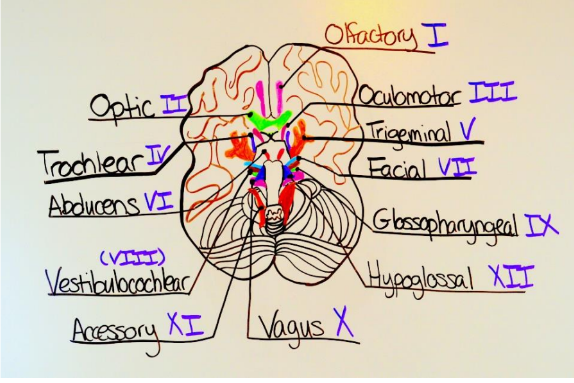
The cranial nerves
I. Olfactory (smell)
II. Optic (vision)
III.Oculomotor (eye movements/ pupil constriction)
IV. Trochlear (eye movements)
V. Trigeminal (skin sensations from the face
VI. Abducens (eye movements)
VII. Facial (taste from anterior 2/3 of tongue, facial expression,
salivation
VIII. Statoacoustic (hearing / equilibrium)
IX. Glossopharyngeal (taste from posterior 1/3 of tongue, swallowing, throat movements during speech
X. Vagus (sensations from neck, control of throat, esophagus, larynx, parasym. nerves of stomach/intestines
XI. Accessory (neck & shoulder movements)
XII. Hypoglossal (control of muscles of the tongue)
The midbrain is the middle of the brain, surrounded & dwarfed by the forebrain
It is also sometimes referred to as
the mesencephalon.
Main parts of the midbrain include the tectum, tegmentum, and substantia nigra.

The Tectum is the roof of the midbrain. The swelling on each side of the tectum are called superior colliculus which process
information for vision and the inferior colliculus which process information for hearing (audition).
The Tegmentum is the intermediate level of the midbrain that covers several other midbrain structures. It contains nucleus
for III & IV. cranial nerves
The Substantia nigra gives rise to the dopamine containing a pathway that facilitates readiness for movement
The forebrain is the top most structure of the brain wherein higher cognitive processes take place.
It is sometimes referred to as the telencephalon. The outer portion of the forebrain is the cerebral cortex, also
called the cerebrum. Under it are other structures like the thalamus and hypothalamus (aka diencephalon).
The Thalamus is where most sensory input is received, then sends it to the cerebral cortex.
The Hypothalamus is a small area at the base of the brain ventral to thalamus. It conveys a message to the pituitary gland, controls feeding, drinking, temperature regulation and sexual behavior.
The forebrain also houses the Basal Ganglia - a group of subcortical structures lateral to the thalamus. It includes three major structures: caudate nucleus, putamen, globus pallidus. In conditions like Parkinson's & Huntington's disease, the basal ganglia deteriorates which result in impaired movement, depression, deficits of memory, reasoning and attention.
The CNS begins its development as a tube surrounding a fluid canal. The canal persists into adulthood as the central canal- a fluid filled channel at the center of the spinal cord, and as the ventricles (4 fluid filled cavities within the brain).
Cells called choroid plexus inside the 4 ventricles produce cerebrospinal fluid (CSF), a clear fluid similar to blood plasma.
Cerebrospinal fluid fills the ventricles, and some of it flows into the central canal of the spinal cord. Most of the CSF goes into the narrow between the brain and thin meninges (membranes that surround the brain & spinal cord)
Meningitis is a condition characterized by inflammation of the meninges. It is very painful because of presence of pain
receptors at this area. CSF cushions the brain and provides buoyancy.
Hydrocephalus is a condition that happens when the flow of CSF is obstructed, CSF is accumulated within the ventricles increasing pressure on the brain, causing the spread of skull bones.
The cells of the cerebral cortex are gray matter, and their axons extending inward are the white matter. Neurons in each hemisphere communicate with neurons in the corresponding part of the other hemisphere through a bundle of axons called corpus callosum.
The Cerebral Cortex is where the highest mental functions, such as thinking and planning, take place. It is divided into
two hemispheres, a wrinkled surface of the cerebral cortex each hemisphere is subdivided into four regions or four lobes.
The Frontal lobe
The frontal lobe contains the primary motor cortex involved in the control of movements and the prefrontal cortex which receives information from all the sensory system and plays a role in working memory.
Further this lobe is associated to planning, judgment, adapting to new situations, flexibility of behavior and personality.
Prefrontal lobotomies (perfromed in the 1940-50s) is done by cutting the
prefrontal cortex from other parts of the cortex. It resulted in tamer behaviors, apathy, loss of ability to plan and/or take initiative, memory disorders, distractibility and loss of emotional expression.
The Temporal Lobe
The temporal lobe is located on the lateral portion of each hemisphere. It processes auditory information and complex visual
information (such as the perception of movement, recognition of faces)
The Wernicke's area (on the left temporal lobe) is responsible for understanding spoken language - a tumor in a temporal lobe may cause elaborate visual & auditory hallucinations - plays a role in emotional and motivational behaviors.
The Parietal Lobe
The parietal lobe lies between the occipital lobe & central sulcus. It contains the primary somatosensory cortex which processes touch sensations. This lobe is involved in registering spatial location, attention and motor control.
The Occipital Lobe
The occipital lobe is located at the posterior end of the cortex and it contains the primary visual cortex. A stroke or wound in this area can cause cortical blindness.