Muscle Physiology
SKELETAL MUSCLE
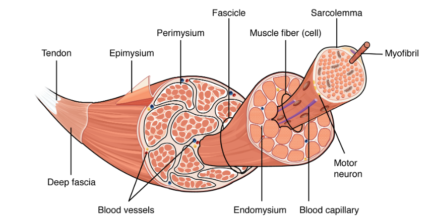
bundles called “fascicles”, fascicles have bundles called “fibers”, and fibers have muscle cells called “myofibrils”
Muscle strength based on the number of myofibrils and sarcomeres
BIG PARTS TO SMALL PARTS
Deep fascia: connective tissue that surrounds muscle groups with common actions
Epimysium “Above muscle”: sheet that separates individual muscles (ex. Italian sausage casing)
Perimysium “around muscle”: around fascicles and divide muscles into parts
Endomysium “Inside muscle”: separate fascicles into fibers
Sarcomeres “flesh part”: basic functional unit of muscle, contract for muscle contraction
electrical impulses cause them to shorten
10K sarcomeres: 1 myofibril
sarcolemma: specialized endomysium layer that surrounds myofibrils, connects to them through T tubules
separates sarcoplasm from interstitial fluids
Sarcoplasmic reticulum: bunch up arounf the T tubule, web of myofibrils, T tubles pinch the SR
ZONES: A , Z disk/line, M line, density of filaments give striations (darker bands = myosin and actin) (lighter bands (actin OR myosin)
Conservability of Muscles
Contractility: shortening muscle cells
Excitability: responsiveness
Extensibility: lengthening of muscle cells
Elasticity: ability to bring muscle back to OG length
Sarcomere movement
myofilaments slide to shorten muscle (contraction), fast process
Muscle Synapses: chemical and electrical synapses (chemical ones cause electrical ones to be produced)
chemical synapse: abundant, relies on neurotransmitters
electrical synapses: rare, locked together with gap junctions
Plasma membranes in neurons and muscle cells are excitable
can carry electrical currents
resting potential -60mV and -90mV
Sarcomere Contraction
watch a video since you were late this day
Sarcomeres shorten and pull on the things next to them (ec. muscles, tendon)
called “muscle tension”
muscles are not regulated by the body
VIDEO NOTES
Sarcomere regions:
A bands- dark bands
H zone: in the A band, split by M line
I band- light bands
Z disc
Myosin: thick filament, extend across A band, connect at M line
Actin: thin filament, extends across I band into A band, subunits have active sites where myosin can bind, tropomyosin (spiral strands) blocks these sites
elastic filaments containing titin
Myosin protein: globular ATP/actin binding sites, makes cross bridges
Troponin: has 3 peptides
binds actin
binds tropomyosin
binds calcium
Sarcoplasmic reticulum: tubules around a myofibril, regulate calcium (aids muscle contraction) through storage and secretion
T-tubules: sit where A and I bands meet, help signals reach all portions of muscle cell
SLIDING FILAMENT MODEL
Nervous system stimulates muscle fibers
myosin heads (thick fil.) bind with actin filaments (thin fil.)
myosin head pulls actin filaments to center of sarcomere, shortening it
Z discs move to M line
I bands shorten
H band disappears
A bands get closer
NEUROMUSCULAR JUNCTION
Axon terminals: area where motor neuron meets the muscle
Synaptic Cleft: space where junctional folds form on postsynaptic membrane, accepts acetylcholine with receptors
Sodium Potassium pump\
PHASES OF TWITCH
Latent Twitch: beginning of contraction, from 0s to time contraction begins
Contraction Phase: tension increases, myosin-actin bridges form
Relaxation phase: Ca2+ levels drop, tension decreases, myosin-actin bridges detach
Faster in an eye muscle than a calf muscle
TENSION TYPES
Peak Tension: most amount of tension, regulated by motor unit + cell bodies
Treppe: Muscles slowly increase tension over 30-50 muscle stimulations, doesn’t hit 0, “staircase” in German
Wave Summation: twitches do not cause relaxation and increase in intensity over time
Incomplete Tetanus: near peak tension, rapid cycles of relaxation and contraction, “holding up heavy objects”
Complete Tetanus: complete tension from constant stimulation, no relaxation bc bridge cannot relax; bacterial toxins that cause tetanus exhibits similar effects
Recuruitment: exciting other motot units, smooth and steady increase in tension, max #of motor units uased during tetanus, short time frame, make less than max tension, motoor units relax in rotation
CARDIAC MUSCLE
intercalated discs: contain gap junctions: a channel that conducts current between cells
desmosomes: anchors cells during contractions
SMOOTH MUSCLE
generally in arteries and GI tract
cells divde fast
Vasocosities
Phasic Contraction: rapid
Tonic Contraction: slow, controlled contraction and relaxation
Stress-Relaxation: mechanical stress causes contraction, immediately followed by relaxation
SPINAL CORD REFLEXES
Reflex: rapid, automatic responses to stimuli, involves dorsal root ganglion, ignore brain
Muscle stretch: one side stretches, other side is inhibited
flexor reflex
crossed extension reflex: simultaneous, opposing activity to maintain posture
Pain » dorsal root ganglia» spinal cord» Interneuron relays signals »effector muscles
SAME DAVE (Sensory/Dorsal, Afferent, Ventral, Efferent)
Main Classifications of Muscle Contraction
isometric: muscle doesn’t change length and makes enough tension to remain still, many muscles do this (holding weight)
Isotonic: change the length but keep constant tension (actually exercising)
concentric: muscles shorten and tension increases to exceed the force of gravity (to be able to lift weight and hold it)
eccentric: lengthening muscles and decreasing tension (to be able to lower the weight and hold it)
Slow Oxidative Muscles: contract slowly, use aerobic repiration to make ATP, red, high fatigue resistance (endurance)
Fast Oxidative Muscles: contract quickly, aerobic respiration to make ATP, pink, moderate fatigue resistance (no particular exercise)
Fast Glycolytic Muscles: contract quickly, anaerobic glycolysis, white, poor fatigue resistance
FO + FG5 v SO is determined by genes
FO:FG ratio can change with exercise (makes FG act like FO)