transport in humans
identify main blood vessels to and from the heart, lungs, liver, kidneys
To the heart,
Pulmonary veins
Upper vena cava (blood from the head, neck and arms)
Lower vena cava (blood from the rest of the body)
From the heart,
pulmonary arteries
Aorta
To the lungs,
pulmonary artery
From the lungs,
pulmonary veins
To the kidney,
renal artery
From the kidney,
renal vein
To the liver,
hepatic portal vein (from the small intestine)
Hepatic artery
From the liver,
hepatic vein
Relate structure of arteries, veins and capillaries to their functions
Arteries
Thick elastic walls, withstand the high blood pressure in the artery
Elasticity, enables artery wall to stretch and recoil, helps to push the blood in spurts along the artery, gives rise to the pulse
Veins
Walls of the vein is not as thick and muscular as the arteries, blood pressure is lower than in the arteries
Contains less elastic tissue
Have valves, to prevent the backflow of blood
Capillaries
Walls are partially permeable, enable certain substances to diffuse quickly through them
Numerous branches, provide a large surface area for the exchange of substances between blood and the tissue cells
Numerous branches, cross-sectional area of blood vessels increase, lowers the blood pressure, flow of blood slows down giving more time for the exchange of substances
Walls made up of only a single layer of flattened cells, shorten diffusion distance between the exchange of substances between the blood and tissue cells
Describe the transfer of materials between capillaries and tissue fluid
tissue fluid: transports dissolved substances between the tissue cells and the blood capillaries
dissolved food substances and oxygen diffuse from the blood in the blood capillaries into the tissue fluid, then they move into the cells
Metabolic waste products diffuse from the cell into the tissue fluid, then transferred through the blood capillaries walls into the blood
State the components of blood and their roles in transport and defence
RBC
Haemoglobin for oxygen transport
Haemoglobin binds reversibly with oxygen to form oxyhaemoglobin
When oxygen conc. is low, oxyhaemoglobin releases its oxygen to tissue cells
plasma
Contains mainly water and substances such as glucose, salts, proteins, amino acids, fats, vitamins, hormones, excretory products such as urea, red and white blood cells
Transports:
blood cells around the body
Nutrients from the small intestine to other parts of the body
Excretory products from organs where they are produced to excretory organs for removal
Hormones from endocrine glands to target organs
WBC
Phagocytes & lymphocytes
Phagocytes
Perform Phagocytosis, where it engulfs and destroys foreign particles such as bacteria
Lymphocytes
produces antibodies
Antibodies,
recognise foreign particles
Destroy disease-causing organisms such as bacteria and viruses
Causes bacteria to clump together for easy ingestion by phagocytes
Neutralise the toxins produced by bacteria
platelets
when blood vessels are damaged, platelets are activated
Release an enzyme thrombokinase which converts plasma protein prothrombin into thrombin
Thrombin converts soluble fibrinogen into insoluble fibrin threads
Fibrin them forms a mesh across the damaged surface and traps red blood cells, forming a clot
clot seals the wound, preventing excess loss of blood, preventing foreign particles from entering the bloodstream
List the different ABO blood group and describe all possible combinations for the donor and recipient in blood transfusion
blood group | A | B | AB | O |
antigen on RBC | antigen a | antigen b | antigen a & b | no antigen |
antibody in plasma | antibody b | antibody a | no antibodies | antibodies a & b |
recipients blood group | antibodies in recipients plasma | donors blood group: A | donors blood group: B | donors blood group: AB | donors blood group: O (universal donor) |
A | b | no clumping | clumping | clumping | no clumping |
B | a | clumping | no clumping | clumping | no clumping |
AB (universal recipient) | no antibodies | no clumping | no clumping | no clumping | no clumping |
O | a & b | clumping | clumping | clumping | no clumping |
agglutination: where antibodies in the blood plasma of recipient would react with the antigens of red blood cells of donor
Describe the structure and function of the heart in terms of muscular contraction and the working of valves
Function
Supply the body and its organs with blood
Structure
Atria
Right and left atrium
Receives blood from the veins
Have comparatively thin muscular walls, since they only force blood into the ventricles that lie directly below them, does not require high pressure
Ventricles
right and left ventricle
Have comparatively thick muscular walls
Left ventricle is thicker walls, pumps blood around the whole body and requires high pressure
Right ventricle has thinner walls, only pumps blood to the lungs, which is close to the heart
Median septum
muscular wall that separated the left and right sides of the heart, run down the middle of the heart
Prevents the mixing of deoxygenated blood in the right side with oxygenated blood in the left side
Mixing them will reduce the amount of oxygen carried to the rest of the body
Tricuspid valves
prevents backflow of blood from the right ventricle to the right atrium
Bicuspid valve
prevents backflow of blood from the left ventricle to the left atrium
Aortic valve (semi-lunar valve in the aorta)
prevents backflow of blood from the aorta to the left ventricle
Pulmonary valve (semi-lunar valve in the pulmonary artery)
prevents backflow of blood from the pulmonary artery to the right ventricle
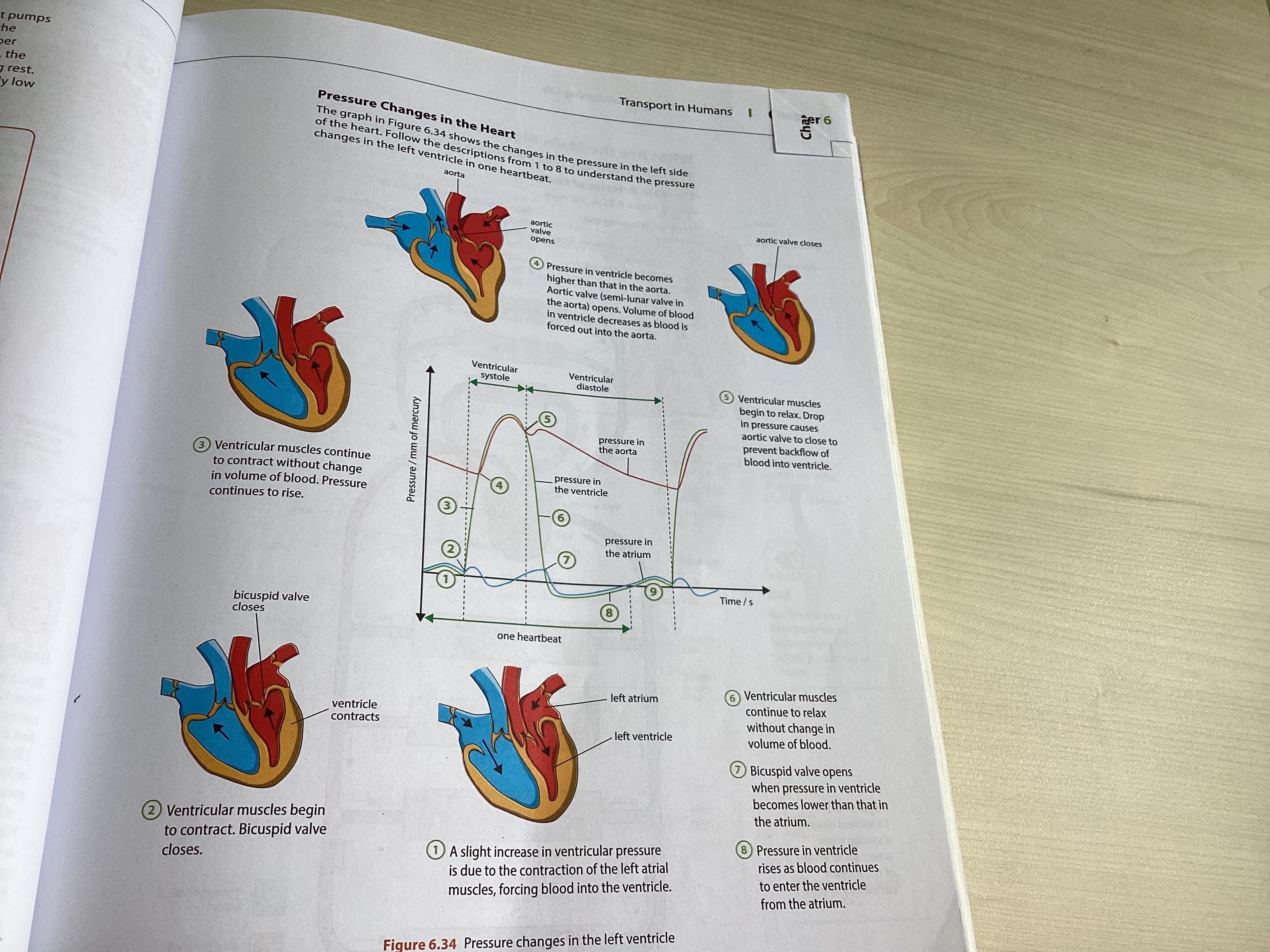
outline the cardiac cycle in terms of what happens during systole and diastole
Systole
Ventricular and atrial muscles contract
Diastole
Ventricular and atrial muscles relax
Cardiac cycle
Atrial muscles contract (systole), forcing blood into the ventricles
After a short pause, ventricle muscles contract (systole), causing a rise in pressure inside the ventricles. The rise in pressure causes the bicuspid and tricuspid valves to close and prevent backflow of blood back into the atria. This produces a loud ‘lub’ sound. The semi-lunar valves open, blood flows from the right and left ventricles into the pulmonary artery and the aorta respectively.
Ventricular muscles relax (diastole), causing a fall in pressure. The semi-lunar valves will close to prevent the backflow of blood from the pulmonary artery and the aorta to the ventricles. This produces a softer ‘dub’ sound. The bicuspid and tricuspid valves open and blood flow from the atria into the ventricles.
Ventricles must contract again (systole). The whole cycle repeats again

Describe coronary heart disease in terms of the occlusion of coronary arteries and list the possible causes and preventative measures
Fatty substances such as cholesterol and saturated fats are deposited on the inner surface of the coronary arteries
Narrows the lumen, increases blood pressure
Rough inner surface
Increase the risk of blood clot being formed in the artery
The supply of blood and oxygen to the heart muscles may be cut off
Muscles of the heart is unable to carry out aerobic respiration to release energy for activities of the muscle cells
Heart muscle cells may be damaged, heart attack occurs
Possible causes
Unhealthy diet
diet high in cholesterol, saturated fats and salt contents
Increases the risk of high blood pressure and heart attack
Smoking
contains CO and nicotine
Increase the risk of coronary heart disease
Nicotine increases blood pressure and the risk of blood clotting in the arteries
Genetic factor
high blood pressure and high blood cholesterol can run in the family
Age
the risk of heart attack increases with age
Sedentary lifestyle
lack of exercise and being inactive lead to the build-up of fatty deposits that block the arteries
Increased risk of getting coronary heart disease
Preventative measures
Healthy diet. Animal fats in the diet should be substituted with polyunsaturated plant fats, which do not stick to the inner surfaces of arteries. May lower the cholesterol levels in blood
Smoking should be avoided. Contains CO and nicotine which increases the risk of getting coronary heart disease
Regular physical exercise. It has long-term beneficial effects in the circulatory system. Strengthens the heart and maintains the elasticity of the arterial walls. The risk of hypertension and high blood pressure can be greatly reduced.