6.1-6.4 Cells of the Nervous System
6.1 Structure and Maintenance of Neurons
Nervous System Divisions:
Central Nervous System (CNS): Brain and spinal cord; processes information, coordinates responses.
Peripheral Nervous System (PNS): Nerves connect CNS to body; carries sensory info to CNS, relays motor commands from CNS.
Neurons
Neurons: Functional units; generate and transmit electrical signals.
Generate action potentials: rapid changes in electrical potential across membrane.
Electrical signals trigger neurotransmitter release for communication across synapse.
Neurons integrate inputs from other neurons for complex decisions.
Glial Cells
Glial cells: Non-neuronal cells supporting neurons with structure, insulation, and nutrition.
Essential for neuron function, but don't directly participate in electrical communication.
Neuron Structure
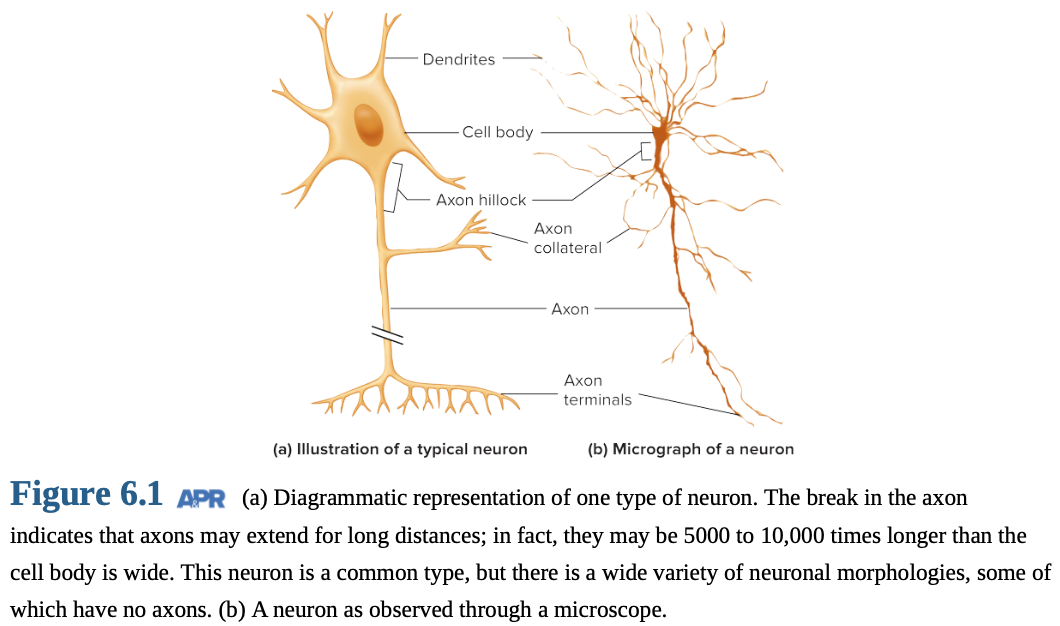
Neurons: cell body (soma), dendrites, and axons for receiving, integrating, and transmitting signals.
Cell Body (Soma): Nucleus and ribosomes for protein synthesis, genetic information.
Dendrites: Receive incoming information; increase surface area.
Dendritic spines: Increase surface area for receiving signals; form synapses.
Axon: Carries outgoing signals.
Length: microns to over a meter.
Axon Hillock (Initial Segment): Generates electrical signals.
Collaterals: Branches of the axon.
Axon Terminal: Releases neurotransmitters; forms synapse.
Varicosities: bulging areas that release chemical messengers along axon.
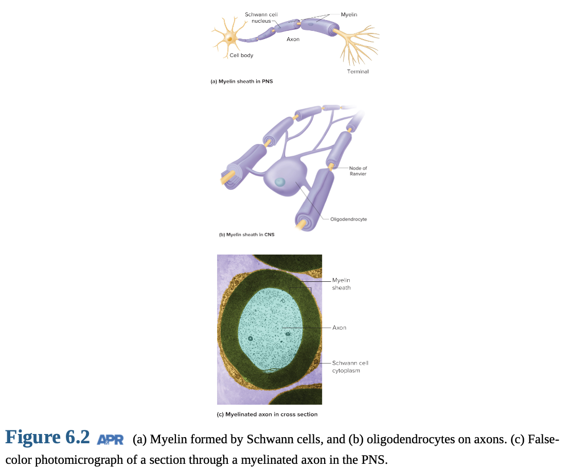
Myelin Sheath
Myelin sheaths: Insulate axons (20-200 layers of modified plasma membrane); speeds up signal conduction.
Oligodendrocytes: Glial cells in CNS; myelinate up to 40 axons.
Schwann Cells: Glial cells in PNS; myelinate 1-1.5 mm segments.
Nodes of Ranvier: Spaces between myelin sections; axon plasma membrane exposed.
Myelin speeds up signal conduction and conserves energy; saltatory conduction.
Axonal Transport
Axonal transport moves materials between cell body and axon terminals (up to 1 meter).
Microtubules: "Rails" for transport.
Kinesins: Anterograde transport (cell body to axon terminals).
Transports nutrients, enzymes, mitochondria, neurotransmitter vesicles.
Dyneins: Retrograde transport (axon terminals to cell body).
Transports recycled vesicles, growth factors, chemical signals.
Retrograde transport: Route for harmful agents (tetanus, herpes, rabies, polio) to invade CNS.
6.2 Functional Classes of Neurons
Neuron Classes:
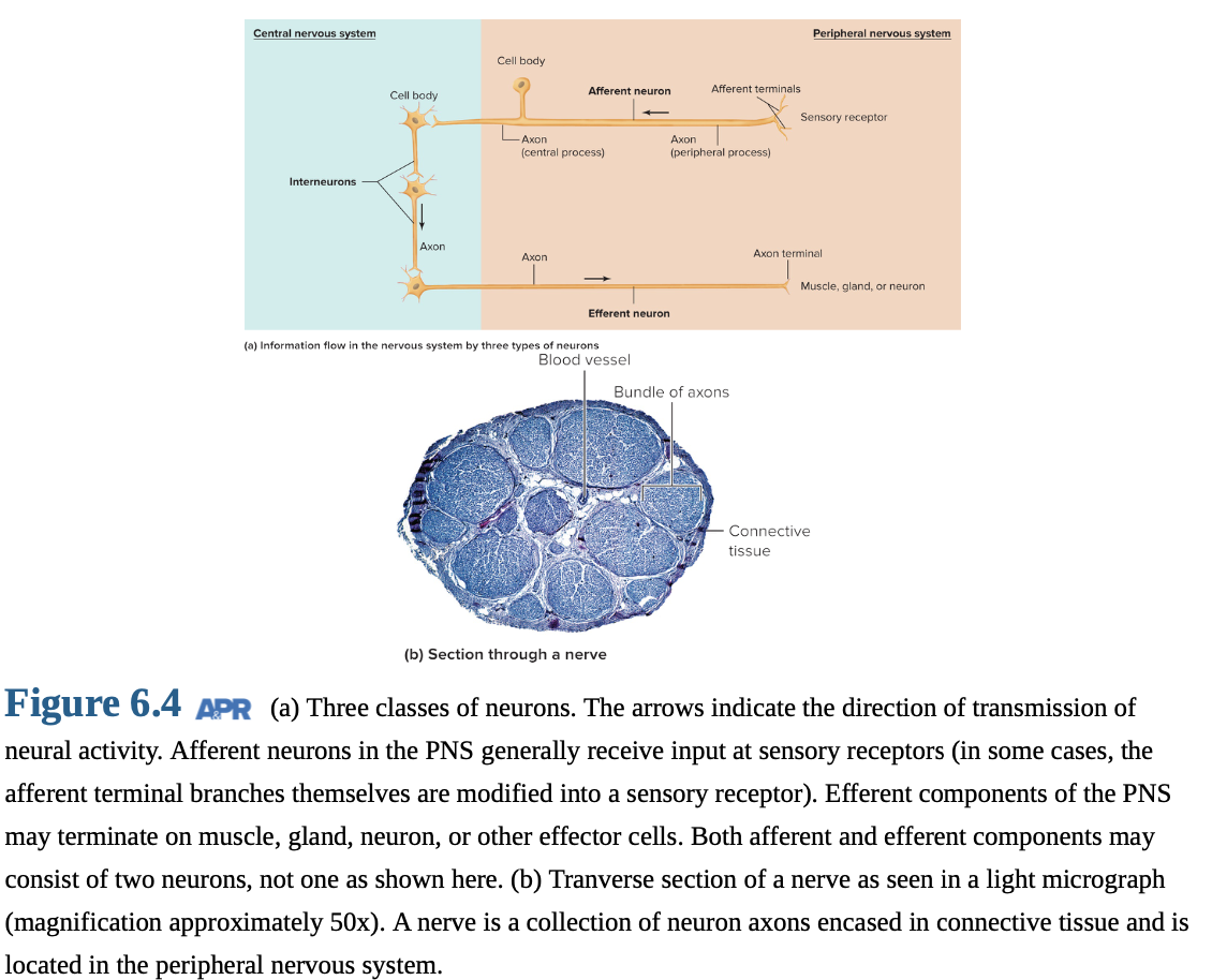
Afferent Neurons
Efferent Neurons
Interneurons
For each afferent neuron, ~10 efferent neurons and ~200,000 interneurons.
Most neurons are interneurons.
Afferent Neurons
Afferent neurons: Transmit information from tissues/organs to CNS; carry sensory information.
Sensory receptors respond to physical/chemical changes.
Receptor region: specialized membrane or separate cell.
Single process divides into peripheral and central processes.
Efferent Neurons
Efferent neurons: Convey information from CNS to effector cells; carry motor commands.
Effector cells: muscle, gland, or other cell types.
Cell bodies/dendrites in CNS, axons extend to periphery.
Interneurons
Interneurons: Connect neurons within CNS; form complex circuits.
99% of all neurons; critical for higher-level processing.
Number varies with action complexity; knee-jerk vs. memory recall.
Nerves
Nerves: Afferent/efferent neuron axons in PNS; myelin, connective tissue, and blood vessels.
Table 6.1 Characteristics of Three Classes of Neurons
I. Afferent Neurons
Transmit information into CNS from receptors.
Single process splits: long peripheral (PNS axon), short central (CNS axon).
II. Efferent Neurons
Transmit information out of CNS to effector cells.
Cell body in CNS; most of axon in PNS.
III. Interneurons
Function as integrators and signal changers.
Integrate afferent/efferent neurons into reflex circuits.
Lie entirely within CNS.
> 99% of all neurons.
Synapses
Synapse: Junction where one neuron alters electrical/chemical activity of another; communication sites.
Signal via neurotransmitters.
Neurotransmitters bind to receptors.
Presynaptic Neuron: Conducts signal toward synapse.
Postsynaptic Neuron: Conducts signal away from synapse.
Could have thousands of synaptic junctions on surface.
many pre synaptic neurons can affect it

6.3 Glial Cells
Glial cells: > half of cells in human CNS; essential for nervous system function.
Provide physical/metabolic support to neurons.
Can divide throughout life.
Primary Types of Glial Cells
Oligodendrocytes: Form myelin sheath of CNS axons.
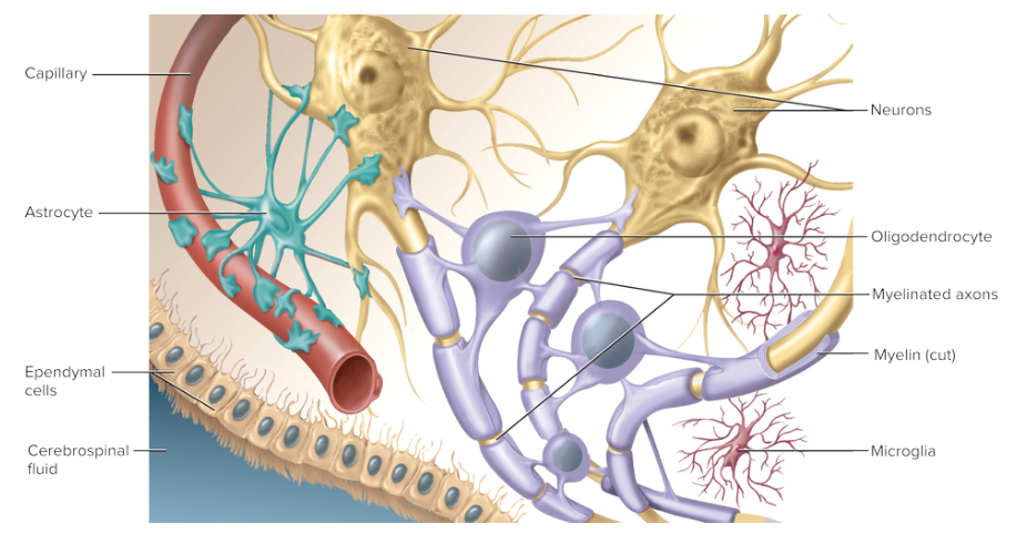
Astrocytes:
Regulate extracellular fluid composition.
removing potassiom ions + neurotransmitters around synapses
Stimulate tight junctions for blood-brain barrier.
Provide metabolic support.
provide glucose, remove secreted metabolic waste (ammonia)
Guide CNS neurons in embryos; secrete growth factors to stimulate nuronal growth.
Have neuron-like characteristics; participate in signaling.
ion channels
receptors for certain neurotransmitters + enzymes for processing them
capability of generating weak electrical responses
possible information signaling role
Microglia:
Macrophage-like
Perform immune functions in CNS.
Contribute to synapse remodeling and plasticity.
Ependymal Cells:
Line fluid-filled cavities in brain/spinal cord.
Regulate production/flow of cerebrospinal fluid.
Schwann Cells:
PNS glial cells; produce myelin sheath.
Similar properties to CNS glia.
6.4 Neural Growth and Regeneration
Growth and Development of Neurons
Nervous system development: Begins with stem cell division into neurons/glia.
Neuronal daughter cells: Differentiate, migrate, and extend axons/dendrites.
Growth cone: Tip of extending axon; finds correct route and target.
Axon guidance: Influenced by attracting, supporting, deflecting molecules.
Cell adhesion molecules: On glia and embryonic neurons.
Neurotrophic factors: Soluble growth factors.
Synapse formation: Occurs upon reaching target.
Early neural development: Vulnerable to alcohol, drugs, radiation, malnutrition, viruses.
Zika virus in moms → microcephaly in babies
Neuron and synapse degeneration: 50-70% undergo apoptosis.
Function: Refines connectivity.
Memory: May explain lack of early childhood memories (before age 4).
Plasticity: Brain's ability to modify structure and function.
Stimuli: Exercise and cognitive activities.
Mechanism: Remodeling synaptic connections; new neurons.
Critical time windows: Vary with neural system.
Visual pathways: Impaired if no visual stimulation between 1-2 years.
Language learning: Easier in youth, slower after adolescence.
Mature CNS: Basic circuits remain, but synaptic contacts change throughout life.
Neurogenesis: Production of new neurons continues in some brain regions.
Stimuli: Cognitive stimulation and exercise.
Antidepressants: Effectiveness may depend on new neuron production.
Regeneration of Axons
Axon repair: Possible outside CNS if cell body is unaffected.
Process: Axon segment separates and degenerates, growth cone grows from cell body
Rate: 1 mm per day.
Example: Thumb sensation may take 2 years to restore after shoulder injury.
Spinal injuries: Typically crush tissue, causing oligodendrocyte apoptosis and demyelination.
CNS axon regeneration: Limited; no significant return of function.
Reasons: Differences in CNS neurons, inhibitory factors from glia.
Evolutionary pressure: Limits growth to maintain network architecture.