0.0(0)
Explore Top Notes Note
Note Studied by 193 peopleNoteStudied by 8 peopleNoteStudied by 157 peopleNoteStudied by 154 peopleNoteStudied by 2 peopleNoteStudied by 21 people
AP HUG Unit 6 Urbanization Topics
Principles and Elements of Interpersonal Communication (ch2)
1.1 Periodic Table
Chapter 1: An Introduction to the Human Body
Matter - PS.2
CHAPTER 8: BASIC CONCEPTS OF CHEMICAL BONDING
Lecture 15 – Articulation, Hearing.docx
Lecture 16 – Articulation, Hearing
14.03.24
- Mandible muscles (restricted to elevation and depression)
- Mandibular elevators
- Masseter – elevates mandible
- Temporalis – elevates mandible (and retracts)
- Medial pterygoid – elevates mandible
- Mandibular depressors
- Digastricus (anterior and posterior) – depress mandible
- Mylohyoid – depresses mandible if hyoid fixed
- Geniohyoid – depresses mandible if hyoid fixed
- Platysma
- Mandibular elevators
- Q: how can I identify the two big mandibular muscles masseter and temporalis?
- By touching your face and side of the head while at the same time elevating and depressing the jaw
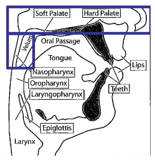
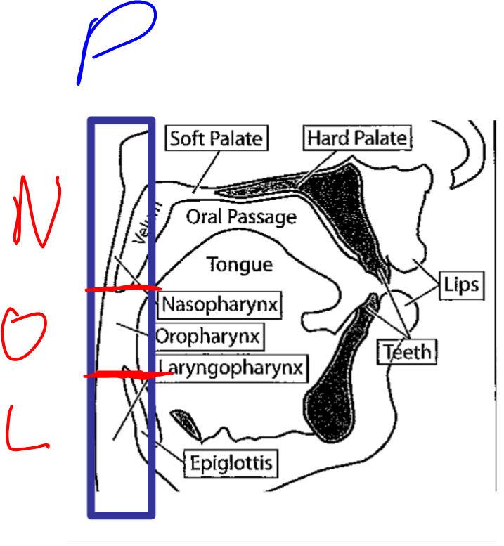
- Velum
- Referred to as the soft palate
- Performs wide range of movements
- Production of most speech sounds
- Must remain in a moderately elevated position (for non-nasal sounds)
- Non-nasal speech: velum generally remains closed (velum raised) (=most of the time)
- Nasality
- Hypernasality – excessive nasal resonance due to inadequately closing velum
- Hyponasality – absence absence of nasalized speech in nasal sounds such as /m/ and /n/ due to inadequate velopharyngeal (VP) opening
- Raised= closed=active/contracted muscles, lowered=open=relaxed
- Velum is closed for non-nasalized speech
- Muscle function for closing: Levator veli palatini (most important one)
- Assimilation – some nasal quality to adjacent phonemes
- High pressure consonants (fricatives and stops) require greater velo[haryngeal closure
- Hard and soft palate are endowed with sensors
- Uvula is part of the velar structure (hangs from the end of the soft palate)
- Referred to as the soft palate
- Can one actively move the uvula?
- You can actively move the velum which moves the uvula but you can’t move the uvula itself?
- = yes. We don’t entirely know why we have a muscle but probably to help push uvula backwards/sideways out of the way of food if you are swallowing food that is hard, large, spiky so you don’t damage it
- Uvula muscularis
- Don’t use it for speech though
- Nasal tract and velum
- Upper part of the nasopharynx merges into the nasal tract and ends at the nose
- Biological functions
- Warming of the inhaled air while breathing
- All surfaces are covered by mucous membranes
- Coupling to the oral tract
- Connection to oropharynx is achieved by lowering the velum 🡪 this process opens the velopharyngeal port (VPP)
- Sound propagation
- The nasal tract cannot produce speech sounds by itself 🡪 but in connection to the rest of the vocal tract (connected or not connected) it can alter acoustic properties of speech sounds considerably 🡪 coupling nasal tract is achieved by lowered or raised velum (lowered velum = nasal cavity coupled; raised velum = nasal cavity not coupled 🡪 not active in speech production)
- Nearly no sound energy leaves the nasal tract 🡪 due to small openings plus high damping (reason: the mucous walls)
- Rather the nasal cavity “draws” energy away from the sounds that are produced and modified in the oral tract
- Velum muscles
- Muscles of velum compress to elevate the velar structure to completely separate oral and nasal areas 🡪 this preventing the nasal cavity to be coupled to the oral cavity
- Muscles are compressed during most speaking time and when swallowing
- Muscles not compressed only when using a few speech sounds in English – nasals
- In other languages velum is opened for both nasal phonemes and nasalized vowels
- The pharynx
- The top of the larynx joins with the esophagus at the laryngopharynx (area above the larynx)
- The most superior part of the epiglottis marks the beginning of the oropharynx
- The area above the velum is the nasopharynx
- The pharynx is a four-way crossing: nasal tract, oral tract, larynx and esophagus
- Esophagus is used during swallowing and eating (epiglottis closed)
Anatomy and physiology of Hearing
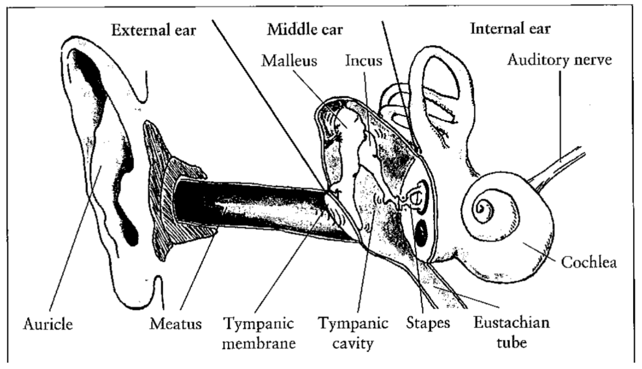
- The ear
- The hearing organ “ear”) is composed of a series of structures:
- The external ear
- The middle ear
- The internal ear
- The neural impulses registered by the sensory cells are then analyzed by the brain
- Two balance systems: visual and semicircular canals. You get dizzy when they are disturbed / not agreeing
- The hearing organ “ear”) is composed of a series of structures:
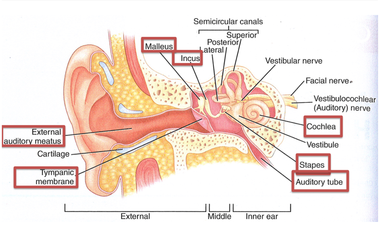
- 1. The external ear
- Everyday use of the word “ear” means technically the external ear
- External ear consists of
- Auricle (pinna)
- Meatus (ear canal)
- Tympanic membrane (ear drum)
- task of the auricle: localize the sound source (important: evolution)
- tympanic membrane works like a loudspeaker or microphone membrane
- Hardly relevant for hearing – just used to sensing directionality of sounds, otherwise you can hear perfectly fine if it is cut off
- External audiotry meatus
- External ear canal
- 7mm in diameter and 2.5 cm long
- This generates resonance frequencies at 3400 Hz
- From an acoustic point of view: ear canal is a filter that amplifies frequencies between 2kHz and 5 kHz
- Terminates at the tympanic membrane
- Two-thirds of ear canal houses in bone (osseus portion)
 One-third of ear canal composed of cartilaginous parts
- Resonating cavity that contributes to hearing
- Determine resonant frequency
- Outer third-line with hair cells and cerum (ear wax) – protects by trapping dirt and insects
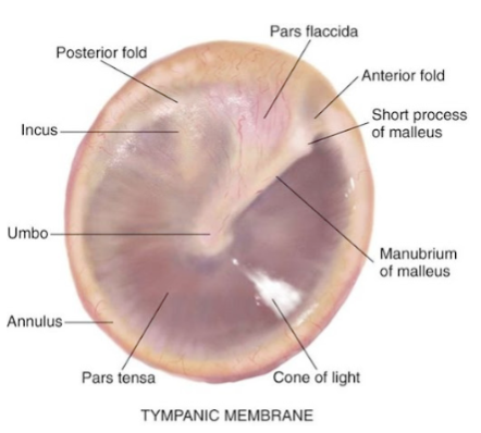
- Tympanic membrane
- Also known as the ear drum
- Separates the middle ear from the outer ear
- Oval shaped, 10 mm in diameter
- Thin three layered sheets of tissue
- Landmarks
- Umbo – point of attachment for malleus, middle ear bone – location is cone of light (reflects light from otoscope)
- Responsible for initiating mechanical impedance-matching process of middle ear
- first layer: outer (cuticular) layer
- second layer: intermitted (fibrous) layer
- third layer: inner (mucous) layer
- 2. The middle ear
- The middle ear consists of the tympanic cavity
- This cavity contains the smallest moving bones of the human body – the ossicles:
- Malleus (hammer): touches the tympanic membrane and transmits to
- The incus (anvil) which transmits to
- Stapes (stirrup) which transmits to internal ear (oval window)
- Malleus and stapes are attached to muscles (may attenuate to transmission of sound by these bones
- This cavity contains the smallest moving bones of the human body – the ossicles:
- The stapes connect directly to the internal ear through the oval window 🡪 transmission of stapes movement to the lymphatic fluid inside the internal ear
- The middle ear consists of the tympanic cavity
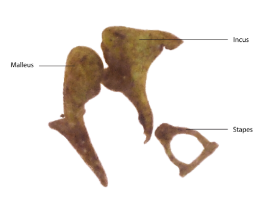 Ossicles
- Malleus
- Largest of the ossicles
- 9mm long and weighs only 25 mg
- Provides point of attachment with tympanic membrane
- Bulk of bone is the head of caput
- Incus
- Shaped like an anvil
- Weighs 20 gm and is around 7mm long
- Provides intermediate link of ossicular chain
- Incus and malleus articulate by means of a saddle joint
- Stapes (stirrup)
- Third bone of the ossicular chain
- Weights 4 mg with an area of 3.5 mm^2
- Helps to transmit sound vibrations from eardrum to oval window
- Articulation of the incus and stapes of ball and socket type
- Ossicular chain is held in place by ligaments
- Malleus
- Tympanic muscles
- Muscles of middle ear attached to ossicles
- Smallest muscles of human body
- Stapedius muscle
- Imbedded in posterior wall of middle ear
- Pulls stapes posteriorly
- Tensor tympani
- Pulls malleus anterior and medial
- Stapedius muscle
0.0(0)
Explore Top NotesNoteStudied by 193 peopleNoteStudied by 8 peopleNoteStudied by 157 peopleNoteStudied by 154 peopleNoteStudied by 2 peopleNoteStudied by 21 people
AP HUG Unit 6 Urbanization Topics
Principles and Elements of Interpersonal Communication (ch2)
1.1 Periodic Table
Chapter 1: An Introduction to the Human Body
Matter - PS.2
CHAPTER 8: BASIC CONCEPTS OF CHEMICAL BONDING