Lecture 2: Gene & Genetic Disease
DNA:
genes are composed of dna
dna consists of:
deoxyribose (pentose sugar)
phosphate molecule
4 nitrogenous bases
pyrimidines: cytosine and thymine
they have 1 ring
purines: adenine and guanine
they have 2 rings
proteins:
a gene is a section of dna that codes for a protein
a protein consists of 1 or more polypeptide chain
each polypeptide chain is composed of amino acids
there are 20 possible amino acids
they are directed by codons (sequences of nitrogenous bases)
dna replication:
dna should replicate itself accurately during cell division
untwisting and unzipping of the dna strand allows for replication
the now single strand of dna acts as a template
complementary base pairing is done by dna polymerase
adenine goes with thymine and cytosine goes with guanine
mutation:
a mutation is any inherited alteration of genetic material
base pair mutation- 1 base pair is substituted for another
silent substitution- a type of base pair mutation that doesn’t result in an amino acid change bc the new codon codes for the same amino acid as the old one
ex. rna codons guu, guc, gua, and gug all code for the amino acid valine
frameshift mutation- an insertion or deletion of 1 or more (but not a multiple of 3) base pairs
causes a change in the entire ‘reading frames’
the most dangerous type of mutation
spontaneous mutation- a mutation that occurs in the absence of exposure to known mutagens
mutational hotspots- areas of the chromosomes that have high mutation rates
ex. a cytosine base followed by a guanine is known to account for a disproportionately large percentage of disease causing mutations
mutagens- agents known to increase the frequency of mutations
ex. radiation and chemicals
transcription:
rna is synthesized from the dna template
results in the formation of messenger rna (mrna)
mrna moves out of the nucleus and into the cytoplasm
once in the cytoplasm, mrna is translated into a polypeptide chain
(theres a pic, not putting it here, slide 14)
translation:
process by which rna directs the synthesis of a polypeptide
site of protein synthesis is the ribosome
trna contains a sequence of nucleotides (anticodon) complementary to the triad of nucleotides on the mrna strand (codon)
the ribosome moves along the mrna sequence to translate the amino acid sequence
(another pic, slide 16)
chromosomes:
somatic cells:
contain 46 chromosomes (23 pairs)
diploid cells (the chromosomes occur in pairs)
gametes (egg or sperm)
contain 23 chromosomes
haploid cells (one member of each chromosome pair)
meiosis- formation of haploid cells from diploid cells
autosomes- the first 22 of the 23 pairs of chromosomes in humans
the 2 members of each chromosome are virtually identical and thus said to be homologous
sex chromosomes- the last pair of chromosomes
in females, its a homologous pair (XX)
in males, it is a nonhomologous pair (XY)
karyotype- an ordered display of chromosomes
chromosome aberrations:
euploid cells- cells that have a multiple of the normal number of chromosomes
haploid and diploid cells are (normal) euploid forms
polyploid cells- when a euploid cell has more than the diploid number of chromosomes
triploidy- a zygote having 3 copies of each chromosome (69 total)
tetraploidy- 4 copies of each (92 total)
both triploid and tetraploid fetuses don’t survive
aneuploidy- a somatic cell that doesn’t have a multiple of 23 chromosomes (not all of them are either extra or less, only some)
trisomic cell- a cell that has 3 copies of 1 chromosome (aka trisomy)
monosomy- the presence of only 1 copy of any chromosome
monosomy is often lethal, but infants can survive with trisomy of some chromosomes
its better to have extra than less
disjunction- normal separation of chromosomes during cell division
nondisjunction- failure of homologous chromosomes or sister chromatids to separate normally during meiosis or mitosis
usually the cause of aneuploidy
if occurs in meiosis 1:
2 daughter cells will have 2 chromosomes, 2 will have 0 instead of 1 each
if occurs in meiosis 2:
1 daughter cell will have 2 chromosomes, 1 will have 0 (other 2 will probably be normal if they went through normal separation)
autosomal aneuploidy:
trisomy can occur for any chromosome but the most common ones are 13, 18, and 21
partial trisomy- only an extra portion of a chromosome is present in each cell
chromosome mosaics- only some cells of the body have trisomies
down syndrome- trisomy 21
the best known ex of aneuploidy
1:800 live births
intellectually disabled (assume that everytime it says this, it should be green bc i’m lazy), low nasal bridge, protruding tongue, poor muscle tone
risk increases with maternal age
sex chromosome aneuploidy:
one of the most common is trisomy x
this is a female that has 3 x chromosome
called ‘metafemales’ (i can’t find any mention online of it being called that except for in the history section of Wikipedia and it just says it was proposed and then criticized (by another guy involved) so… maybe don’t use this???)
symptoms are variable: sterility (my research shows that this isn’t usual though), menstrual irregularity, and/or intellectual disability
symptoms worsen with each additional x
turner syndrome:
females with only 1 x chromosome
characteristics:
absence of ovaries (which obviously causes sterility)
short stature (~4’7”)
webbing of the neck
edema
underdeveloped breasts
wide nipples
high number of aborted fetuses (i’m assuming spontaneously??)
the x is usually inherited from the mother
Klinefelter syndrome:
individuals with at least 2 x’s and 1 y
characteristics:
male appearance
develop female-like breasts
small testes
sparse body hair
long limbs
some individuals can be xxxy and xxxxy. the abnormalities will increase with each x
alterations in chromosome structure:
chromosome breakage:
if a chromosome break does occur, physiological mechanisms will usually repair the break, but the breaks often heal in a way that alters the structure of the chromosome
agents of chromosome breakage- ionizing radiation, chemicals, and viruses
breakage or loss of DNA
cri du chat syndrome (cry of the cat):
deletion of the short arm of chromosome 5
causes low birth weight, intellectual disability, and microcephaly
duplication
presence of a repeated gene or gene sequence
rare occurrence
less serious consequences bc its better to have more genetic material than less
duplication in the same region as cri du chat causes intellectual disabilities but no physical abnormalities
inversions
2 breaks on a chromosome followed by a reinsertion (but the reinsertion is reversed so the gene order is reversed)
usually occurs from a breakage that gets reversed during reattachment (what do you mean ‘usually’??? how else would it happen???)
abcdefg may become abedcfg
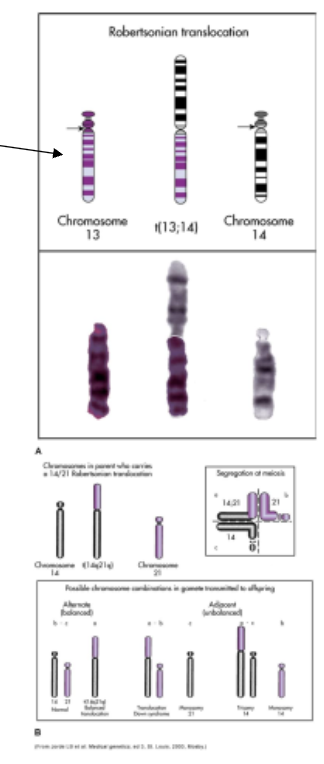
translocations
the interchanging of material between nonhomologous chromosomes
translocation occurs when 2 chromosomes break and the segments are rejoined in an abnormal arrangement
the 2 long arms of nonhomologous chromosomes fuse at the centromere (???)
fragile sites
fragile sites are areas on chromosomes that develop distinctive breaks or gaps when cells are cultured
no apparent relationship to disease
fragile x syndrome
site on the long arm of the x chromosome
associated with intellectual disability; second in occurrence to down syndrome (? like this is the second most common cause of intellectual disability??)
higher incidence in males because they have only 1 x chromosome
honestly i have no clue what any of this is trying to say
genetics:
gregor mendel- Austrian monk, garden pea experiments, mendelian traits
locus- position of a gene along a chromosome
allele- a different form of a particular gene at a given locus
ex. Hgb A vs Hgb S
polymorphism- locus that has 2 or more alleles that occur with appreciable frequency
homozygous- loci on a pair of chromosomes that have identical genes
ex. o blood type has oo genes (same allele)
heterozygous- loci on a pair of chromosomes have different genes
ex. ab blood type has a and b genes on a pair of loci (different alleles)
genotype- the genetic makeup of an organism
‘what they have’
phenotype- the observable, detectable, or outward appearance of the genetics of an organism
‘what they demonstrate’
ex of genotype vs phenotype: a person with the a blood type could be aa or ao. a is the phenotype, aa or ao would be the genotype
dominant allele- the allele that is observable when 2 alleles are found together
recessive allele- the allele that isn’t observable when 2 alleles are found together
in genetics, the dominant allele is represented by a capital letter and the recessive by a lowercase letter
co-dominant alleles- alleles that when put together are both dominant and both observable
carrier- one that has a (recessive) disease gene but is phenotypically normal
this is bc for one to demonstrate a recessive disease, they must inherit 2 recessive genes
ex. Ss = sickle cell anemia carrier vs. ss = demonstrates sickle cell disease
pedigrees:
used to study genetic disorders within families
summarizes family relationships and shows which members of a family are affected by genetic disease
begins with 1 individual in the family (the proband)
pedigree symbols key:
white circle- normal female
white square- normal male
white diamond- (normal) sex unknown
single bar between a square and a circle- indicates mating
roman numerals by side- indicates the generation of the individuals in that row
number under an individual- indicates their place in the birth order of that immediate family
(birth order is also indicated by the order of the individuals from left to right)
bar from mate line to a straight line with other lines from it- parents and children (multiple children)
if there is only 1 parent listed, it means the other parent is normal or of no significance to the analysis
if the line from the bar to the mate line is like an upside down v, the resulting children are fraternal twins
if that line is like an upside down v and has a line that connects them (like an uppercase a), the twins are identical
if there is only one child, there will be a line from the mating line that leads to the child
double bar between a square and a circle- close relatives who mated
circle/square with a number in it- shorthand for noting number and sex of children. the number in the symbol indicates the number of children of that sex
darkened circle/square- affected individual
if there is an arrow by this symbol, they are the individual that led to the analysis
square/circle that is half darkened, half white- autosomal heterozygous recessive (aka a carrier)
circle with a dot in it- x linked carrier
carrier individuals with Klinefelter syndrome will be a square with a dot and a note indicating they have Klinefelter
circle/square with diagonal line crossing through- individual is dead
it says a circle with a ^ strikethrough indicates a stillborn but that didn’t make sense to me so i looked up what the stillborn symbol was and i found that a triangle represents a pregnancy not carried to term (and passed i think) (w a note under saying at what week the miscarriage occurred)
i think a live pregnancy would be a circle/square/diamond with a p in it
other symbols found through research:
circle/square with brackets around it- individual was adopted
if the line leading to them is dashed, they were adopted into the family
if the line leading to them is reg, they were adopted out of the family
single-gene disorders:
recurrence risk- the probability that parents of a child with a genetic disease will have another child with the same disease
recurrence risk of an autosomal dominant trait- when 1 parent is affected by an autosomal dominant disease and the other is normal, the occurrence and recurrence risks for each child is 50%
autosomal dominant disorder- abnormal allele is dominant, normal allele is recessive, and the genes exist on a pair of autosomes
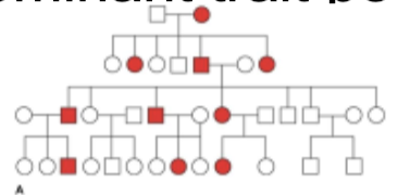
autosomal dominant traits:
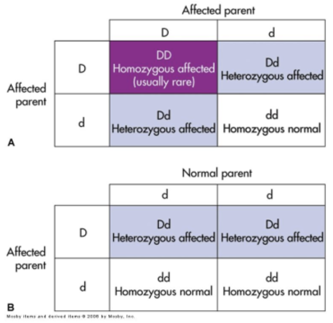
autosomal dominant trait pedigree:
penetrance:
the percentage of individuals with a specific genotype who also express the expected phenotype
incomplete penetrance- individual who has the gene for a disease but does not express the disease
doesn’t that just make them a carrier??
single-gene disorders: (again ig…)
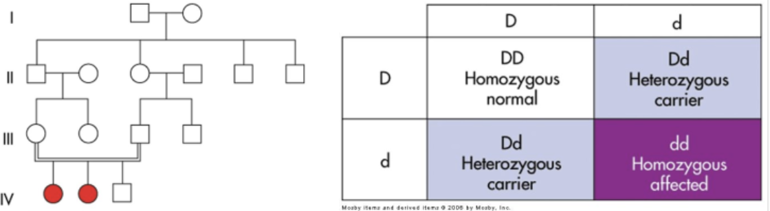
autosomal recessive disorder:
abnormal allele is recessive and a person must be homozygous (recessive) for the abnormal trait to express the disease
the trait usually appears in the children, not the parents, and it affects the genders equally bc it is present on a pair of autosomes
autosomal recessive disorder recurrence risk:
recurrence risk of an autosomal dominant trait (???)
when 2 parents are carriers of an autosomal recessive disease, the occurrence and recurrence risks for each child are 25%
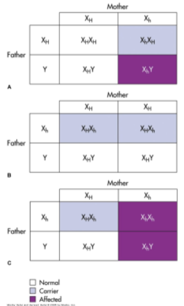
sex-linked disorders:
the y chromosome contains only a few dozen genes, so most sex linked traits are located on the x chromosome and are said to be x linked
sex linked (x linked) disorders are usually expressed by males bc females have another x chromosome to mask the abnormal gene
i think that would only be true if it was recessive bc if its dominant, then females have 2 chances to get it
looked it up: females are more common but less severely affected
x linked recessive:
most x linked disorders are recessive
affected males cannot transmit the genes to sons, but they can to all daughter
sons of female carriers have a 50% risk of being affected