BIO-1040 Cumulative Final Exam Review Flashcards
Organization of the Human Body
Definitions:
Anatomy: Study of the structure of the human body.
Physiology: Study of the function of the human body.
Levels of Organization:
Chemicals.
Cells.
Tissues.
Organs.
Systems.
Organism.
Homeostasis:
Maintaining a stable internal environment.
Feedback Systems:
Components:
Receptor (sensor): Monitors a controlled condition.
Control center: Determines the set point and responds to changes.
Effector: Produces a response to alter the controlled condition.
Types:
Negative feedback: Reverses a change in a controlled condition; fluctuates around a set point.
Positive feedback: Reinforces or strengthens a change in a controlled condition; change in the same direction.
Anatomical Terminology:
Body regions.
Directional terms.
Planes and sections.
Body cavities.
Introductory Chemistry
Introduction to Chemistry:
Elements and atoms.
Ions, molecules, and compounds.
Chemical Bonds:
Ionic bonds.
Covalent bonds.
Hydrogen bonds.
Chemical Reactions:
Forms of energy and chemical reactions.
Synthesis reactions.
Decomposition reactions.
Exchange reactions.
Reversible reactions.
Chemical Compounds and Life Processes:
Inorganic Compounds:
Water and its properties.
Acids, bases, and salts.
The concept of pH is based on the concentration of hydrogen ions.
Buffer systems.
Organic Compounds:
Carbohydrates.
Lipids:
Triglycerides.
Phospholipids.
Steroids.
Proteins and enzymes.
Nucleic acids—DNA, RNA, ATP.
Cells
Main Cell Components:
Plasma membrane.
Cytoplasm—cytosol and organelles.
Nucleus.
Plasma Membrane—selectively permeable
Structure:
Phospholipid bilayer.
Cholesterol.
Membrane proteins.
Transportation:
Regulates what enters and leaves the cell.
Passive Transport Processes:
Diffusion.
Facilitated diffusion.
Osmosis.
Active Transport Processes:
Use energy of ATP to “push” a substance against or up its concentration gradient (pump changes shape).
Transport in Vesicles:
Active transport.
Transport in vesicles—phagocytosis, endocytosis, and exocytosis.
Cytoplasm—all the components between the plasma membrane and the nucleus
Cytosol:
Fluid substance containing cytoskeleton (microfilaments, intermediate filaments, and microtubules).
Organelles:
Centrosome.
Cilia and flagella.
Ribosomes.
Endoplasmic reticulum—smooth and rough.
Golgi complex.
Lysosomes.
Mitochondria.
Nucleus—control center of the cell; contains the DNA
Nucleolus.
Nuclear membrane—has pores.
DNA/Chromosome Structure:
Protein Synthesis:
Transcription—DNA is copied into an RNA message.
Translation—mRNA is translated to make a protein.
Cell Division:
Interphase—DNA doubles.
Mitosis.
Cytokinesis.
Tissues
Tissue Types:
Use Every Child Makes Noise to remember the four types.
Epithelia:
Be able to identify the different types.
General Characteristics:
Features, locations, and functions.
Classification:
Number of layers—simple, stratified, and pseudostratified.
Cell shape—squamous, cuboidal, columnar, and transitional.
Glandular epithelium.
Connective:
Be able to identify the different types.
General Characteristics:
Features, locations, and functions.
Connective Tissue Cells:
Fibroblasts.
Macrophages.
Plasma cells.
Mast cells.
Adipocytes.
Extracellular Matrix:
Ground substance.
Fibers—collagen, elastic, and reticular.
Types:
Loose connective tissue—areolar, adipose, reticular.
Dense connective tissue—dense regular, dense irregular, elastic.
Cartilage—hyaline, fibrocartilage, elastic cartilage.
Bone.
Liquid connective tissue—blood and lymph.
Muscle:
Be able to identify the different types.
General Characteristics:
Features, locations, and functions.
Types:
Skeletal.
Cardiac.
Smooth.
Nervous:
Be able to identify.
General Characteristics:
Features, locations, and functions.
Cell Types:
Neurons—cell body, dendrites, and axon.
Neuroglia.
The Integumentary System
Skin
Epidermis:
Cells—keratinocytes, melanocytes, Langerhans cells, and tactile epithelial cells.
Layers—stratum basale, spinosum, granulosum, lucidum, corneum.
Dermis:
Dermal papillae.
Meissner’s corpuscle.
Free nerve endings—pain and temperature.
Hypodermis/Subcutaneous Layer (not a true skin layer):
Adipose and areolar tissue.
Blood vessels.
Pacinian Corpuscle.
Skin Color:
Melanin.
Carotene.
Hemoglobin.
Glands
Sebaceous
Sudoriferous
Ceruminous
Functions
Regulate body temperature
Protection
Sensation
Excretion and absorption
Vitamin D synthesis
The Skeletal System
Functions of Bone
Support and protect
Assists in movement by providing attachment sites for muscles
Storage of minerals, calcium, and phosphate
Production of blood cells in red bone marrow
Triglyceride storage in the yellow bone marrow
Types of Bones
Long, short, flat, irregular bones
Macroscopic Parts of Long Bone
Diaphysis
Epiphysis with epiphyseal plate for growth
Metaphysis
Articular Cartilage
Periosteum
Medullary Cavity—yellow bone marrow
Endosteum
Microscopic Parts of Long Bone (Histology)
Matrix of water, collagen fibers, mineral salts
Cells—osteoprogenitor cells, osteoblasts, osteocytes, and osteoclasts
Structure of Compact Bone
Haversian system or osteon
Canals—Volkmann, Haversian
Lamellae
Lacunae
Canaliculi
Structure of Spongy Bone
Trabeculae
Red bone marrow
Hormones of Bone Metabolism and Calcium Homeostasis
Parathyroid—parathyroid hormone
Kidneys—calcitriol
Thyroid gland—calcitonin, thyroxine, and triiodothyronine
Anterior pituitary—human growth hormone
Pancreas—insulin
Axial Skeleton
Be able to identify the following bones
Skull—frontal, temporal, parietal, sphenoid, occipital, zygomatic, maxilla, mandible, hyoid
Vertebrae—cervical, thoracic, lumbar, sacrum, coccyx
Thorax—sternum and ribs
Appendicular Skeleton
Be able to identify the following bones
Pectoral girdle and upper limb—clavicle, scapula, humerus, radius, ulna, carpals, metacarpals, and phalanges
Pelvic girdle and lower limb—hip bones, femur, patella, tibia, fibula, tarsals, metatarsals, and phalanges
Muscles
Review of Muscle Tissue
Skeletal
Cardiac
Smooth
Functions of Muscle
Produce body movements
Maintain posture
Regulate organ volume
Moves substances within body
Produce heat
Skeletal Muscle
Histology of a Muscle Cell
Sarcolemma
Transverse (T) tubules
Sarcoplasm
Mitochondria
Myoglobin
Sarcoplasmic reticulum
Myofibrils
Thin filaments—actin, troponin, and tropomyosin
Thick filaments—myosin
Other Components
Tendons—attach muscle to bone (connective tissue)
Blood supply—capillaries deliver oxygen and remove wastes of ATP production
Nerves—neurons instruct muscle cells to contract at the neuromuscular junction (NMJ)
Contraction—The Sliding-Filament Mechanism
Contraction
Impulse in the axon causes release of acetylcholine into synaptic cleft
Acetylcholine binds to receptors in the sarcolemma of the muscle, causing sodium to flood into the muscle
Get action potential (impulse) in muscle
Muscle action potential travels along sarcolemma and T tubules, which stimulates release of calcium from the sarcoplasmic reticulum
Calcium binds to troponin, and troponin-tropomyosin moves away from the myosin-binding sites on actin
Myosin breaks down ATP and attaches to actin, forming cross-bridges
The cross-bridges rotate to release ADP, sliding the thin filaments past the thick filaments during the power stroke
Relaxation
Acetylcholinesterase (AChE) breaks down acetylcholine within the synaptic cleft
Muscle action potential ceases
Active transport pumps calcium back into the sarcoplasmic reticulum
Tropomyosin-troponin complex recovers binding sites on actin
Muscles of the Body
Be able to identify the following muscles
Muscles that move the face and head—occipitofrontalis, orbicularis oris, zygomaticus major, sternocleidomastoid
Muscles of the abdominal wall—rectus abdominis, external oblique, internal oblique
Muscle used during breathing—diaphragm
Muscles that move the arm—deltoid, trapezius, latissimus dorsi, pectoralis major, biceps brachii, triceps brachii
Muscles that move the leg—rectus femoris, sartorius, gracilis, gluteus maximus, semitendinosus, semimembranosus, biceps femoris, gastrocnemius
Nervous System
Functions
Sensory
Integrative
Motor
Divisions of the Nervous System
Central Nervous System (CNS)
Brain
Spinal cord
Peripheral Nervous System (PNS)—all nervous tissue outside the CNS
Somatic nervous system
Autonomic nervous system
Sympathetic division—epinephrine and norepinephrine mediate “fight-or-flight” response
Parasympathetic division—acetylcholine mediates “rest and digest” response
Enteric nervous system
Nervous Tissue
Neurons—conduct nerve impulses
Cell body
Dendrites—receive nervous impulses
Axons—take impulses away from cell body
Axon hillock
Myelin sheath for insulation
Nodes of Ranvier
Synaptic end bulbs hold neurotransmitters
Neuroglia—support, nourish, and protect neurons
CNS—oligodendrocytes
PNS—Schwann cells
White and gray matter
Action Potentials (Nerve Impulses)
Resting membrane potential (-70
ormal mV)Generation of Action Potential
Depolarization—inside of cell becomes more positive than outside
If depolarization reaches threshold (-55
ormal mV) an action potential is fired; all-or-none phenomenonSodium voltage-gated channels open—sodium rushes in and membrane potential hits +30
ormal mV
Repolarization—membrane potential is restored
Voltage-gated potassium channels open, and potassium rushes out
Membrane potential is returned to -70
ormal mV and potassium channels close
Refractory period
Conduction
Continuous conduction occurs in unmyelinated neurons—slow
Saltatory conduction occurs in myelinated neurons—FAST!
Synaptic Transmission—how one neuron communicates with another
Presynaptic Neuron
Nerve impulse opens voltage-gated calcium channels, and calcium floods into presynaptic neuron
Neurotransmitter is released
Postsynaptic Neuron
Neurotransmitters bind to receptors on postsynaptic neuron
Sodium channels open, depolarization occurs, and another action potential is fired
Central Nervous System
Meninges (Coverings)
Dura mater
Arachnoid mater
Pia mater
Spinal Cord—be able to identify the parts
Structure
Central canal
Posterior (dorsal) root ganglion
Anterior (ventral) root
Gray horns
White columns
Reflex Arc
sensory information conducted by sensory neurons in the posterior root
motor information conducted by motor neurons in the anterior root
motor neuron acts on effector
Brain—be able to identify the parts and know functions
Cerebrospinal fluid and ventricles
Brain Stem
Medulla oblongata
Pons
Midbrain
Diencephalon
Thalamus
Hypothalamus
Pineal gland
Cerebellum
Cerebrum
Lobes—frontal, parietal, occipital, and temporal
Functional areas—sensory, motor, and association areas
Eye and Ear
The Eye
Be able to identify the following parts
Parts of the Eye
Cornea
Sclera
Conjunctiva
Choroid
Ciliary body—process (aqueous humor) and muscle
Lens
Iris
Vitreous chamber—vitreous humor
Retina—rods, cones, fovea centralis
Optic disk and optic nerve
Light Passage and Image Formation
Refraction
Photopigments—rhodopsin
Bipolar cells
Ganglion cells
Optic nerve
Optic chiasma
Thalamus
Occipital lobe
The Ear
Be able to identify the following parts
Auditory Pathway
Auricle
External auditory canal
Tympanic membrane
Malleus
Incus
Stapes
Cochlea
Organ of Corti
Vestibulocochlear nerve
Temporal lobe
Physiology of Equilibrium
Dynamic equilibrium—semicircular canals
Static equilibrium—saccule and utricle
The Endocrine System
Functions
Regulation of:
reproductive systems
glandular secretions
chemical composition of body fluids
metabolism
smooth and cardiac muscle contraction
some immune system activities
growth and development
circadian rhythms
Hormones
* Definition
* Mechanism of hormonal action
* Lipid soluble
* Water soluble—cAMP is the second messenger
* Regulation of hormone release
* nervous system
* chemical changes in blood
* other hormones
* Endocrine glands and their hormones; The ones you are responsible for are listed with the organ system on which it acts (e.g. calcitonin in the skeletal system, erythropoietin in blood, etc.)
Blood
Functions
Transport
oxygen, carbon dioxide, and metabolic wastes
nutrients from digestive tract
hormones and heat
Regulation
body temperature
pH via buffers
keeps liquid content high
Defense
attack pathogens
antibodies
blood clotting
Components
Plasma
water—92%
proteins
albumin
globulins
fibrinogen
Formed Elements—give the function of each of the following cells
red blood cells (erythrocytes)—no nucleus or organelles
hemoglobin
life cycle and recycling of the breakdown products
erythropoiesis—secretion of erythropoietin by kidneys
anemia and detection by hematocrit
White blood cells (leukocytes)
granulocytes—neutrophils, basophils, eosinophils
agranulocytes—monocytes (macrophages) and lymphocytes
Platelets (thrombocytes)—fragmentation of megakaryocyte
Hemostasis
vascular spasm
platelet plug formation
clot formation
one clotting factor activates the next in a cascade of events that require calcium
fibrinogen ultimately converted to fibrin threads
Problems—thrombus and embolus
Blood Typing
Be able to determine blood typing results
ABO System
Rh System
The Cardiovascular System: Heart and Blood Vessels
The Heart
Location
Pericardium
Heart wall
epicardium
myocardium
endocardium
Chambers, valves, and vessels of the heart
be able to identify these
right atrium
receives blood from superior and inferior vena cava and coronary sinus
contracts, sending blood through the tricuspid valve on its way to the right ventricle
right ventricle
contracts to send blood through the pulmonary semilunar valve and out into the pulmonary trunk
the pulmonary trunk becomes the pulmonary arteries, which carry blood to the lung capillaries
left atrium
receives blood returning from the lung capillaries via the pulmonary veins
contracts, sending blood through the bicuspid (mitral) valve on its way to the left ventricle
left ventricle
contracts to send blood through the aortic semilunar valve and out into the aorta
Associated structures
chordae tendineae
papillary muscles
Coronary circulation
coronary arteries
coronary veins
Blood flow through the heart
Conduction system and the electrocardiogram (ECG)
Sinoatrial node (pacemaker)
spontaneously fires an action potential and atria contract
P wave
Atrioventricular node
receives impulse from SA node
sends impulse through bundle of His, bundle branches, and into Purkinje fibers and ventricles contract; QRS complex
ventricular repolarization is seen as the T wave
Blood vessels
Arteries
take blood away from heart
be able to identify these major arteries: brachiocephalic, right & left common carotid, right & left subclavian, aorta, pulmonary trunk & arteries, right & left renal, right & left femoral, right & left popliteal, right & left external & internal iliac, right & left axillary, right & left brachial, right & left radial, and right & left ulnar
Arterioles
Capillaries—allows diffusion of gases and nutrients
Venules
Veins
return blood to the heart
venous return controlled by valves, skeletal muscle contraction, and respiratory movements
be able to identify these major veins: superior & inferior vena cava, pulmonary veins, right & left external jugular, right & left subclavian, right & left brachiocephalic, right & left common iliac, right & left internal iliac, right & left external iliac, right & left renal, right & left femoral, right & left greater saphenous, right & left popliteal, right & left axillary, right & left brachial, right & left radial, and right & left ulnar
Diseases
Atherosclerosis and the role of cholesterol
Myocardial infarction
Lymphatic and Immune Systems
Lymphatic System
Functions
return excess tissue fluid (lymph) to bloodstream
lymph vessels transport dietary lipids into blood
defend against disease
Vessels
a lot like veins
valves, skeletal muscle contraction, and respiratory movements aid lymph movement
flow—lymph capillaries to lymph vessels, through nodes, to right lymphatic duct or thoracic duct, and to subclavian veins
Organs
red bone marrow—both B and T lymphocytes made here; B lymphocytes mature here
thymus—site of T lymphocyte maturation
tonsils
lymph nodes
spleen
Immune System
needed for the body to defend itself
Innate immunity
first line of defense (barriers to entry)
second line of defense (Internal defenses)
antimicrobial substances (e.g. complement system)
inflammatory response—redness, warmth, swelling, and pain
phagocytes
fever
Adaptive immunity
T cells and cell-mediated immunity
antigen presenting cell presents antigen to T cell
T cell recognizes foreign antigen with the help of a T helper cell (clonal selection)
T cell divides to produce memory T cells, which produce long lasting immunity, and cytotoxic T cells, which directly attack foreign cell (clonal expansion)
B cells and antibody-mediated immunity
B cells recognize antigen with the help of a T helper cell (clonal selection)
B cell divides into memory B cells, which produce long lasting immunity, and plasma cells that produce antibody (clonal expansion)
Antibodies attack invader
The Respiratory System
Respiratory processes
* breathing/ventilation
* external respiration
* internal respiration
* cellular/aerobic respirationOrgans
be able to identify the following structures
Nose/nasal cavities
pseudostratified ciliated columnar epithelium
sinuses
Pharynx
nasopharynx
oropharynx
laryngopharynx
Glottis
covered by epiglottis during swallowing
Larynx
contains vocal cords for production of sound
thyroid cartilage
Trachea
lined with pseudostratified ciliated columnar
held open by hyaline cartilage
Bronchi
lead into left and right lung
Bronchioles
branches of bronchi; constrict during asthma attack
Lungs
lobes
alveoli
type I cells—simple squamous epithelium for diffusion
type II cells—secrete surfactant
macrophages
membranes—visceral and parietal pleura
Breathing Mechanics
* Boyle’s Law
* Muscle contraction and regulation
* Inhalation/inspiration
1. inspiratory center in medulla oblongata active
2. external intercostals and diaphragm contract
* Exhalation/expiration (quiet)
1. inspiratory center inactive
2. diaphragm and external intercostals relaxGas exchange
* External Respiration—gas exchange between alveoli and blood
* Internal Respiration—gas exchange between blood and tissues
The Digestive System
Functions
* Ingestion
* Digestion
* chemical
* mechanical
* Absorption
* EliminationOrgans of the digestive system
* be able to identify the following
* Mouth
* hard and soft palate
* uvula
* tongue and cheeks
* salivary glands
* parotid, submandibular, sublingual
* saliva composed of mucus, salivary amylase, and lysozyme
* teeth—incisors, canines, premolars, and molars
* Pharynx and esophagus
* swallowing
* peristalsis
* Stomach
* mechanical and chemical digestion
* structure
* rugae
* sphincters—pyloric and cardiac
* secretions—mucus, pepsinogen, hydrochloric acid, and gastrin
* Small intestine
* parts—duodenum, jejunum, ileum
* secretions—secretin and cholecystokinin (CCK)
* functions
* digestion
* absorption via villi, microvilli, lacteals
* Pancreas
* Exocrine secretions
* amylase digests sugars
* trypsin digests proteins
* lipase digests fats
* RNAse and DNAse digest nucleotides
* bicarbonate is buffer
* endocrine function—insulin and glucagon
* regulated by secretin and CCK
* Liver
* functions
* regulates glucose levels
* makes bile
* removes and detoxifies poisons
* makes plasma proteins
* regulates cholesterol levels
* Gallbladder
* stores bile and releases it into small intestine
* Large intestine
* structure
* cecum, appendix, colon (ascending, transverse, descending, sigmoid), and rectum
* functions
* water absorption, bacteria, vitamin production, formation and expulsion of feces
The Urinary System
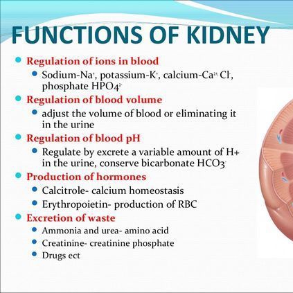
Functions of the urinary system
* Regulate blood ion levels—sodium, potassium, calcium, chloride, and phosphate
* Regulate blood pH by secreting hydrogen ions and reabsorbing bicarbonate
* Regulate blood volume
* Regulate blood pressure
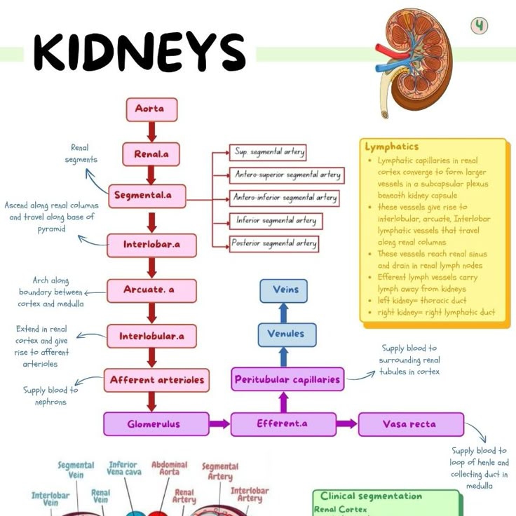
* Produce hormones—erythropoietin and calcitriol
* Excrete wastes—ammonia, urea, and drugsLocation and structure of the kidneys
be able to identify the following structures
Location—retroperitoneal
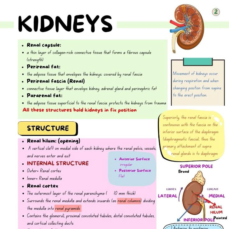
Renal capsule
Renal cortex
Renal medulla
Renal pyramids
Renal columns
Renal calyces—major calyx and minor calyx
Renal pelvis
Nephron
glomerulus and Bowman’s capsule (renal corpuscle)
proximal convoluted tubule
descending loop of Henle
ascending loop of Henle
distal convoluted tubule
collecting duct
Functions of the Nephron
* Filtration—occurs at renal corpuscle due to blood pressure
* Reabsorption and Secretion
* occur in the convoluted tubules, loop of Henle, and the collecting duct
* affected by diuretics (caffeine and alcohol)Elimination of Urine
* Ureters
* Urinary bladder—transitional epithelium
* Urethra—internal and external sphincter
The Reproductive Systems
Male Reproductive System
be able to identify the following
Organization of the Human Body
Definitions:*
Anatomy: Study of the structure of the human body. Includes:
Gross Anatomy: Study of large, visible structures.
Microscopic Anatomy: Study of structures at the microscopic level.
Developmental Anatomy: Study of structural changes from conception to adulthood.
Physiology: Study of the function of the human body. Includes:
Neurophysiology: Study of the nervous system's function.
Cardiovascular Physiology: Study of the heart and blood vessels' function.
Exercise Physiology: Study of how the body functions during exercise.
Levels of Organization:*
Chemicals (atoms, molecules).
Cells—basic structural and functional units.
Tissues—groups of cells performing similar functions (e.g., epithelial, connective, muscle, nervous).
Organs—two or more tissue types performing specific functions (e.g., heart, kidney, stomach).
Systems—related organs with a common function (e.g., digestive system, respiratory system).
Organism—all systems functioning together.
Homeostasis:*
Maintaining a stable internal environment despite changes in external conditions.
Dynamic equilibrium: Conditions fluctuate slightly around a set point.
Feedback Systems:*
Components:
Receptor (sensor): Monitors a controlled condition and sends input to the control center.
Control center: Determines the set point and evaluates input, generates output commands.
Effector: Receives output from the control center and produces a response to alter the controlled condition.
Types:
Negative feedback: Reverses a change in a controlled condition; fluctuates around a set point. Examples: body temperature regulation, blood glucose levels.
Positive feedback: Reinforces or strengthens a change in a controlled condition; change in the same direction. Examples: blood clotting, childbirth.
Anatomical Terminology:*
Body regions: Anterior and posterior aspects.
Directional terms: Superior vs. inferior, anterior vs. posterior, medial vs. lateral, proximal vs. distal.
Planes and sections: Sagittal, frontal (coronal), transverse (horizontal) planes.
Body cavities: Dorsal (cranial and vertebral) and ventral (thoracic and abdominopelvic) cavities.
Introductory Chemistry
Introduction to Chemistry:*
Elements and atoms: Basic building blocks of matter; elements are pure substances composed of one type of atom.
Ions, molecules, and compounds: Ions are charged atoms; molecules are formed by atoms sharing electrons; compounds are substances containing two or more different elements.
Chemical Bonds:*
Ionic bonds: Transfer of electrons between atoms.
Covalent bonds: Sharing of electrons between atoms.
Hydrogen bonds: Weak attraction between partially charged atoms in polar molecules.
Chemical Reactions:*
Forms of energy and chemical reactions: Energy is the capacity to do work; reactions involve breaking and forming chemical bonds.
Synthesis reactions: A + B \rightarrow AB.
Decomposition reactions: AB \rightarrow A + B.
Exchange reactions: AB + CD \rightarrow AD + BC.
Reversible reactions: A + B \leftrightarrow AB.
Chemical Compounds and Life Processes:*
Inorganic Compounds:*
Water and its properties: High heat capacity, excellent solvent, participates in chemical reactions.
Acids, bases, and salts: Acids release hydrogen ions (H^+); bases release hydroxide ions (OH^-); salts are formed by the reaction between acids and bases.
The concept of pH is based on the concentration of hydrogen ions: pH scale ranges from 0 to 14; pH < 7 is acidic, pH > 7 is basic, pH = 7 is neutral.
Buffer systems: Resist changes in pH; maintain acid-base balance.
Organic Compounds:*
Carbohydrates: Provide energy; include sugars, starches, and cellulose. Monosaccharides, disaccharides, polysaccharides.
Lipids:*
Triglycerides: Energy storage, insulation, and protection.
Phospholipids: Major component of cell membranes.
Steroids: Cholesterol, hormones (e.g., estrogen, testosterone).
Proteins and enzymes: Structural components, enzymes catalyze biochemical reactions.
Nucleic acids—DNA, RNA, ATP: DNA stores genetic information; RNA involved in protein synthesis; ATP provides energy for cellular activities.
Cells
Main Cell Components:*
Plasma membrane: Outer boundary of the cell.
Cytoplasm—cytosol and organelles: Cytosol is the fluid portion, organelles are specialized structures within the cell.
Nucleus: Control center of the cell; contains the DNA.
Plasma Membrane—selectively permeable*
Structure:*
Phospholipid bilayer: Forms the basic structure of the membrane.
Cholesterol: Provides stability to the membrane.
Membrane proteins: Integral and peripheral proteins; involved in transport, communication, and enzymatic activities.
Transportation:*
Regulates what enters and leaves the cell.
Passive Transport Processes:*
Diffusion: Movement of molecules from an area of high concentration to an area of low concentration.
Facilitated diffusion: Movement of molecules across the membrane with the help of transport proteins.
Osmosis: Movement of water across the membrane from an area of high water concentration to an area of low water concentration.
Active Transport Processes:*
Use energy of ATP to “push” a substance against or up its concentration gradient (pump changes shape).
Transport in Vesicles:*
Active transport.
Transport in vesicles—phagocytosis, endocytosis, and exocytosis: Phagocytosis (cell eating), endocytosis (cell drinking), exocytosis (cell secretion).
Cytoplasm—all the components between the plasma membrane and the nucleus*
Cytosol:*
Fluid substance containing cytoskeleton (microfilaments, intermediate filaments, and microtubules).
Organelles:*
Centrosome: Organizes microtubules; important in cell division.
Cilia and flagella: Involved in movement; cilia are short and numerous, flagella are long and singular.
Ribosomes: Site of protein synthesis.
Endoplasmic reticulum—smooth and rough: Smooth ER synthesizes lipids, rough ER synthesizes proteins.
Golgi complex: Modifies, sorts, and packages proteins.
Lysosomes: Contain digestive enzymes; break down cellular waste.
Mitochondria: Site of cellular respiration; produce ATP.
Nucleus—control center of the cell; contains the DNA*
Nucleolus: Site of ribosome synthesis.
Nuclear membrane—has pores: Regulates movement of substances into and out of the nucleus.
DNA/Chromosome Structure:*
Chromosomes are composed of DNA and proteins; contain genes that determine traits.
Protein Synthesis:*
Transcription—DNA is copied into an RNA message in the nucleus.
Translation—mRNA is translated to make a protein in the cytoplasm.
Cell Division:*
Interphase—DNA doubles: Cell prepares for division.
Mitosis: Nuclear division; prophase, metaphase, anaphase, telophase.
Cytokinesis: Cytoplasmic division.
Tissues
Tissue Types:*
Use Every Child Makes Noise to remember the four types.
Epithelia:*
Be able to identify the different types.
General Characteristics:*
Features, locations, and functions.
Classification:*
Number of layers—simple, stratified, and pseudostratified.
Cell shape—squamous, cuboidal, columnar, and transitional.
Glandular epithelium: Endocrine and exocrine glands.
Connective:*
Be able to identify the different types.
General Characteristics:*
Features, locations, and functions.
Connective Tissue Cells:*
Fibroblasts: Produce fibers and ground substance.
Macrophages: Engulf bacteria and cellular debris.
Plasma cells: Produce antibodies.
Mast cells: Release histamine; involved in inflammation.
Adipocytes: Store fat.
Extracellular Matrix:*
Ground substance: Supports and binds cells; provides medium for exchange.
Fibers—collagen, elastic, and reticular: Collagen provides strength, elastic provides flexibility, reticular provides support.
Types:*
Loose connective tissue—areolar, adipose, reticular: Areolar is widely distributed, adipose stores fat, reticular forms supporting framework.
Dense connective tissue—dense regular, dense irregular, elastic: Dense regular in tendons and ligaments, dense irregular in dermis, elastic in walls of arteries.
Cartilage—hyaline, fibrocartilage, elastic cartilage: Hyaline in articular cartilage, fibrocartilage in intervertebral discs, elastic cartilage in ear.
Bone: Compact and spongy bone.
Liquid connective tissue—blood and lymph: Blood transports nutrients and waste, lymph transports fluids and immune cells.
Muscle:*
Be able to identify the different types.
General Characteristics:*
Features, locations, and functions.
Types:*
Skeletal: Voluntary, striated.
Cardiac: Involuntary, striated.
Smooth: Involuntary, non-striated.
Nervous:*
Be able to identify.
General Characteristics:*
Features, locations, and functions.
Cell Types:*
Neurons—cell body, dendrites, and axon.
Neuroglia: Support and protect neurons.
The Integumentary System
Skin*
Epidermis:*
Cells—keratinocytes, melanocytes, Langerhans cells, and tactile epithelial cells: Keratinocytes produce keratin, melanocytes produce melanin, Langerhans cells are immune cells, tactile epithelial cells are touch receptors.
Layers—stratum basale, spinosum, granulosum, lucidum, corneum: Stratum basale is the deepest layer, stratum corneum is the outermost layer.
Dermis:*
Dermal papillae: Increase surface area for exchange of nutrients and waste.
Meissner’s corpuscle: Touch receptor.
Free nerve endings—pain and temperature.
Hypodermis/Subcutaneous Layer (not a true skin layer):
Adipose and areolar tissue.
Blood vessels.
Pacinian Corpuscle: Pressure receptor.
Skin Color:*
Melanin: Brown pigment produced by melanocytes.
Carotene: Yellow-orange pigment.
Hemoglobin: Red pigment in blood.
Glands*
Sebaceous: Secrete sebum (oil).
Sudoriferous: Secrete sweat.
Ceruminous: Secrete cerumen (earwax).
Functions*
Regulate body temperature: Through sweat glands and blood vessels.
Protection: Physical barrier against pathogens and UV radiation.
Sensation: Touch, pressure, pain, and temperature receptors.
Excretion and absorption: Sweat excretes waste, skin absorbs some substances.
Vitamin D synthesis: Activated by UV radiation.
The Skeletal System
Functions of Bone*
Support and protect: Provides framework for the body and protects organs.
Assists in movement by providing attachment sites for muscles: Muscles pull on bones to produce movement.
Storage of minerals, calcium, and phosphate: Bones store and release minerals to maintain homeostasis.
Production of blood cells in red bone marrow: Hematopoiesis.
Triglyceride storage in the yellow bone marrow: Energy reserve.
Types of Bones*
Long, short, flat, irregular bones.
Macroscopic Parts of Long Bone*
Diaphysis: Shaft of the long bone.
Epiphysis with epiphyseal plate for growth: End of the long bone; epiphyseal plate allows bone to lengthen.
Metaphysis: Region between diaphysis and epiphysis.
Articular Cartilage: Covers joint surfaces; reduces friction and absorbs shock.
Periosteum: Outer covering of bone; involved in bone growth and repair.
Medullary Cavity—yellow bone marrow: Contains fat.
Endosteum: Lines the medullary cavity; involved in bone remodeling.
Microscopic Parts of Long Bone (Histology)*
Matrix of water, collagen fibers, mineral salts: Provides strength and flexibility.
Cells—osteoprogenitor cells, osteoblasts, osteocytes, and osteoclasts: Osteoprogenitor cells are stem cells, osteoblasts build bone, osteocytes maintain bone, osteoclasts break down bone.
Structure of Compact Bone*
Haversian system or osteon: Basic structural unit of compact bone.
Canals—Volkmann, Haversian: Volkmann's canals connect Haversian canals.
Lamellae: Concentric rings of bone matrix.
Lacunae: Spaces containing osteocytes.
Canaliculi: Small channels connecting lacunae.
Structure of Spongy Bone*
Trabeculae: Irregular lattice-like structure; provides strength and reduces weight.
Red bone marrow: Fills spaces between trabeculae; site of hematopoiesis.
Hormones of Bone Metabolism and Calcium Homeostasis*
Parathyroid—parathyroid hormone: Increases blood calcium levels.
Kidneys—calcitriol: Increases calcium absorption in the intestines.
Thyroid gland—calcitonin, thyroxine, and triiodothyronine: Calcitonin decreases blood calcium levels, thyroxine and triiodothyronine regulate metabolism.
Anterior pituitary—human growth hormone: Stimulates bone growth.
Pancreas—insulin: Promotes bone formation.
Axial Skeleton*
Be able to identify the following bones
Skull—frontal, temporal, parietal, sphenoid, occipital, zygomatic, maxilla, mandible, hyoid.
Vertebrae—cervical, thoracic, lumbar, sacrum, coccyx.
Thorax—sternum and ribs.
Appendicular Skeleton*
Be able to identify the following bones
Pectoral girdle and upper limb—clavicle, scapula, humerus, radius, ulna, carpals, metacarpals, and phalanges.
Pelvic girdle and lower limb—hip bones, femur, patella, tibia, fibula, tarsals, metatarsals, and phalanges.
Muscles
Review of Muscle Tissue*
Skeletal: Voluntary, striated.
Cardiac: Involuntary, striated.
Smooth: Involuntary, non-striated.
Functions of Muscle*
Produce body movements: Skeletal muscles contract to move bones.
Maintain posture: Stabilize body position.
Regulate organ volume: Smooth muscle contraction controls organ size.
Moves substances within body: Cardiac muscle pumps blood, smooth muscle moves food.
Produce heat: Muscle contraction generates heat.
Skeletal Muscle*
Histology of a Muscle Cell*
Sarcolemma: Plasma membrane of a muscle cell.
Transverse (T) tubules: Invaginations of the sarcolemma; transmit action potentials.
Sarcoplasm: Cytoplasm of a muscle cell.
Mitochondria: Produce ATP.
Myoglobin: Stores oxygen in muscle cells.
Sarcoplasmic reticulum: Stores calcium ions.
Myofibrils: Contractile units of muscle cells.
Thin filaments—actin, troponin, and tropomyosin: Actin binds to myosin, troponin and tropomyosin regulate muscle contraction.
Thick filaments—myosin: Binds to actin and generates force.
Other Components*
Tendons—attach muscle to bone (connective tissue).
Blood supply—capillaries deliver oxygen and remove wastes of ATP production.
Nerves—neurons instruct muscle cells to contract at the neuromuscular junction (NMJ).
Contraction—The Sliding-Filament Mechanism*
Contraction
Impulse in the axon causes release of acetylcholine into synaptic cleft
Acetylcholine binds to receptors in the sarcolemma of the muscle, causing sodium to flood into the muscle
Get action potential (impulse) in muscle
Muscle action potential travels along sarcolemma and T tubules, which stimulates release of calcium from the sarcoplasmic reticulum
Calcium binds to troponin, and troponin-tropomyosin moves away from the myosin-binding sites on actin
Myosin breaks down ATP and attaches to actin, forming cross-bridges
The cross-bridges rotate to release ADP, sliding the thin filaments past the thick filaments during the power stroke
Relaxation
Acetylcholinesterase (AChE) breaks down acetylcholine within the synaptic cleft
Muscle action potential ceases
Active transport pumps calcium back into the sarcoplasmic reticulum
Tropomyosin-troponin complex recovers binding sites on actin
Muscles of the Body*
Be able to identify the following muscles
Muscles that move the face and head—occipitofrontalis, orbicularis oris, zygomaticus major, sternocleidomastoid.
Muscles of the abdominal wall—rectus abdominis, external oblique, internal oblique.
Muscle used during breathing—diaphragm.
Muscles that move the arm—deltoid, trapezius, latissimus dorsi, pectoralis major, biceps brachii, triceps brachii.
Muscles that move the leg—rectus femoris, sartorius, gracilis, gluteus maximus, semitendinosus, semimembranosus, biceps femoris, gastrocnemius.
Nervous System
Functions*
Sensory: Detect stimuli.
Integrative: Process information.
Motor: Initiate responses.
Divisions of the Nervous System*
Central Nervous System (CNS)*
Brain.
Spinal cord.
Peripheral Nervous System (PNS)—all nervous tissue outside the CNS
Somatic nervous system: Voluntary control of skeletal muscles.
Autonomic nervous system*
Sympathetic division—epinephrine and norepinephrine mediate “fight-or-flight” response.
Parasympathetic division—acetylcholine mediates “rest and digest” response.
Enteric nervous system: Regulates digestive system.
Nervous Tissue*
Neurons—conduct nerve impulses
Cell body: Contains nucleus and organelles.
Dendrites: Receive nervous impulses.
Axons: Take impulses away from cell body
Axon hillock: Cone-shaped region connecting axon to cell body.
Myelin sheath for insulation: Increases speed of impulse conduction.
Nodes of Ranvier: Gaps in myelin sheath.
Synaptic end bulbs hold neurotransmitters.
Neuroglia—support, nourish, and protect neurons
CNS—oligodendrocytes: Form myelin sheath in CNS.
PNS—Schwann cells: Form myelin sheath in PNS.
White and gray matter: White matter contains myelinated axons, gray matter contains cell bodies and unmyelinated axons.
Action Potentials (Nerve Impulses)*
Resting membrane potential (-70
ormal mV).Generation of Action Potential
Depolarization—inside of cell becomes more positive than outside
If depolarization reaches threshold (-55
ormal mV) an action potential is fired; all-or-none phenomenonSodium voltage-gated channels open—sodium rushes in and membrane potential hits +30
ormal mV
Repolarization—membrane potential is restored
Voltage-gated potassium channels open, and potassium rushes out
Membrane potential is returned to -70
ormal mV and potassium channels close
Refractory period
Conduction*
Continuous conduction occurs in unmyelinated neurons—slow.
Saltatory conduction occurs in myelinated neurons—FAST!
Synaptic Transmission—how one neuron communicates with another
Presynaptic Neuron
Nerve impulse opens voltage-gated calcium channels, and calcium floods into presynaptic neuron
Neurotransmitter is released
Postsynaptic Neuron
Neurotransmitters bind to receptors on postsynaptic neuron
Sodium channels open, depolarization occurs, and another action potential is fired
Central Nervous System*
Meninges (Coverings)
Dura mater
Arachnoid mater
Pia mater
Spinal Cord—be able to identify the parts
Structure
Central canal
Posterior (dorsal) root ganglion
Anterior (ventral) root
Gray horns
White columns
Reflex Arc
sensory information conducted by sensory neurons in the posterior root
motor information conducted by motor neurons in the anterior root
motor neuron acts on effector
Brain—be able to identify the parts and know functions
Cerebrospinal fluid and ventricles
Brain Stem
Medulla oblongata
Pons
Midbrain
Diencephalon
Thalamus
Hypothalamus
Pineal gland
Cerebellum
Cerebrum
Lobes—frontal, parietal, occipital, and temporal
Functional areas—sensory, motor, and association areas
Eye and Ear
The Eye*
Be able to identify the following parts
Parts of the Eye*
Cornea
Sclera
Conjunctiva
Choroid
Ciliary body process (aqueous humor) and muscle
Lens
Iris
Vitreous chamber—vitreous humor
Retina—rods, cones, fovea centralis
Optic disk and optic nerve
Light Passage and Image Formation*
Refraction
Photopigments—rhodopsin
Bipolar cells
Ganglion cells
Optic nerve
Optic chiasma
Thalamus
Occipital lobe
The Ear*
Be able to identify the following parts
Auditory Pathway*
Auricle
External auditory canal
Tympanic membrane
Malleus
Incus
Stapes
Cochlea
Organ of Corti
Vestibulocochlear nerve
Temporal lobe
Physiology of Equilibrium*
Dynamic equilibrium—semicircular canals
Static equilibrium—saccule and utricle
The Endocrine System
Functions*
Regulation of:
Reproductive systems
Glandular secretions
Chemical composition of body fluids
Metabolism
Smooth and cardiac muscle contraction
Some immune system activities
Growth and development
Circadian rhythms
Hormones
Definition
Mechanism of hormonal action
Lipid soluble
Water soluble—cAMP is the second messenger
Regulation of hormone release
nervous system
chemical changes in blood
other hormones
Endocrine glands and their hormones; The ones you are responsible for are listed with the organ system on which it acts (e.g. calcitonin in the skeletal system, erythropoietin in blood, etc.)
Blood
Functions*
Transport*
oxygen, carbon dioxide, and metabolic wastes
nutrients from digestive tract
hormones and heat
Regulation*
body temperature
pH via buffers
keeps liquid content high
Defense*
attack pathogens
antibodies
blood clotting
Components*
Plasma*
water—92%
proteins*
albumin
globulins
fibrinogen
Formed Elements—give the function of each of the following cells
red blood cells (erythrocytes)—no nucleus or organelles
hemoglobin
life cycle and recycling of the breakdown products
erythropoiesis—secretion of erythropoietin by kidneys
anemia and detection by hematocrit
White blood cells (leukocytes)
granulocytes—neutrophils, basophils, eosinophils
agranulocytes—monocytes (macrophages) and lymphocytes
Platelets (thrombocytes)—fragmentation of megakaryocyte
Hemostasis*
vascular spasm
platelet plug formation
clot formation*
one clotting factor activates the next in a cascade of events that require calcium
fibrinogen ultimately converted to fibrin threads
Problems—thrombus and embolus
Blood Typing*
Be able to determine blood typing results
ABO System
Rh System
The Cardiovascular System: Heart and Blood Vessels
The Heart*
Location
Pericardium
Heart wall*
epicardium
myocardium
endocardium
Chambers, valves, and vessels of the heart be able to identify these
right atrium receives blood from superior and inferior vena cava and coronary sinus
contracts, sending blood through the tricuspid valve on its way to the right ventricle
right ventricle contracts to send blood through the pulmonary semilunar valve and out into the pulmonary trunk
the pulmonary trunk becomes the pulmonary arteries, which carry blood to the lung capillaries
left atrium receives blood returning from the lung capillaries via the pulmonary veins
contracts, sending blood through the bicuspid (mitral) valve on its way to the left ventricle
left ventricle contracts to send blood through the aortic semilunar valve and out into the aorta
Associated structures
chordae tendineae
papillary muscles
Coronary circulation*
coronary arteries
coronary veins
Blood flow through the heart*
Conduction system and the electrocardiogram (ECG)*
Sinoatrial node (pacemaker)*
spontaneously fires an action potential and atria contract P wave
Atrioventricular node*
receives impulse from SA node; sends impulse through bundle of His, bundle branches, and into Purkinje fibers and ventricles contract
QRS complex ventricular repolarization is seen as the T wave
Blood vessels*
Arteries*
take blood away from heart
be able to identify these major arteries: brachiocephalic, right & left common carotid, right & left subclavian, aorta, pulmonary trunk & arteries, right & left renal, right & left femoral, right & left popliteal, right & left external & internal iliac, right & left axillary, right & left brachial, right & left radial, and right & left ulnar
Arterioles
Capillaries—allows diffusion of gases and nutrients
Venules
Veins*
return blood to the heart
venous return controlled by valves, skeletal muscle contraction, and respiratory movements
be able to identify these major veins: superior & inferior vena cava, pulmonary veins, right & left external jugular, right & left subclavian, right & left brachiocephalic, right & left common iliac, right & left internal iliac, right & left external iliac, right & left renal, right & left femoral, right & left greater saphenous, right & left popliteal, right & left axillary, right & left brachial, right & left radial, and right & left ulnar
Diseases*
Atherosclerosis and the role of cholesterol
Myocardial infarction
Lymphatic and Immune Systems
Lymphatic System*
Functions*
return excess tissue fluid (lymph) to bloodstream
lymph vessels transport dietary lipids into blood
defend against disease
Vessels*
a lot like veins
valves, skeletal muscle contraction, and respiratory movements aid lymph movement
flow—lymph capillaries to lymph vessels, through nodes, to right lymphatic duct or thoracic duct, and to subclavian veins
Organs*
red bone marrow—both B and T lymphocytes made here; B lymphocytes mature here
thymus—site of T lymphocyte maturation
tonsils
lymph nodes
spleen
Immune System*
needed for the body to defend itself
Innate immunity*
first line of defense (barriers to entry)
second line of defense (Internal defenses)*
antimicrobial substances (e.g. complement system)
inflammatory response—redness, warmth, swelling, and pain
phagocytes
fever
Adaptive immunity*
T cells and cell-mediated immunity
antigen presenting cell presents antigen to T cell
T cell recognizes foreign antigen with the help of a T helper cell (clonal selection)
T cell divides to produce memory T cells, which produce long lasting immunity, and cytotoxic T cells, which directly attack foreign cell (clonal expansion)
B cells and antibody-mediated immunity
B cells recognize antigen with the help of a T helper cell (clonal selection)
B cell divides into memory B cells, which produce long lasting immunity, and plasma cells that produce antibody (clonal expansion)
Antibodies attack invader
The Respiratory System
Respiratory processes
breathing/ventilation
external respiration
internal respiration
cellular/aerobic respiration
Organs*
be able to identify the following structures
Nose/nasal cavities*
pseudostratified ciliated columnar epithelium
sinuses
Pharynx*
nasopharynx
oropharynx
laryngopharynx
Glottis*
covered by epiglottis during swallowing
Larynx*
contains vocal cords for production of sound
thyroid cartilage
Trachea*
lined with pseudostratified ciliated columnar
held open by hyaline cartilage
Bronchi*
lead into left and right lung
Bronchioles
branches of bronchi; constrict during asthma attack
Lungs*
lobes
alveoli*
🧠 Organization of the Human Body
1. Define anatomy and physiology. How are they related?
2. Place the following in order from simplest to most complex: organ, tissue, cell, organism, system.
3. Describe the components and purpose of a negative feedback loop. Give an example.
4. What are the directional terms for “toward the front of the body” and “toward the midline”?
---
⚗ Introductory Chemistry
1. What is the difference between an ionic and a covalent bond?
2. Identify the 4 major types of organic macromolecules and their building blocks.
3. Explain the function of a buffer system in the human body.
4. What type of reaction breaks down molecules into smaller units?
---
🔬 Cells
1. Describe the structure and function of the plasma membrane.
2. Compare passive and active transport. Give an example of each.
3. What is the function of the mitochondria? Of the ribosomes?
4. Outline the steps of protein synthesis: transcription and translation.
5. List the phases of mitosis in order and describe what happens in each.
---
🧵 Tissues
1. Name the four major tissue types. What mnemonic helps you remember them?
2. What are the structural differences between simple and stratified epithelia?
3. Name three types of muscle tissue and where each is found.
4. What cells are found in connective tissue and what are their functions?
---
🧴 Integumentary System
1. Name and describe the five layers of the epidermis.
2. What are the three pigments that contribute to skin color?
3. List three functions of the skin.
4. What is the difference between sebaceous and sudoriferous glands?
---
🦴 Skeletal System
1. What are the macroscopic parts of a long bone?
2. Describe the role of osteoblasts, osteocytes, and osteoclasts.
3. What hormones regulate blood calcium levels?
4. Identify the bones of the axial vs. appendicular skeleton.
---
💪 Muscular System
1. What proteins are involved in muscle contraction?
2. Describe the sliding filament theory.
3. What causes a muscle to contract at the neuromuscular junction?
4. Name three muscles involved in arm movement.
---
🧠 Nervous System
1. What is the resting membrane potential of a neuron?
2. Compare sympathetic and parasympathetic divisions.
3. Name the major parts of the brain and their functions.
4. Outline the steps of an action potential.
---
👁👂 Eye and Ear
1. Trace the pathway of light through the eye to the brain.
2. What is the function of rods and cones?
3. What structures are involved in maintaining equilibrium in the ear?
---
🩸 Endocrine System
1. What is the difference between lipid-soluble and water-soluble hormones?
2. What hormones are involved in regulating calcium and where are they produced?
3. Describe how hormones are regulated by negative feedback.
---
🩸 Blood
1. What are the components of blood plasma?
2. Describe the function of red blood cells, white blood cells, and platelets.
3. What is erythropoiesis and what triggers it?
4. What blood types can safely donate to each other?
---
❤ Cardiovascular System
1. Trace the flow of blood through the heart.
2. What causes the “lub-dub” sounds of the heart?
3. What is the role of coronary arteries?
4. How does the conduction system of the heart control heartbeats?
---
🧬 Lymphatic and Immune Systems
1. What is the function of lymph nodes?
2. What are the differences between innate and adaptive immunity?
3. How do B cells and T cells function differently?
---
🌬 Respiratory System
1. Explain Boyle’s Law and how it relates to breathing.
2. What muscles are used in quiet inhalation?
3. What is the difference between external and internal respiration?
4. What are the roles of Type I and Type II alveolar cells?
---
🍽 Digestive System
1. What enzymes are produced by the pancreas and what do they digest?
2. Trace the path of food from the mouth to the anus.
3. What is the role of the liver in digestion?
4. What structures increase surface area for absorption in the small intestine?
---
🚽 Urinary System
1. What are the three main functions of the nephron?
2. Describe the parts of a nephron and their functions.
3. What hormones influence kidney function?
4. How does the urinary system regulate pH?
---
🧬 Reproductive System
1. Describe the structure and function of the testes and ovaries.
2. What is the hormonal control of the menstrual cycle?
3. Trace the path of sperm from production to ejaculation.
4. What is the function of the corpus luteum?
---