Unit 2 Part 2 Bio
A phenotype is a trait or characteristic of an individual.
The alleles an individual has for genes that are linked to that trait is called the genotype.
Some traits are linked to only one gene. However, most human traits are far more complex.
A trait that is linked to many genes is called a polygenic trait.
Polygenic traits exhibit a wide range of possible phenotypes because various genes interact with each other. The expression of the trait depends on the genotypes of multiple genes. Different individuals may have different genotypes for each gene that is linked.
The phenotype distribution of a polygenic trait can be visualized graphically:
Predicting inheritance of polygenic traits is difficult because of the interaction of multiple genes. For traits that are linked to only one gene, we can make predictions about inheritance if we know the genotypes of both parents.
Remember that a genotype for a trait consists of 2 alleles.
These alleles are located on chromosomes.
One allele for a trait is carried on a chromosome that was inherited from an individual’s mother and the other allele for a trait is carried on the same type of chromosome that was inherited from an individuals’ father.
For a trait, there are different versions called alleles. There are various types of inheritance that determine how the versions of different traits are expressed in an individual.
We can use letters to represent the alleles for a trait.
TOPIC: HOW CAN WE PREDICT WHICH TRAITS WILL BE INHERITED? (LS.Bio.7.1) | |||||||||||||||
CMS BSCS Alignment: Lesson 10-13 | |||||||||||||||
NOTES | ESSENTIAL CONTENT | ||||||||||||||
| |||||||||||||||
How is Cystic Fibrosis related to high cholesterol? Draw a Punnett square that shows one parent with a heterozygous genotype (Ff) and the other with the homozygous dominant genotype (FF). What is the probability that this couple will have a child with Cystic Fibrosis? | Dominant / Recessive
| ||||||||||||||
What is the connection between Tay-Sachs disease and cholesterol? | Incomplete Dominance
In the case of this disease, individuals with the intermediate phenotype DO NOT have Tay-Sachs. The enzyme produced by the one normal allele is sufficient.
| ||||||||||||||
Which proteins would be “recognized” as normal by the immune system of someone with Blood Type A? B? AB? O? Why are people with Blood Type O considered the universal donors? Why are people with Blood Type AB considered the universal acceptors? | Codominance and Multiple Alleles
| ||||||||||||||
Color-blindness is another sex-linked disorder. Predict what is happening in the cells that detect color in someone that is color-blind. | Sex-Linkage
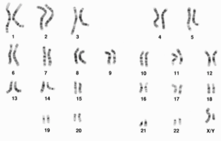 Image from BSCS
| ||||||||||||||
TOPIC: HOW CAN THE INHERITANCE OF A TRAIT IN A FAMILY BE SHOWN? (LS.Bio.7.1) | |
CMS BSCS Alignment: Lesson 10-13 | |
NOTES | ESSENTIAL CONTENT |
| |
Assume the pedigree shown is for the inheritance of Cystic Fibrosis. Label each individual on the pedigree with the correct genotype. |
|
TOPIC: HOW ARE TRAITS PASSED FROM PARENT TO OFFSPRING? | |
CMS BSCS Alignment: Lesson 14-15 | |
NOTES | ESSENTIAL CONTENT |
Draw a simple picture of sperm and egg combining in fertilization. | Sexual Reproduction
|
Write in the correct sequence of nucleotides that will be added to each side of the DNA molecule: G - - C T - - A T - - A G - - C A - - T | DNA Replication
|
Label the diagram of Meiosis with the correct number of chromosomes for a human cell: Draw another simple diagram of fertilization, this time showing the number of chromosomes in the gametes and the zygote. Label each cell on your drawing as haploid or diploid. | Meiosis
|
Nondisjunction
| |
TOPIC: WHY ARE SIBLINGS FROM THE SAME PARENT DIFFERENT? (LS.Bio.6.2) | |
CMS BSCS Alignment: Lesson 16 | |
NOTES | ESSENTIAL CONTENT |
Name a source of genetic variation that has already been discussed. | Genetic Variation
|
The diagram below shows maternal chromosomes and paternal chromosomes in different colors. In the circles provided, show 4 different ways these chromosomes can be sorted during meiosis. | Independent Assortment
|
How can crossing over make it harder to predict the inheritance of a polygenic trait such as high cholesterol? | Crossing Over
|