Cardiac Anatomy and Physiology Notes
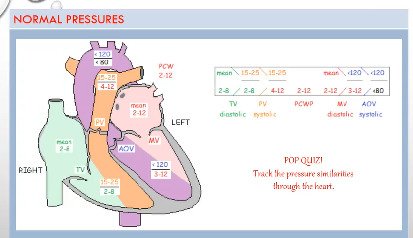
Pressures
PA/PCWP diastolic = LA mean
LA mean = LV diastolic
LV systolic = AO systolic
AO diastolic = LV end-diastolic pressure
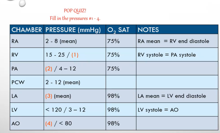
Pop Quiz Answers (Pressures)
2-8
15-25
2-12
Less than 120
Network of Vessels
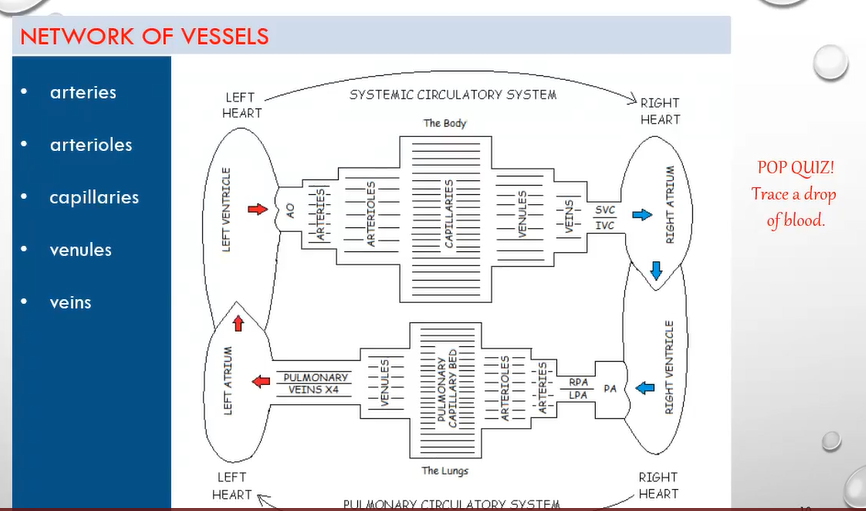
Flowchart shows the path from the right heart to the left, and from the left heart back to the right.
The capillary mesh bed brings arteries, arterioles, venules, and veins together.
Vessel Functions
Vessels transport blood to and from the heart throughout the body.
Arteries, arterioles, and capillaries: deliver oxygen and nutrients to tissues.
Capillaries, venules, and veins: remove carbon dioxide and waste products from tissues.
Arteries
Carry blood away from the heart.
Relatively thick with elasticity.
Maintain pressure in the system to keep blood flow moving forward.
High pressure within the systemic circulatory system presents greater resistance to the heart compared to the pulmonary circulatory system.
Arteries redistribute into smaller vessels called arterioles.
Arterioles
Redistribute further into tiny vessels, the capillaries.
Capillaries
Bridge arterioles and venules.
Tiny vessels that permit the exchange of nutrients, oxygen, waste, and carbon dioxide between the arterial and venous systems.
Venules
Converge to form larger veins.
Veins
Carry blood towards the heart.
Not as thick as arteries and can collapse/expand rapidly.
Pulmonary Circulatory System
Includes the right heart and all pulmonary vessels.
Order of flow:
Right heart to main pulmonary artery (PA).
Main PA bifurcates into left PA (LPA) and right PA (RPA).
Branches into smaller pulmonary arteries, arterioles.
Pulmonary capillary bed.
Venules.
Four pulmonary veins back to the left heart.
Systemic Circulatory System
Includes the left heart and all systemic vessels.
Order of flow from the left heart:
Aorta (AO).
Branches into smaller arteries, arterioles.
Capillaries.
Venules.
Veins.
Superior vena cava (SVC) and inferior vena cava (IVC) back to the right heart.
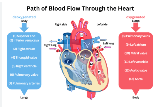
Trace a Drop of Blood
Start in the RA (Right Atrium) through the TV to RV (Right Ventricle) → through the PV (Pulmonary Valve) → PA (Pulmonary Artery) → RPA/LPA (Right/Left Pulmonary Artery) → Arteries → Arterioles → Pulmonary Capillary Bed → Venules → PV (Pulmonary Veins - 4) → LA (Left Atrium) → MV (Mitral Valve) → LV (Left Ventricle) → AOV (Aortic Valve) → AO (Aorta) → Arteries → Arterioles → Capillaries → Venules → Veins → SVC/IVC (Superior/Inferior Vena Cava) → RA (Right Atrium).
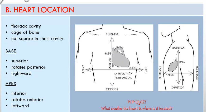

Heart Location
Located in the thoracic cavity.
Cradled in a cage of bone (sternum and vertebral column), cartilage, and muscle.
Just to the left of the midline of the mediastinum.
Above the diaphragm, with the lungs on either side.
Does not sit squarely in the chest cavity.
Terminology for Understanding Heart Position
Anterior (Ventral): Front of the body.
Posterior (Dorsal): Back of the body.
Superior (Basal): Towards the head, higher, or above.
Inferior (Diaphragmatic): Towards the feet, below, or lower.
Medial: Towards the midline.
Lateral: Away from the midline.
Proximal: Near the origin.
Distal: Away from the origin.
Visualizing the Heart in the Chest Cavity
Base (Top of the Heart):
Most superior.
Rotates posterior and in a rightward direction.
Apex (Bottom of the Heart):
Most inferior.
Rotates anterior and in a leftward direction.
Pop Quiz: Heart Location
What cradles the heart & where is it located?
The heart is cradled in a cage of bone (sternum, vertebral column), cartilage, and muscle, just left of the midline of the mediastinum, above the diaphragm, with lungs on each side.
Pericardium and Heart Wall
Pericardium
The heart is encased within the pericardium, a fluid-filled sac.
Covers the heart and helps prevent infection and trauma.
Aids in the free pumping motion of the heart.
It is a loose, double-layered sac.
Fibrous pericardium.
Serous pericardium.
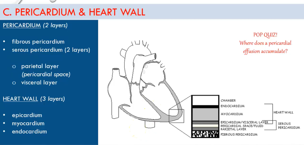
Fibrous Pericardium
The outer layer of the pericardium.
Serous Pericardium
The inner layer of the pericardium.
Consists of two layers:
Parietal layer: Lines the inside of the fibrous pericardium.
Visceral layer: Adheres to the outside of the heart (also known as the epicardium, the outermost layer of the heart).
Pericardial Space
Sits between the visceral and parietal layers and contains clear fluid released by the serous membrane.
The fluid acts as a lubricant during contraction (systole) and relaxation (diastole) of the heart.
Normally contains 10-30 ml of fluid.
In certain disease states, the pericardial space can fill with additional fluid, known as pericardial effusion.
Heart Walls
The heart wall consists of three layers:
Epicardium.
Myocardium.
Endocardium.
Epicardium
The outermost layer of the heart wall.
Covers the surface of the heart and extends to the great vessels.
Also known as the visceral layer of the serous pericardium.
Myocardium
The central layer of the heart wall.
Thick and muscular.
Composed of striated muscle fibrils that contract.
This layer is responsible for the heart's pumping action, facilitating the movement of blood throughout the circulatory system.
Endocardium
The innermost layer of the heart, it provides a smooth surface for blood flow and reduces friction during the contraction and relaxation of the heart.
The innermost layer of the heart, providing a smooth lining for the chambers and valves, and playing a crucial role in preventing blood clots.