knowt logo

Lecture 10: Alteration in pulmonary function

  • alterations of pulmonary function:

    • pulmonary disease is often classified as

      • acute or chronic

      • obstructive or restrictive

      • infectious or noninfectious

  • signs and symptoms of pulmonary disease:

    • pulmonary disease have many signs and symptoms (the most common symptoms are cough and dyspnea)

    • dyspnea

      • subjective sensation of uncomfortable breathing and feeling of unable to get enough air

      • orthopnea

        • dyspnea when a person is lying down

        • orthopnea is generally relieved by sitting up in a forward leaning position

      • paroxysmal nocturnal dyspnea is caused by fluid in the lungs

      • dyspnea causes

        • decreased ph, increased PaCO2, an decreased PaO2

        • decreased blood flow to the medulla oblongata

          • causes alternating periods of deep and shallow breathing

        • fatigue of the intercostal muscles and diaphragm

    • abnormal breathing patterns

      • kussmaul respirations (hyperpnea)- characterized as a slightly increased ventilatory rate; large, tidal volumes; and no expiratory pause

      • cheyne-stokes respirations- characterized by alternating deep and shallow breathing

    • hypoventilation- inadequate alveolar ventilation in relation to metabolic demand

    • hyperventilation- alveolar ventilation exceeds metabolic demand - lungs remove co2 faster - respiratory alkalosis can result

      • hyperventilation causes hypocapnia (reduced co2 in blood, not necessarily bad in short term)

    • cough- important reflex

      • acute cough

      • chronic cough

    • hemoptysis- coughing blood

    • cyanosis is bluish discoloration of the skin and mucous membrane caused by

      • increasing amount or desaturated or reduced hemoglobin in blood

      • usually develops when 5g of Hb is desaturated

    • pain- caused by pulmonary disorders

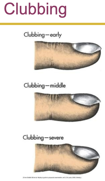
    • clubbing- the selective bulbous enlargement of the distal segment of a digit and is commonly associated w diseases that interfere with oxygenation of the blood

    • abnormal sputum- color, consistency, odor, and amount
  • conditions caused by pulmonary disease or injury:

    • hypercapnia- increased co2 in arterial blood caused by hypoventilation

    • hypoxemia- reduced oxygenation of arterial blood

      • high altitudes may produce hypoxemia through decreased inspired oxygen

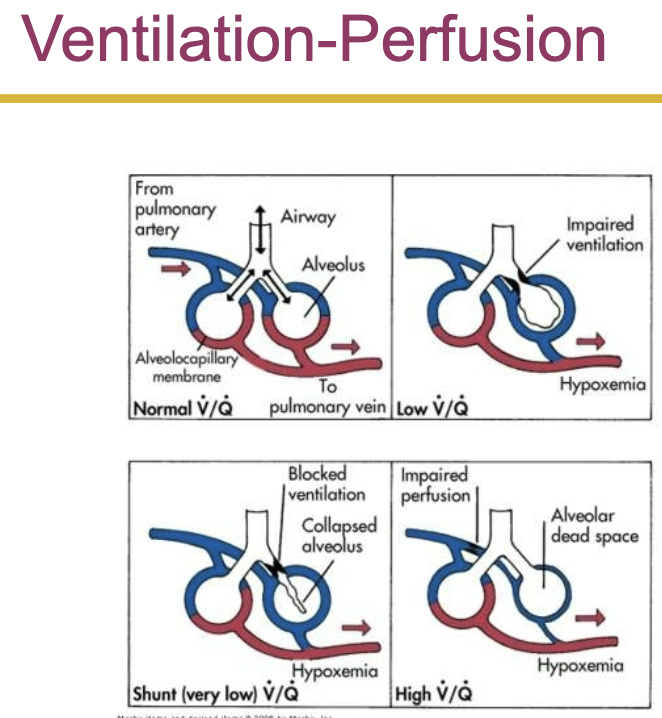
      • hypoxemia is most commonly caused by abnormal ventilation-perfusion ratio

      • pulmonary emboli causes alveolar dead space- alveolar is ventilated but not perfused

    • acute respiratory failure- inadequate gas exchange (hypoxemia or hypercapnia)

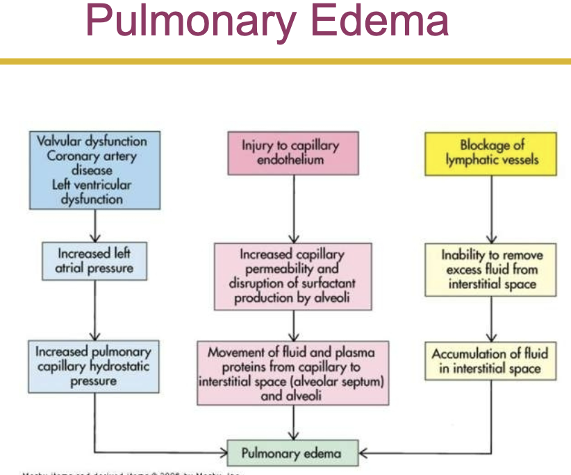
    • pulmonary edema

      • excess water in the lungs

      • edema and pulmonary fibrosis cause hypoxemia by impairing diffusion through the alveolocapillary membrane

      • the most common cause of edema is left sided heart failure

      • pulmonary edema usually begins when pulmonary capillary wedge pressure or left atrial pressure is 20mmhg
    • aspiration

      • passage of fluid and solid particles into the lungs due to impaired swallowing mechanism and cough reflex

    • atelectasis- collapse of lung tissue

      • compression atelectasis- caused by external pressure

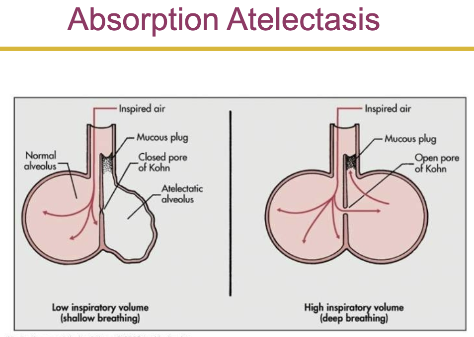
      • absorption atelectasis- the collapse of lung tissue caused from the lack of collateral ventilation through the pores of Kohn

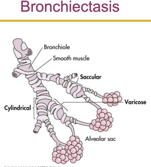
    • bronchiectasis

      • persistent abnormal dilation of the bronchi

      • in varicose bronchiectasis, constrictions and dilations deform the bronchi
    • bronchiolitis

      • inflammatory obstruction of the small airways

      • most common in children

      • occurs in adults w chronic bronchitis, in association with a viral infection, or with inhalation of toxic gasses

    • bronchiolitis obliterans

      • late stage fibrotic disease of the airways

      • can occur w all causes of bronchiolitis

  • pleural abnormalities:

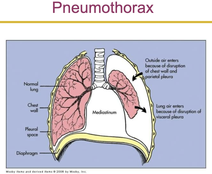
    • pneumothorax- the presence of gas or air in the pleural caused by rapture in visceral pleura

      • open pneumothorax- air pressure in pleural space equals barometric pressure

        • the lung no longer has the negative pressure environment it uses in order to expand

      • tension pneumothorax- involves a site of pleural rupture that acts as a one way valve, permitting air to enter on inspiration, but preventing its escape by closing during expiration

        • air builds up, putting pressure on the lung and compressing it

    • pleural effusion- the presence of fluid in the pleural space

      • transudative effusion- the fluid is watery and diffuses out of the capillaries as a result of increased blood pressure or decreased capillary oncotic pressure

      • exudative effusion- is less watery and contains high concentration of wbcs

    • empyema

      • involves an infected pleural effusion, the presence of pus in the pleural space, and a complication of respiratory infection, usually pneumonia

  • conditions caused by pulmonary disease or injury:

    • pulmonary fibrosis

      • excessive amount of fibrous or connective tissue in the lung

    • chest wall restriction

      • compromised chest wall

        • caused by neuromuscular disease, gross obesity, severe kyphoscoliosis (lateral bending and rotation of the spinal column, w distortion of thoracic cage)

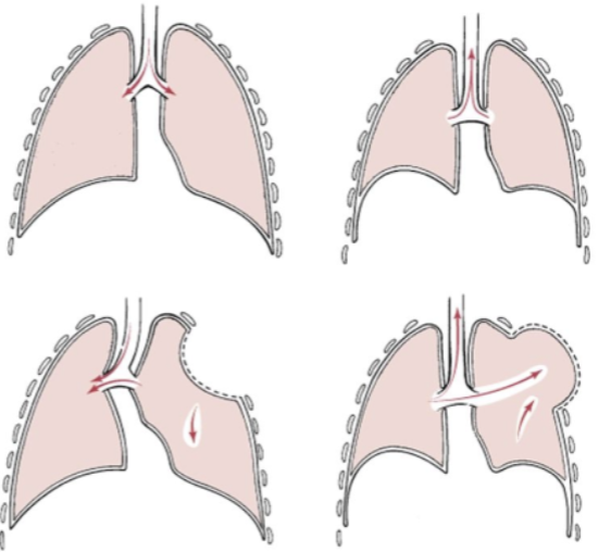
    • (flail chest- when multiple adjacent ribs are fractured in 2+ places which causes a section of the chest to lose stability causing it to move in the opposite direction that it should during breathing (it follows the lungs with pressure, inhalation = negative pressure in flail chest so goes in (instead of forcing air in bc the chest cant move) and vice versa)

    • inhalation disorders

      • exposure to toxic gasses

      • pneumoconiosis- any change in the lungs caused by inhalation or inorganic dust particle

        • silicosis and asbestosis

          • caused by inhalation of inorganic dust particles (ex. silicon)

          • cause inflammation and cellular apoptosis of lung cells

  • pulmonary disorders:

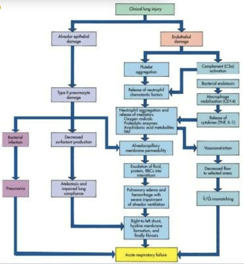
    • acute respiratory distress syndrome (ards)

      • is a fulminant (sudden development or rapid progression) form of respiratory failure characterized by acute lung inflammation and diffuse alveolocapillary injury

      • injury to the pulmonary capillary endothelium

      • inflammation leads to complement cascade and platelet activation

        • complement cascade releases inflammatory mediators such as proteolytic enzymes, oxygen free radicals, prostaglandins, leukotrienes, and platelet activating factor

      • increased capillary permeability causes pulmonary edema

      • surfactant is inactivated and type 2 alveolar cells are impaired, this causes alveoli and respiratory bronchioles to fill w fluid

      • manifestations

        • rapid, shallow breathing

        • respiratory alkalosis

        • dyspnea

        • decreased lung compliance

        • unresponsive hypoxemia

  • obstructive pulmonary disease:

    • in obstructive pulmonary disease either more force is required to expire a given volume of air, or emptying of the lungs is slowed, or both

    • airway obstruction that is worse w expiration

    • common signs and symptoms

      • dyspnea and wheezing

    • common obstructive disorders

      • asthma

      • emphysema

      • chronic bronchitis- caused when h influenzae and streptococcus pneumoniae bacteria become embedded in the airway secretions

        • manifestations: decreased exercise tolerance, wheezing, shortness of breath, and productive cough

    • asthma

      • immunoglobulin e may contribute to the pathophysiology of asthma

        • inflammatory mediators such as histamine, prostaglandins, and leukotrienes are produced in asthma

        • manifestations: inspiratory and expiratory wheezing, dyspnea, nonproductive cough, and tachypnea

        • elimination of the causative agent is the most successful treatment for asthma

  • chronic obstructive pulmonary disease:

    • chronic bronchitis

      • hypersecretion of mucus and chronic productive cough that lasts for at least 3 months of the year and for at least 2 consecutive years

      • inspired irritants increase mucous production and the size and number of mucous glands

      • the mucous is thicker than normal

    • emphysema

      • abnormal permanent enlargement of the gas exchange airways accompanied by destruction of alveolar walls without obvious fibrosis

      • loss of elastic recoil

      • centriacinar emphysema

      • panacinar emphysema

      • manifestations: dyspnea on exertion, use of accessory muscles, tachypnea, and prolonged expiration

  • respiratory tract infections:

    • the most common route of lower respiratory tract infection is aspiration of oropharyngeal secretions

    • tuberculosis

      • caused by mycobacterium tuberculosis

        • an acid fast bacillus that usually infects the lungs but may invade other organs

      • airborne transmission (highly contagious)

      • tubercle formation

        • in tuberculosis, the body wall off the bacilli in a tubercle by stimulating apoptotic infected macrophages that activate cytotoxic cells

      • caseous necrosis

      • positive tuberculin skin test (ppd)

      • treatment: antibiotic (type of drug and duration depends in individuals health history)

    • acute bronchitis

      • acute infection or inflammation of the airways or bronchi

      • commonly follows a viral illness

      • acute bronchitis causes similar symptoms to pneumonia but does not demonstrate pulmonary consolidation and chest infiltrates

    • chronic bronchitis

      • pathophysiology: increase in the number and size of mucous glands and goblet cells in airway epithelium

  • pulmonary vascular disease:

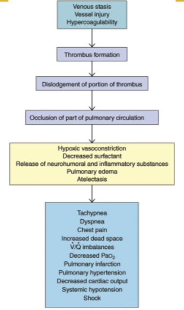
    • pulmonary embolism

      • occlusion of a portion of the pulmonary vascular bed by a thrombus, embolus, tissue fragment, lipids, or an air bubble

      • pulmonary emboli commonly arise from the deep veins in the thigh

      • the impact of embolus depends on the extend of pulmonary blood flow obstruction, the size of affected vessel, the nature of embolus, and the secondary effects

  • cancer:

    • lip cancer

      • most prevalent in men

      • the most common form of lip cancer is exophytic

    • laryngeal cancer

      • increased by the amount of tobacco smoked

      • combination of alcohol and smoking increases the risk of

  • lung cancer:

    • primary lung cancer arises from the bronchi within lungs, therefore called bronchogenic carcinomas

    • bronchogenic carcinomas

    • most common cause is cigarette smoking

      • heavy smokers have a 20 times greater chance of developing lung cancer than nonsmokers

      • smoking is related to cancers of the larynx, oral cavity, esophagus, and urinary bladder

    • environmental or occupational risk factors are also associated with lung cancer

    • other types of lung cancer are classified into small cell lung carcinoma and non small cell lung carcinoma

    • non small cell lung cancer

      • squamous cell carcinoma- slow growing tumor that produces a productive cough and airway obstruction in addition to pneumonia and atelectasis (partial or complete collapse of a lung or lobe of a lung due to the alveoli losing air (either bc they become deflated or filled w fluid))

      • adenocarcinoma

    • small cell carcinoma

      • secretion of antideuretic hormone is the most common paraneoplastic syndrome associated with small cell lung cancer

    • other lung cancers:

      • bronchial carcinoid tumors

      • adenocystic tumors (cylindromas)

      • mucoepidermoid carcinomas

      • mesotheliomas

Lecture 10: Alteration in pulmonary function

  • alterations of pulmonary function:

    • pulmonary disease is often classified as

      • acute or chronic

      • obstructive or restrictive

      • infectious or noninfectious

  • signs and symptoms of pulmonary disease:

    • pulmonary disease have many signs and symptoms (the most common symptoms are cough and dyspnea)

    • dyspnea

      • subjective sensation of uncomfortable breathing and feeling of unable to get enough air

      • orthopnea

        • dyspnea when a person is lying down

        • orthopnea is generally relieved by sitting up in a forward leaning position

      • paroxysmal nocturnal dyspnea is caused by fluid in the lungs

      • dyspnea causes

        • decreased ph, increased PaCO2, an decreased PaO2

        • decreased blood flow to the medulla oblongata

          • causes alternating periods of deep and shallow breathing

        • fatigue of the intercostal muscles and diaphragm

    • abnormal breathing patterns

      • kussmaul respirations (hyperpnea)- characterized as a slightly increased ventilatory rate; large, tidal volumes; and no expiratory pause

      • cheyne-stokes respirations- characterized by alternating deep and shallow breathing

    • hypoventilation- inadequate alveolar ventilation in relation to metabolic demand

    • hyperventilation- alveolar ventilation exceeds metabolic demand - lungs remove co2 faster - respiratory alkalosis can result

      • hyperventilation causes hypocapnia (reduced co2 in blood, not necessarily bad in short term)

    • cough- important reflex

      • acute cough

      • chronic cough

    • hemoptysis- coughing blood

    • cyanosis is bluish discoloration of the skin and mucous membrane caused by

      • increasing amount or desaturated or reduced hemoglobin in blood

      • usually develops when 5g of Hb is desaturated

    • pain- caused by pulmonary disorders

    • clubbing- the selective bulbous enlargement of the distal segment of a digit and is commonly associated w diseases that interfere with oxygenation of the blood

    • abnormal sputum- color, consistency, odor, and amount

  • conditions caused by pulmonary disease or injury:

    • hypercapnia- increased co2 in arterial blood caused by hypoventilation

    • hypoxemia- reduced oxygenation of arterial blood

      • high altitudes may produce hypoxemia through decreased inspired oxygen

      • hypoxemia is most commonly caused by abnormal ventilation-perfusion ratio

      • pulmonary emboli causes alveolar dead space- alveolar is ventilated but not perfused

    • acute respiratory failure- inadequate gas exchange (hypoxemia or hypercapnia)

    • pulmonary edema

      • excess water in the lungs

      • edema and pulmonary fibrosis cause hypoxemia by impairing diffusion through the alveolocapillary membrane

      • the most common cause of edema is left sided heart failure

      • pulmonary edema usually begins when pulmonary capillary wedge pressure or left atrial pressure is 20mmhg

    • aspiration

      • passage of fluid and solid particles into the lungs due to impaired swallowing mechanism and cough reflex

    • atelectasis- collapse of lung tissue

      • compression atelectasis- caused by external pressure

      • absorption atelectasis- the collapse of lung tissue caused from the lack of collateral ventilation through the pores of Kohn

    • bronchiectasis

      • persistent abnormal dilation of the bronchi

      • in varicose bronchiectasis, constrictions and dilations deform the bronchi
    • bronchiolitis

      • inflammatory obstruction of the small airways

      • most common in children

      • occurs in adults w chronic bronchitis, in association with a viral infection, or with inhalation of toxic gasses

    • bronchiolitis obliterans

      • late stage fibrotic disease of the airways

      • can occur w all causes of bronchiolitis

  • pleural abnormalities:

    • pneumothorax- the presence of gas or air in the pleural caused by rapture in visceral pleura

      • open pneumothorax- air pressure in pleural space equals barometric pressure

        • the lung no longer has the negative pressure environment it uses in order to expand

      • tension pneumothorax- involves a site of pleural rupture that acts as a one way valve, permitting air to enter on inspiration, but preventing its escape by closing during expiration

        • air builds up, putting pressure on the lung and compressing it
    • pleural effusion- the presence of fluid in the pleural space

      • transudative effusion- the fluid is watery and diffuses out of the capillaries as a result of increased blood pressure or decreased capillary oncotic pressure

      • exudative effusion- is less watery and contains high concentration of wbcs

    • empyema

      • involves an infected pleural effusion, the presence of pus in the pleural space, and a complication of respiratory infection, usually pneumonia

  • conditions caused by pulmonary disease or injury:

    • pulmonary fibrosis

      • excessive amount of fibrous or connective tissue in the lung

    • chest wall restriction

      • compromised chest wall

        • caused by neuromuscular disease, gross obesity, severe kyphoscoliosis (lateral bending and rotation of the spinal column, w distortion of thoracic cage)

    • (flail chest- when multiple adjacent ribs are fractured in 2+ places which causes a section of the chest to lose stability causing it to move in the opposite direction that it should during breathing (it follows the lungs with pressure, inhalation = negative pressure in flail chest so goes in (instead of forcing air in bc the chest cant move) and vice versa)
    • inhalation disorders

      • exposure to toxic gasses

      • pneumoconiosis- any change in the lungs caused by inhalation or inorganic dust particle

        • silicosis and asbestosis

          • caused by inhalation of inorganic dust particles (ex. silicon)

          • cause inflammation and cellular apoptosis of lung cells

  • pulmonary disorders:

    • acute respiratory distress syndrome (ards)

      • is a fulminant (sudden development or rapid progression) form of respiratory failure characterized by acute lung inflammation and diffuse alveolocapillary injury

      • injury to the pulmonary capillary endothelium

      • inflammation leads to complement cascade and platelet activation

        • complement cascade releases inflammatory mediators such as proteolytic enzymes, oxygen free radicals, prostaglandins, leukotrienes, and platelet activating factor

      • increased capillary permeability causes pulmonary edema

      • surfactant is inactivated and type 2 alveolar cells are impaired, this causes alveoli and respiratory bronchioles to fill w fluid

      • manifestations

        • rapid, shallow breathing

        • respiratory alkalosis

        • dyspnea

        • decreased lung compliance

        • unresponsive hypoxemia

  • obstructive pulmonary disease:

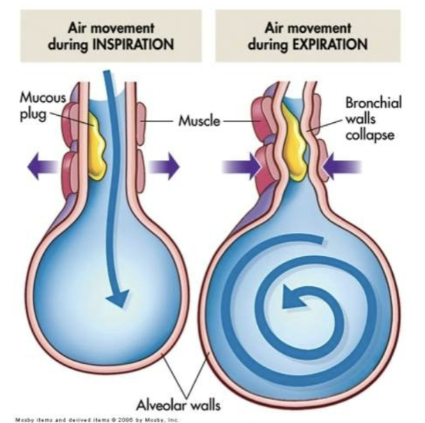
    • in obstructive pulmonary disease either more force is required to expire a given volume of air, or emptying of the lungs is slowed, or both

    • airway obstruction that is worse w expiration

    • common signs and symptoms

      • dyspnea and wheezing

    • common obstructive disorders

      • asthma

      • emphysema

      • chronic bronchitis- caused when h influenzae and streptococcus pneumoniae bacteria become embedded in the airway secretions

        • manifestations: decreased exercise tolerance, wheezing, shortness of breath, and productive cough

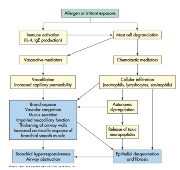
    • asthma

      • immunoglobulin e may contribute to the pathophysiology of asthma

        • inflammatory mediators such as histamine, prostaglandins, and leukotrienes are produced in asthma

        • manifestations: inspiratory and expiratory wheezing, dyspnea, nonproductive cough, and tachypnea

        • elimination of the causative agent is the most successful treatment for asthma
  • chronic obstructive pulmonary disease:
    • chronic bronchitis

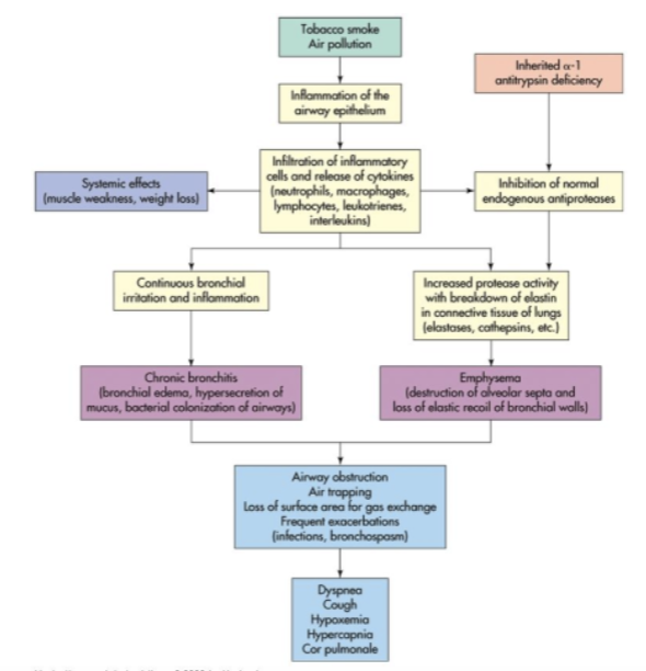
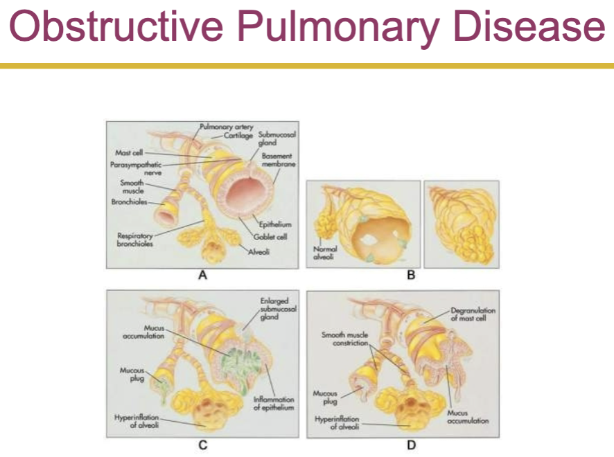
      • hypersecretion of mucus and chronic productive cough that lasts for at least 3 months of the year and for at least 2 consecutive years

      • inspired irritants increase mucous production and the size and number of mucous glands

      • the mucous is thicker than normal

    • emphysema

      • abnormal permanent enlargement of the gas exchange airways accompanied by destruction of alveolar walls without obvious fibrosis

      • loss of elastic recoil

      • centriacinar emphysema

      • panacinar emphysema

      • manifestations: dyspnea on exertion, use of accessory muscles, tachypnea, and prolonged expiration
  • respiratory tract infections:

    • the most common route of lower respiratory tract infection is aspiration of oropharyngeal secretions

    • tuberculosis

      • caused by mycobacterium tuberculosis

        • an acid fast bacillus that usually infects the lungs but may invade other organs

      • airborne transmission (highly contagious)

      • tubercle formation

        • in tuberculosis, the body wall off the bacilli in a tubercle by stimulating apoptotic infected macrophages that activate cytotoxic cells

      • caseous necrosis

      • positive tuberculin skin test (ppd)

      • treatment: antibiotic (type of drug and duration depends in individuals health history)

    • acute bronchitis

      • acute infection or inflammation of the airways or bronchi

      • commonly follows a viral illness

      • acute bronchitis causes similar symptoms to pneumonia but does not demonstrate pulmonary consolidation and chest infiltrates

    • chronic bronchitis

      • pathophysiology: increase in the number and size of mucous glands and goblet cells in airway epithelium

  • pulmonary vascular disease:

    • pulmonary embolism

      • occlusion of a portion of the pulmonary vascular bed by a thrombus, embolus, tissue fragment, lipids, or an air bubble

      • pulmonary emboli commonly arise from the deep veins in the thigh

      • the impact of embolus depends on the extend of pulmonary blood flow obstruction, the size of affected vessel, the nature of embolus, and the secondary effects
  • cancer:

    • lip cancer

      • most prevalent in men

      • the most common form of lip cancer is exophytic

    • laryngeal cancer

      • increased by the amount of tobacco smoked

      • combination of alcohol and smoking increases the risk of

  • lung cancer:

    • primary lung cancer arises from the bronchi within lungs, therefore called bronchogenic carcinomas

    • bronchogenic carcinomas

    • most common cause is cigarette smoking

      • heavy smokers have a 20 times greater chance of developing lung cancer than nonsmokers

      • smoking is related to cancers of the larynx, oral cavity, esophagus, and urinary bladder

    • environmental or occupational risk factors are also associated with lung cancer

    • other types of lung cancer are classified into small cell lung carcinoma and non small cell lung carcinoma

    • non small cell lung cancer

      • squamous cell carcinoma- slow growing tumor that produces a productive cough and airway obstruction in addition to pneumonia and atelectasis (partial or complete collapse of a lung or lobe of a lung due to the alveoli losing air (either bc they become deflated or filled w fluid))

      • adenocarcinoma

    • small cell carcinoma

      • secretion of antideuretic hormone is the most common paraneoplastic syndrome associated with small cell lung cancer

    • other lung cancers:

      • bronchial carcinoid tumors

      • adenocystic tumors (cylindromas)

      • mucoepidermoid carcinomas

      • mesotheliomas

robot