Lecture 3
Summary of Actin Dynamics
Actin filaments undergo dynamic assembly and disassembly, which is crucial for various cellular processes
Actin at the Leading Edge of a Motile Cell
Actin filaments are highly dynamic at the leading edge of motile cells
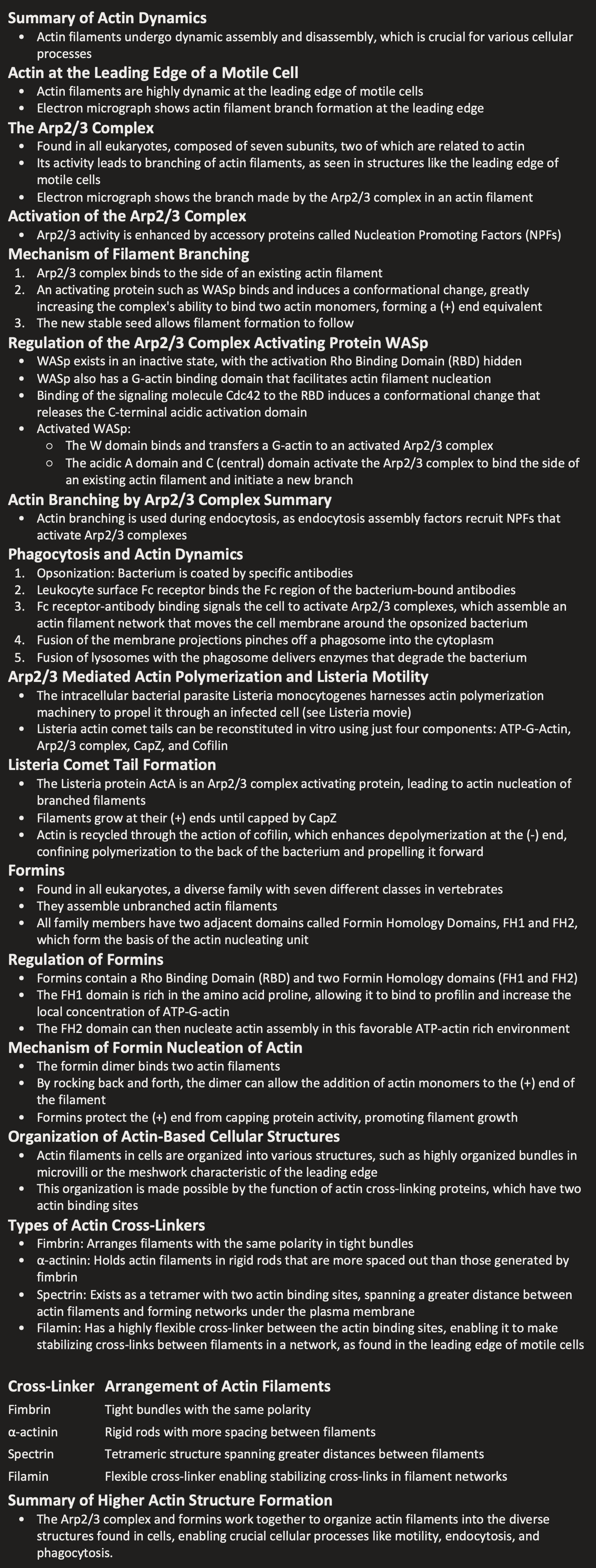
Electron micrograph shows actin filament branch formation at the leading edge
The Arp2/3 Complex
Found in all eukaryotes, composed of seven subunits, two of which are related to actin
Its activity leads to branching of actin filaments, as seen in structures like the leading edge of motile cells
Electron micrograph shows the branch made by the Arp2/3 complex in an actin filament
Activation of the Arp2/3 Complex
Arp2/3 activity is enhanced by accessory proteins called Nucleation Promoting Factors (NPFs)
Mechanism of Filament Branching
Arp2/3 complex binds to the side of an existing actin filament
An activating protein such as WASp binds and induces a conformational change, greatly increasing the complex's ability to bind two actin monomers, forming a (+) end equivalent
The new stable seed allows filament formation to follow
Regulation of the Arp2/3 Complex Activating Protein WASp
WASp exists in an inactive state, with the activation Rho Binding Domain (RBD) hidden
WASp also has a G-actin binding domain that facilitates actin filament nucleation
Binding of the signaling molecule Cdc42 to the RBD induces a conformational change that releases the C-terminal acidic activation domain
Activated WASp:
The W domain binds and transfers a G-actin to an activated Arp2/3 complex
The acidic A domain and C (central) domain activate the Arp2/3 complex to bind the side of an existing actin filament and initiate a new branch
Actin Branching by Arp2/3 Complex Summary
Actin branching is used during endocytosis, as endocytosis assembly factors recruit NPFs that activate Arp2/3 complexes
Phagocytosis and Actin Dynamics
Opsonization: Bacterium is coated by specific antibodies
Leukocyte surface Fc receptor binds the Fc region of the bacterium-bound antibodies
Fc receptor-antibody binding signals the cell to activate Arp2/3 complexes, which assemble an actin filament network that moves the cell membrane around the opsonized bacterium
Fusion of the membrane projections pinches off a phagosome into the cytoplasm
Fusion of lysosomes with the phagosome delivers enzymes that degrade the bacterium
Arp2/3 Mediated Actin Polymerization and Listeria Motility
The intracellular bacterial parasite Listeria monocytogenes harnesses actin polymerization machinery to propel it through an infected cell (see Listeria movie)
Listeria actin comet tails can be reconstituted in vitro using just four components: ATP-G-Actin, Arp2/3 complex, CapZ, and Cofilin
Listeria Comet Tail Formation
The Listeria protein ActA is an Arp2/3 complex activating protein, leading to actin nucleation of branched filaments
Filaments grow at their (+) ends until capped by CapZ
Actin is recycled through the action of cofilin, which enhances depolymerization at the (-) end, confining polymerization to the back of the bacterium and propelling it forward
Formins
Found in all eukaryotes, a diverse family with seven different classes in vertebrates
They assemble unbranched actin filaments
All family members have two adjacent domains called Formin Homology Domains, FH1 and FH2, which form the basis of the actin nucleating unit
Regulation of Formins
Formins contain a Rho Binding Domain (RBD) and two Formin Homology domains (FH1 and FH2)
The FH1 domain is rich in the amino acid proline, allowing it to bind to profilin and increase the local concentration of ATP-G-actin
The FH2 domain can then nucleate actin assembly in this favorable ATP-actin rich environment
Mechanism of Formin Nucleation of Actin
The formin dimer binds two actin filaments
By rocking back and forth, the dimer can allow the addition of actin monomers to the (+) end of the filament
Formins protect the (+) end from capping protein activity, promoting filament growth
Organization of Actin-Based Cellular Structures
Actin filaments in cells are organized into various structures, such as highly organized bundles in microvilli or the meshwork characteristic of the leading edge
This organization is made possible by the function of actin cross-linking proteins, which have two actin binding sites
Types of Actin Cross-Linkers
Fimbrin: Arranges filaments with the same polarity in tight bundles
α-actinin: Holds actin filaments in rigid rods that are more spaced out than those generated by fimbrin
Spectrin: Exists as a tetramer with two actin binding sites, spanning a greater distance between actin filaments and forming networks under the plasma membrane
Filamin: Has a highly flexible cross-linker between the actin binding sites, enabling it to make stabilizing cross-links between filaments in a network, as found in the leading edge of motile cells
Cross-Linker | Arrangement of Actin Filaments |
Fimbrin | Tight bundles with the same polarity |
α-actinin | Rigid rods with more spacing between filaments |
Spectrin | Tetrameric structure spanning greater distances between filaments |
Filamin | Flexible cross-linker enabling stabilizing cross-links in filament networks |
Summary of Higher Actin Structure Formation
The Arp2/3 complex and formins work together to organize actin filaments into the diverse structures found in cells, enabling crucial cellular processes like motility, endocytosis, and phagocytosis.