knowt logo

6.2 - The Neuromuscular System

How Muscle Connects to Bone

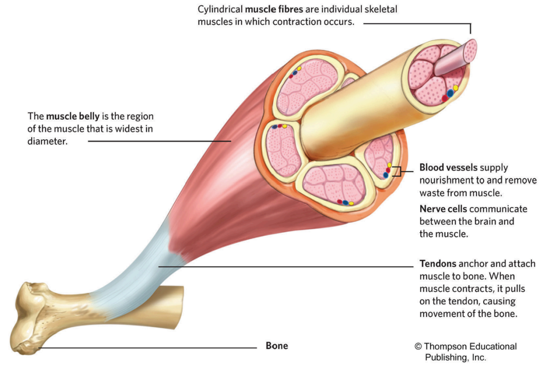
  • Skeletal muscle is attached to the bone either indirectly (via tendons) or directly (when the outer membrane of the muscle attaches to the outer membrane of the bone).

    • The most common of the two ways in which muscles attach to bones is the indirect method (i.e., via tendons)

How Muscle Connects to Bone
The Anatomy of Skeletal Muscle

  • A substantial portion of human body weight is made up of skeletal muscle

    • These are the muscles that are directly involved in movement and locomotion. 

    • The basic unit of skeletal muscle is the individual skeletal muscle fibre or muscle cell.

The Anatomy of Skeletal Muscle (Definitions)

Skeletal muscle fibre: The basic unit of skeletal muscle.

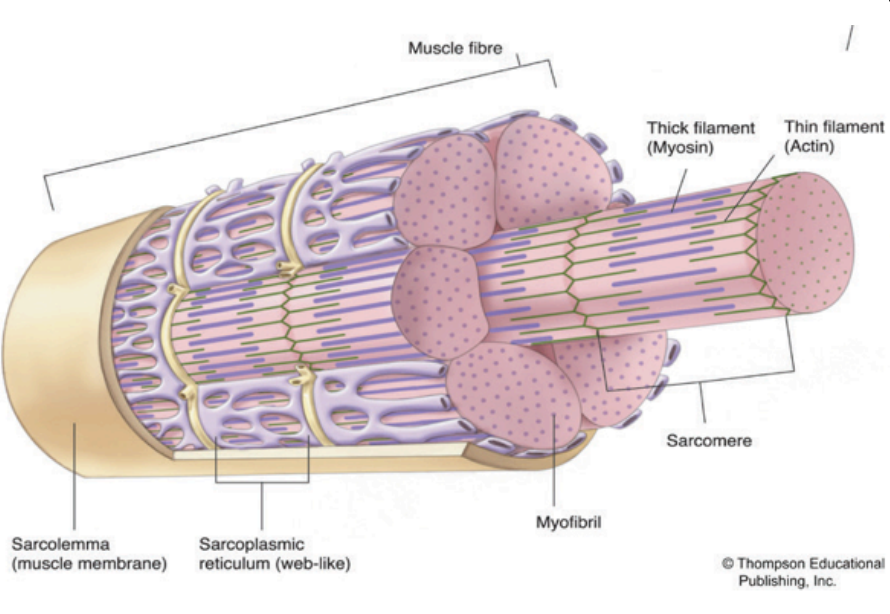
Sarcolemma: A plasma membrane that lies beneath the endomysium, a sheath of connective tissue that surrounds a muscle fibre.

Sarcoplasm: The muscle cell’s cytoplasm, which is contained by the sarcolemma.

Sarcomeres: The units of skeletal muscle containing the cellular proteins myosin and actin.

Sarcoplasmic reticulum: A network of channels in each muscle fibre that transport the electrochemical substances involved in muscle activation.
Looking Outwards

  • Looking outward from the surface of the individual muscle fibre, a sheath of connective tissue (the perimysium) binds groups of muscle fibres together. 

    • These bundles (fasciculi) are bound together by a larger and stronger sheath of tissue (the epimysium) that envelopes the entire muscle.

      • The epimysium then extends beyond the muscle and changes its properties as it becomes one with the tendon. 

    • The tendon extends itself and becomes one with the bone's periosteum. 

      • This connection happens at both attachment sites (at the muscle's origin and its insertion).

Looking Inwards

  • The muscle fiber is surrounded by connective tissue known as the endomysium.

  • The sarcolemma, or muscle cell membrane, encloses the sarcoplasm, the cytoplasm of the muscle cell.

    • The sarcoplasm contains larger amounts of glycogen, myoglobin, calcium, and organelles like mitochondria.

  • Myofibrils, thread-like structures, run along the length of the muscle fiber.

    • Within myofibrils are thick and thin filaments composed of myosin and actin proteins.

      • Sarcomeres, repeating structural units, house myosin and actin.

        • Myosin consists of a head and tail resembling a golf club, with the head having an attachment site for actin.

        • Actin contains troponin (with a calcium-binding site) and tropomyosin (covering the actin-binding site).

  • Troponin (the binding site for calcium) and tropomyosin (the string-looking structure that covers the binding site on actin) act as a swivel-lock mechanism, requiring calcium release for myosin attachment.

    • Muscle contraction occurs as filaments slide across each other, shortening the sarcomere.

  • The sliding filament theory describes muscle contraction at the molecular level.

The Motor Unit

  • Nerves transmit impulses in waves to ensure smooth movements, with each impulse triggering a muscle twitch upon reaching the muscle.

    • A single motor neuron, known as the "motor neuron," can stimulate multiple muscle fibers, collectively referred to as a motor unit, along with its axon pathway.

    • Motor units are categorized into small and large units, where small units control fine motor movements (e.g., eye muscles), while large units govern gross motor movements (e.g., quadriceps).

    • To produce maximal muscle force, all motor units within a muscle or muscle group must be recruited simultaneously, with each unit contracting at the same time.

  • The size of motor units varies, with slow-twitch muscle motor units typically smaller due to fewer muscle fibers compared to fast-twitch motor units.

  • Motor units adhere to the all-or-none principle, meaning when stimulated, they contract to their fullest potential, with all muscle fibers either contracting or none at all.

  1. How do nerves ensure smooth movements?

  2. What is the role of a motor neuron in muscle movement?

  3. What distinguishes small motor units from large motor units?

  4. Why is it necessary for all motor units within a muscle or muscle group to be recruited simultaneously to produce maximal muscle force?

  5. What accounts for the size difference between slow-twitch muscle motor units and fast-twitch motor units?

  6. What is meant by the all-or-none principle in relation to motor units?
The All-or-None Principle

  • Motor units also comply to a rule known as the all-or-none principle (or law).

    • This principle stipulates that, when a motor unit is stimulated to contract, it will do so to its fullest potential.

      • In other words, if a motor unit consists of 10 muscle fibres (or 800 muscle fibres) and they are “turned on,” either all fibres will contract or none will contract.

The Neuromuscular System 

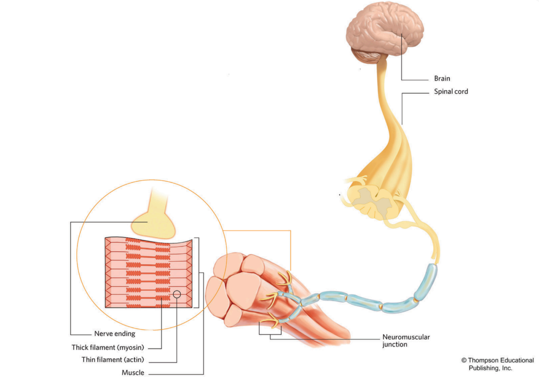
  • The neuromuscular system refers to the connection between the muscular and nervous systems, facilitating nerve impulses from the brain and spinal cord.

    • It involves complex interactions between these systems, working together seamlessly.

  • For example, when preparing to kick a soccer ball, messages are sent from the brain or spinal cord to initiate the action.

    • These messages trigger chemical reactions in the muscles of the leg.

      • Despite involving multiple nerves and muscles, the action of kicking the ball appears automatic and instantaneous.

The Neuromuscular Junction

  • The Neuromuscular Junction (NMJ) is the connection of motor neurons to the muscle fibers, crucial for transmitting signals from the nervous system to the muscular system, leading to muscle contraction.

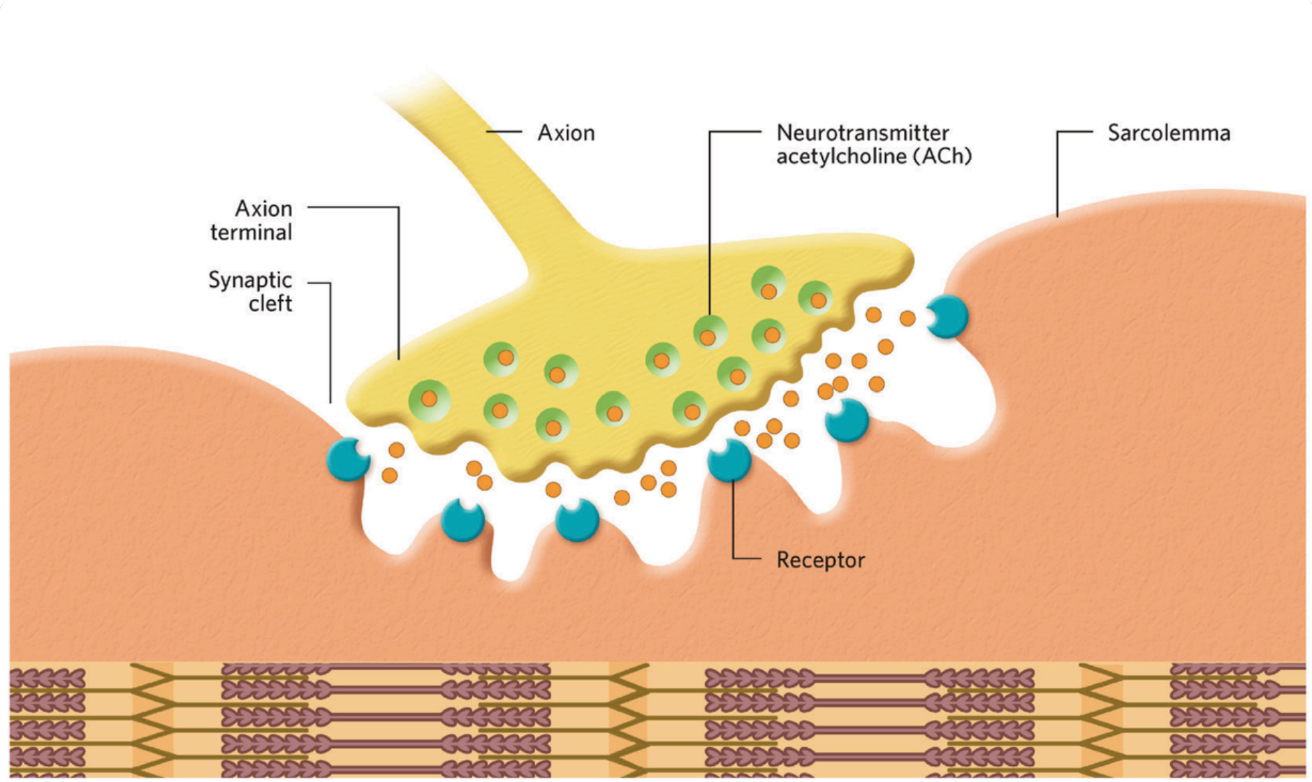
    • It is located where the motor neuron terminal meets the muscle fiber, comprising the motor neuron terminal, synaptic cleft, and motor end plate on the muscle fiber membrane.

  • The process of the NMJ is:

    • The arrival of an action potential at the motor neuron terminal.

    • Depolarization triggers the release of neurotransmitter (acetylcholine - ACh) into the synaptic cleft.

    • ACh binds to receptors on the motor end plate of muscle fiber.

    • The binding of ACh opens ion channels, leading to the depolarization of the muscle fiber membrane.

    • Depolarization grows along muscle fiber membrane (sarcolemma) by transverse tubules (T-tubules).

    • This triggers the release of calcium ions from the sarcoplasmic reticulum (SR).

    • Calcium ions bind to troponin, initiating muscle contraction.

  • The NMJ is essential for both voluntary and involuntary muscle movements, providing precise control over muscle activity.

  • The consistent use and regular practice contribute to the enhancement of neuromuscular and nervous systems, improving efficiency and ability to generate movement.
AP

6.2 - The Neuromuscular System

How Muscle Connects to Bone

  • Skeletal muscle is attached to the bone either indirectly (via tendons) or directly (when the outer membrane of the muscle attaches to the outer membrane of the bone).

    • The most common of the two ways in which muscles attach to bones is the indirect method (i.e., via tendons)

How Muscle Connects to Bone

The Anatomy of Skeletal Muscle

  • A substantial portion of human body weight is made up of skeletal muscle

    • These are the muscles that are directly involved in movement and locomotion. 

    • The basic unit of skeletal muscle is the individual skeletal muscle fibre or muscle cell.

The Anatomy of Skeletal Muscle (Definitions)

Skeletal muscle fibre: The basic unit of skeletal muscle.

Sarcolemma: A plasma membrane that lies beneath the endomysium, a sheath of connective tissue that surrounds a muscle fibre.

Sarcoplasm: The muscle cell’s cytoplasm, which is contained by the sarcolemma.

Sarcomeres: The units of skeletal muscle containing the cellular proteins myosin and actin.

Sarcoplasmic reticulum: A network of channels in each muscle fibre that transport the electrochemical substances involved in muscle activation.

Looking Outwards

  • Looking outward from the surface of the individual muscle fibre, a sheath of connective tissue (the perimysium) binds groups of muscle fibres together. 

    • These bundles (fasciculi) are bound together by a larger and stronger sheath of tissue (the epimysium) that envelopes the entire muscle.

      • The epimysium then extends beyond the muscle and changes its properties as it becomes one with the tendon. 

    • The tendon extends itself and becomes one with the bone's periosteum. 

      • This connection happens at both attachment sites (at the muscle's origin and its insertion).

Looking Inwards

  • The muscle fiber is surrounded by connective tissue known as the endomysium.

  • The sarcolemma, or muscle cell membrane, encloses the sarcoplasm, the cytoplasm of the muscle cell.

    • The sarcoplasm contains larger amounts of glycogen, myoglobin, calcium, and organelles like mitochondria.

  • Myofibrils, thread-like structures, run along the length of the muscle fiber.

    • Within myofibrils are thick and thin filaments composed of myosin and actin proteins.

      • Sarcomeres, repeating structural units, house myosin and actin.

        • Myosin consists of a head and tail resembling a golf club, with the head having an attachment site for actin.

        • Actin contains troponin (with a calcium-binding site) and tropomyosin (covering the actin-binding site).

  • Troponin (the binding site for calcium) and tropomyosin (the string-looking structure that covers the binding site on actin) act as a swivel-lock mechanism, requiring calcium release for myosin attachment.

    • Muscle contraction occurs as filaments slide across each other, shortening the sarcomere.

  • The sliding filament theory describes muscle contraction at the molecular level.

The Motor Unit

  • Nerves transmit impulses in waves to ensure smooth movements, with each impulse triggering a muscle twitch upon reaching the muscle.

    • A single motor neuron, known as the "motor neuron," can stimulate multiple muscle fibers, collectively referred to as a motor unit, along with its axon pathway.

    • Motor units are categorized into small and large units, where small units control fine motor movements (e.g., eye muscles), while large units govern gross motor movements (e.g., quadriceps).

    • To produce maximal muscle force, all motor units within a muscle or muscle group must be recruited simultaneously, with each unit contracting at the same time.

  • The size of motor units varies, with slow-twitch muscle motor units typically smaller due to fewer muscle fibers compared to fast-twitch motor units.

  • Motor units adhere to the all-or-none principle, meaning when stimulated, they contract to their fullest potential, with all muscle fibers either contracting or none at all.

  1. How do nerves ensure smooth movements?

  2. What is the role of a motor neuron in muscle movement?

  3. What distinguishes small motor units from large motor units?

  4. Why is it necessary for all motor units within a muscle or muscle group to be recruited simultaneously to produce maximal muscle force?

  5. What accounts for the size difference between slow-twitch muscle motor units and fast-twitch motor units?

  6. What is meant by the all-or-none principle in relation to motor units?

The All-or-None Principle

  • Motor units also comply to a rule known as the all-or-none principle (or law).

    • This principle stipulates that, when a motor unit is stimulated to contract, it will do so to its fullest potential.

      • In other words, if a motor unit consists of 10 muscle fibres (or 800 muscle fibres) and they are “turned on,” either all fibres will contract or none will contract.

The Neuromuscular System 

  • The neuromuscular system refers to the connection between the muscular and nervous systems, facilitating nerve impulses from the brain and spinal cord.

    • It involves complex interactions between these systems, working together seamlessly.

  • For example, when preparing to kick a soccer ball, messages are sent from the brain or spinal cord to initiate the action.

    • These messages trigger chemical reactions in the muscles of the leg.

      • Despite involving multiple nerves and muscles, the action of kicking the ball appears automatic and instantaneous.

The Neuromuscular Junction

  • The Neuromuscular Junction (NMJ) is the connection of motor neurons to the muscle fibers, crucial for transmitting signals from the nervous system to the muscular system, leading to muscle contraction.

    • It is located where the motor neuron terminal meets the muscle fiber, comprising the motor neuron terminal, synaptic cleft, and motor end plate on the muscle fiber membrane.

  • The process of the NMJ is:

    • The arrival of an action potential at the motor neuron terminal.

    • Depolarization triggers the release of neurotransmitter (acetylcholine - ACh) into the synaptic cleft.

    • ACh binds to receptors on the motor end plate of muscle fiber.

    • The binding of ACh opens ion channels, leading to the depolarization of the muscle fiber membrane.

    • Depolarization grows along muscle fiber membrane (sarcolemma) by transverse tubules (T-tubules).

    • This triggers the release of calcium ions from the sarcoplasmic reticulum (SR).

    • Calcium ions bind to troponin, initiating muscle contraction.

  • The NMJ is essential for both voluntary and involuntary muscle movements, providing precise control over muscle activity.

  • The consistent use and regular practice contribute to the enhancement of neuromuscular and nervous systems, improving efficiency and ability to generate movement.

robot