External Anatomy of Toads
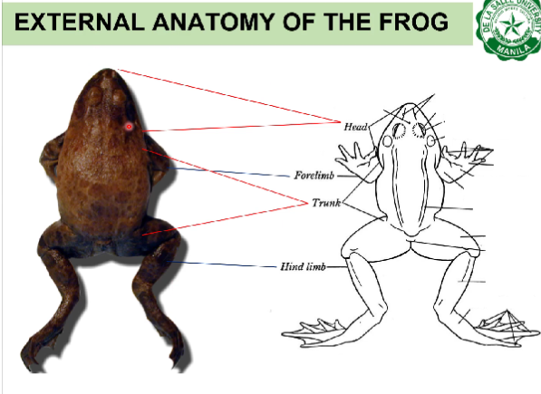
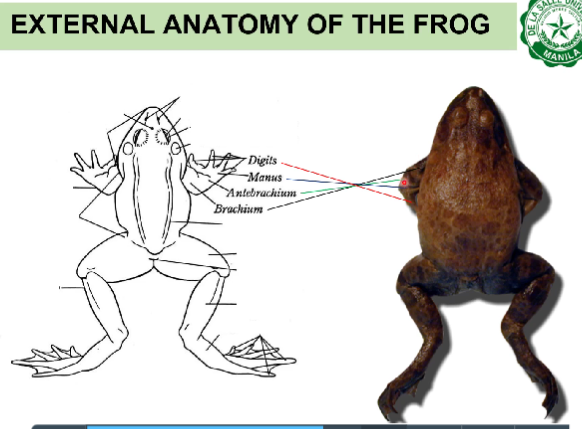
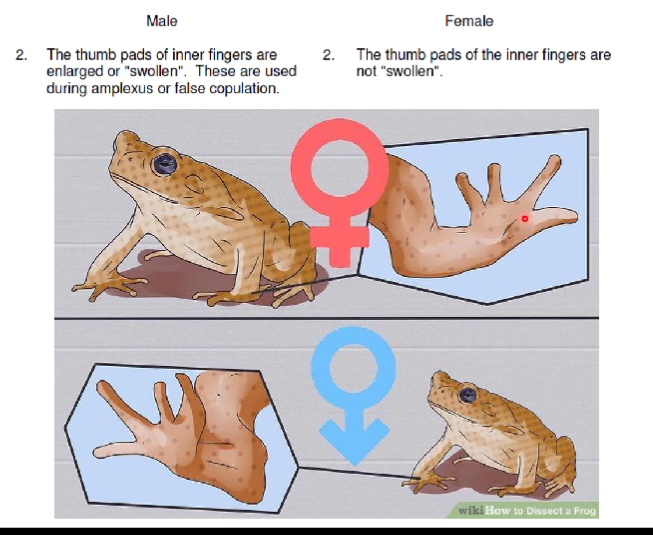
Frogs and toads belong to the order Anura, the largest and the most diverse group of living amphibians. They are commonly used in the general zoology laboratory because of their ready availability. The large bullfrogs and toads are the favorite for dissection because they represent well the body parts and structures of vertebrates. Objectives At the end of the activity, the student should be able to: 1. Describe the histology of frog’s/ toad’s skin 2. Identify the external orientations of a toad 3. Identify the external structures in toad Materials Compound light microscope Dissecting pan Probe Prepared slide of toad’s skin Preserved toad Procedure A. Histology of the Skin Study a cross section of a toad’s skin. The skin of the toad, similar to the frog, is divided into two major layers, the outer epidermis and the inner dermis. These are further subdivided into sublayers, which should be studied with both the low and high powers of the microscopes. The epidermis is a stratified epithelium consisting of two sublayers of epithelial cells. The stratum corneum is the outermost stratified layer of squamous epithelial tissue of flattened, dead cells. This is the thin hardened or cornified layer constantly removed when the toad sheds its skin (ecdysis or molting). The innermost sublayer is the stratum germinativum, consisting of living and growing layer of stratified epithelial tissue. The cuboidal to columnar cells continuously divide and replace the constantly shed off stratum corneum of the skin. At the junction of epidermis and dermis are chromatophores, specialized connective tissue which contains pigments. Specifically, they contain a row of black pigment containing cells called melanophores which are in large part responsible for the color of the skin. The dermis, or inner layer of skin, is divided into two sublayers: the stratum laxum/ spongiosum and the stratum compactum. The stratum laxum/ spongiosum is the outer sublayer immediately below the stratum germinativum. It consists of loosely arranged connective tissue and blood vessels. It is the layer where the body of cutaneous glands are situated, thus giving it a spongy appearance. Numerous glands are found in this layer. The glands are of two types- the poison glands (larger glands) possess a thin epithelial wall, and the smaller and more numerous, the mucous glands, have thicker walls. The inner layer of the dermis is the stratum compactum. It consists of layers of white fibrous connective tissue which are compactly arranged. Label the photomicrograph in the Activity Sheet. B. External Anatomy of the Toad Get a preserved toad; rinse it well with water to remove excess chloroform. Study its external features and identify the different parts as described below: 1. The toad exhibits bilateral symmetry- it can be differentiated into two mirrored portions, namely, right and left halves. The regions of the body include: (a) anterior- head end; (b) posterior- tail end; (c) dorsal- back side; and (d) ventral- belly side. Identify these four regions of the body in man. The fact that the main axis of toad is horizontal, while that of man is vertical might cause confusion. Indicate their relative position. The body of the toad is divided into three planes according to how the body is divided: (a) frontal- into dorsal and ventral sections; (b) sagittal (longitudinal)- into right and left sections; (c) transverse (cross)- into anterior and posterior sections. With reference to location relative to the center of the body, the following terms are used: (a) medial- near the middle; (b) lateral- to the sides; (c) proximalpart of a structure that is nearer the origin; and (d) distal- part farther from the origin. 2. The axial region is composed of the head and the trunk. Note that the two are broadly joined by a very short neck thereby restricting movement of the head. The most anterior portion of the head is the pointed snout. Locate a pair of openings at the tip of the snout. These are the external nares. At the dorsolateral portion of the head, posterior to the exterior nares are the bulging eyes. It has upper and lower eyelids and a third, a transparent nictitating membrane which keeps the eyes moist when underwater. Posterolateral to the eye is an ovoid region of tightly drawn skin, the tympanic membrane (eardrum) covering the auditory canal. The frog and toad do not have external ear, only the middle and internal ears. Behind the tympanic membrane at each side of the head is a large swelling called paratoid gland/ poison gland. Locate the cloacal aperture at the most posterior part of the trunk, on the dorsal side. 3. The appendicular region is made up of the forelimbs (anterior pair) and the hindlimbs (posterior pair). The paired appendages vary in form and function. The forelimbs are short and consist of four main parts: the upper arm, forearm, wrist/carpus, and hand/manus which bear four digits and a rudimentary thumb called prepollux. The hindlimbs are long and consist of the thigh, shank, ankle/tarsus, and foot/pes. These bear five digits and a sixth rudimentary toe called prehallux. Note the webbed toes. 4. The skin of toad has a warty appearance on the dorsal side while the ventral side has none. Note the dorsal-ventral difference in coloration. 5. Label all structures mentioned above in the photograph provided. C. External Sex Determining Characteristics of the toad Determine the sex of the toad based on the following tabulated distinguishing external characteristics: MALE FEMALE 1. The skin on the ventral side of the head near the angles of the jaw has a distinct dark pigmentation due to concentration of pigments. 1. The pigments are diffused; hence, there is no distinct pigmentation at this area. 2. The thumb pads of inner fingers are enlarged or “swollen”. These are used during amplexus or false copulation. 2. The thumb pads of the inner fingers are not “swollen”. 3. Sexually mature male frogs are generally smaller than female. 3. Sexually mature female frogs are relatively bigger than male