The Brain: Neuroplasticity and Tools of Discover 1.4a
1.4-1 Why are psychologists concerned with human biology?
Phrenology: studying bumps on the skull and now a debunked practice
Biological psychologists use advanced technology to study links between biological (genetic, neural, hormonal) and psychological processes
Researchers discovered:
among the body’s cells are neurons that conduct electricity and “Talk to one another by sending chemical messages across a synapse
our experiences wire our adaptive brain
specific brain systems serve specific functions
we integrate info processed in these brain systems to construct our experiences of sights and sounds, meanings and memories, pain and passion
we are each a system composed of subsystems that are in turn composed of even smaller systems
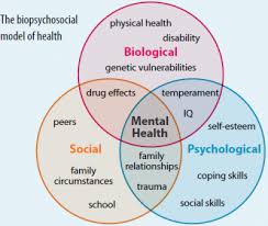
the biopsychosocial approach uses three levels on analysis: biological, psychological, and social-cultural
Biological: Genetics, brain mechanisms, and hormonal influences.
Psychological: Emotions, learned experiences, and cognitive processing.
Social-Cultural: Influence of culture, relationships, and social environments
The Power of Neuroplasticity
your brain is constantly changing as it adjusts to new experiences (neuroplasticity)
Ex: If a pianist practices 45 min a day on the piano their motor learning-related brain areas will grow
Main Idea: Neuroplasticity is how humans are able to adapt, this leads to growth in systems of the brain. Think of how much the world changed in 50 years and how it’ll continue to change
Tools of Discovery: Having our Head Examined
we think with our brain, by releasing billions of neurotransmitter molecules across trillions of synapses
damage to one side of the brain caused numbness or paralysis on the opposite side
suggesting that the body’s right side if wired to the brain’s left and vice versa
damage to the back of the brain disputed vision
damage to the left front part of the brain produced speech difficulties
now, scientists can selectively lesion (destroy) tiny clusters of brain cells, observing brain function
now, neuroscientists can stimulate brain parts (electrically, chemically, or magnetically) and note the effects
optogenetics: a technique that allows neuroscientists to control the activity of individual neurons
EEG (electroencephalogram): amplified readout of waves of electrical activity sweeping across the brain’s surface
recorded through a shower-cap-like hat filled with electrodes covered with conductive gel
researchers lack direct access to the brain but can present a stimulus repeatedly and have a computer filter out brain activity unrelated to the stimulus
Good for measuring brain activity but lacks detailed spatial resolution
MEG (magnetoencephalography): measures magnetic fields from the brain’s natural electrical activity
to isolate the brain’s magnetic fields, researchers create special rooms that cancel out other magnetic signals
participants sit underneath a head coil that’s like a salon hair dryer
participants complete activities that send tens of thousands of neurons that generate electrical pulses, which creates a magnetic field
CT (computed tomography) scan: examines the brain by taking X ray photographs that reveal brain damage
able to see inside the living brain
PET (positron emission tomography): depicts brain activity by showing each brain area’s consumption of sugar glucose
active neurons eat glucose
after a person receives temporarily radioactive glucose, the PET scan tracks the gamma rays released by this “food for thought” as a task is performed
PET scan “hot spots” show the most active brain areas as the person does math calculation, daydreams, looks at images of people
MRI (magnetic resonance imaging): uses magnetic fields and radio waves to produced computer-generated images of soft tissue
show brain anatomy
patient’s head is put into a magnetic field, which aligns spinning atoms in brain molecules
then radio wave pulse momentarily disorients the atoms
after atoms return to normal spin, they emit signals that provide detailed pictures of soft tissues
Ex: scans revealed larger than average neural area in left hemisphere of musicians who display perfect pitch
fMRI (functional MRI): a technique for revealing blood and flow, and brain activity by comparing succecsive MRI scans
where the brain is especially active, blood goes
by comparing successive MRI scans, researchers can watch as specific brain areas activate, showing increased oxygen-laden blood flow
Provides detailed images of brain activity but requires the subject to remain still
Understanding the tools and techniques used in neuroscience helps us appreciate how our adaptive and complex brain works. Neuroplasticity, for example, highlights the brain's resilience and potential for growth, while imaging technologies like fMRI and PET scans help us visualize brain activity and its links to behavior