IB BIO Test 3
D2.1.1 + D2.1.12
Cell division
New cells (daughter cells) are produced by division of preexisting cells
Nucleosome:structural unit made up of DNA wound around a center of histone proteins
Sister chromatids:identical copies of chromatids are held together by cohesin
Kinetochore: connects chromosomes to microtubules
Mersitam: cells that can divide and grow (roots/shoots)
Chromatids: one copy of chromosomes
Chromatins: decondensed chromosomes
Purpose
Growth: increase size of the body (ex. Lengthening roots)
Cell replacement: production of cells to replace those with a limited lifespan (ex. Replacing skin cells that get damaged)
Tissue repair: healing after loss or damaged tissues (ex. Wounds need healing)
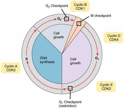
D2.1.13 Phases of the cell cycle
Cells need time to move through the cycle so that it moves from one phase to the next; cancer results when cells divide when they shouldn't
Interphase
G1: The phase after mitosis and before DNA replication where each chromosome is a single DNA molecule; active growth phase
S: All DNA in the nucleus is replicated; two identical pairs of DNA; synthesis of new DNA
G2: growth may resume during this phase while the cell is preparing for mitosis; such as synthesis of proteins
G0: a nondividing state; cell here are alive and perform their role but wont divide again
Differentiated bone cells (osteocytes)
Skeletal muscle fibre cells
Neurons
Cell division:
Mitosis: division of the nucleus
Early Prophase: Microtubules are growing from the centriole; chromosomes become shorter
Late Prophase: Spindle microtubules extend; each chromosomes consists of two identical sister chromatids each with a centromere and kinetochore
Metaphase: chromosomes move to equator; spindle microtubules attached to kinetochore with sister chromatids attached to opposite poles
Anaphase: sister chromatids separated so each is now a separate chromosome; kinetochores shorten spindle microtubules
Early Telophase: all chromosomes reach poles and membrane forms; spindle microtubules break down
Late Telophase: chromosomes uncoil
Cytokinesis: division of cytoplasm and organelles;Mitochondria and chloroplasts also grow and divide here
D2.1.15
Control of the cell using cyclins
Cell cycle checkpoints (G1, M, G2)
Checks for errors or defects before proceeding to the next stage of the cell cycle
Ex. correct amount of chromosomes
If it doesn't repair it may be killed, fixed, or sent to G0
G1: Checks if the cell is the correct size and checks for DNA damage ( if it doesn't pass it moves to G0)
G2: Checks DNA has be correctly replicated
M: Checks that all sister chromatids are all attached to the spindle microtubules
Cyclins + Cyclin dependent kinase
Proteins that control the cell cycle’s progression through the checkpoints (regulatory proteins)
concentration cycle moves up and down as the cell progresses through the cell cycle
4 different types of cyclins (D E, A, and B)
Cyclins bind to kinase enzymes or cyclin dependent kinases (CDKs), activating them, the kinases phosphorylate other proteins in the cell activating them, these proteins then perform tasks specific to the phase of the cell cycle it is in.
Low concentration cyclin the CDk will not be active and the cell cycle will freeze
D2.1.16
Consequence of mutations in genes that control the cell cycle
Initiation of Tumor formation
Cause a mutation to the DNA in a gene that control the cell’s progression through the cell cycle
1) random errors in DNA replication
2) mutagen
Tumors originate from a single cell that loses control of it cell cycle which is inherited by its daughter cells
Mutagens
Are anything that permanently changes genetic material
1) Radiation: high energy (UV, X-ray)
2) Chemicals: carcinogens that interact directly with DNA or produce mutagenic compounds (Cigarettes, Benzoyl peroxide, nitrates)
3) Infectious agents: virus or bacteria cause DNA damage or reduce efficiency of DNA repair systems (HPV and Helicobacter Pylori)
Cell cycle control genes
Proto-oncogenes:
Concerned with control of the cell cycle, such as the genes that code for cyclin proteins
Normally help cells grow
Tumor suppressor genes:
Code for proteins that prevent uncontrolled cell division
Are normal genes that slow down cell division, repair DNA mistakes, or tells cells when to die (apoptosis)
Oncogene
When a proto-oncogenes mutate it creates this cancer causing allele
Permanently activated even when they aren't supposed to be ; Cells grows out of control and causes cancer
Mutated Tumor suppressor gene
Cells grows out of control and causes cancer; losses that defense
Cancer correlates with age
Results from the accumulation of multiple mutations to a cell’s genes that control the cell cycle
Multiple hit hypothesis: cells must acquire a series of mutations leading to unrestrained cell growth and division
Elephants like humans have lots of cells and live a long time yet don't die as frequently to cancer; this is because they have a zombie genes that causes cell apoptosis do cells die before they become cancerous
D2.1.17 Tumors and rates of growth
Benign Tumor: cells in the tumor adhere to each other and remain in a single mass; do not cause cancer
Malignant tumors: cells in the tumor can detach and invade neighboring tissues, lymph vessels or blood vessels; cause cancer
Cancer symptoms:
Fatigue, Lump, Skin changes, Weight changes
Cancer developed in four stages
1) Initiation: normal cell transformed into cancerous cell as a result of mutations
2) Promotions: the cancerous cell divides, making a large # of daughter cells containing the mutations (primary tumor)
3) Progression: cancerous cells become aneuploid (have the wrong # of chromosomes) and begin to invade surrounding tissues
4) Metastasis: cancer cells break away from the primary tumor (First Formed), travel through blood/ lymph system, from new tumors (secondary tumors) in other parts of the body
Mitotic Index (MI)
Ratio of percentage of cells in a sample undergoing mitosis relative to the total number of cells in the sample
Larger the MI, higher rate of division by cells in the simple
MI = # of cells in mitosistotal # of cells100
Uses of MI: predicts how rapidly the tumor will grow
Diagnosis: higher MI relative to a tissue specific standard more likely a tissue is cancerous
Treatment: stop cell division, so if cancer treatment is working means fewer cells in mitosis and MI would decrease; predicts how rapidly the tumor will grow
B2.1.1 + B.1.12
Phospholipid bilayer
Lipid Bilayers : membrane barrier (sheet-like bilayers) separating the interior from its surrounding
Lipids
hydroxyl groups, fourlinked hydrocarbon rings, a hydrocarbon tail
Unique structure (bent shape)
Amphipathic
Hydrophilic head composed of polar hydroxyl groups and charged parts of lipids
Hydrophobic tails composed of nonpolar parts/ hydrocarbon chains and other amphipathic lipids
Membrane Cholesterol ( Animals)
Acts to modulate membrane fluidity and permeability to some solutes
Plants have (Sterols)
Cholesterol interests into the bilayer of phospholipids
Amphipathic (contains both hydrophilic and hydrophobic parts)
B2.1.11
Membrane fluidity
Greater the temperature:greater the fluidity/ lower viscosity/ less densely packed/ won't hold shape/ too permeable
Lower temperature: high viscosity/ densely packed/ more rigid/ may break/ not permeable
Importance of Membrane Fluidity
Enables molecules to diffuse through the membrane
Facilitate the interactions between proteins (for cell signaling)
Enable membranes to fuse with each other during vesicle formation (endocytosis and exocytosis)
Ensure an even distribution of membrane molecules between daughter cells during cytokinesis
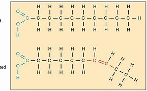
Phospholipids structure affects membrane fluidity
Phospholipids molecules can vary in their tail length and degree of tail saturation
Fatty acids and membrane fluidity
Satuaturared: fatty acids do not have double bonds between adjacent carbon atoms; straight tail
Press closely together making a dense and viscous membrane
Stronger intermolecular forces causing them to have higher melting points
Ex. Arabidopsis plant grows at higher temps has an increase in saturation of fatty acids
Unsaturated: fatty acids have one or more double bonds. Leads to a bend in the molecule
Have kinks in their tails preventing close packing; keeping space between them; Helps increase fluidity of the membrane
Weaker intermolecular forces causing them to have lower melting points
Ex. Plants (Chickpea) increase in fatty acids at low temperatures
The saturation of membrane lipids can vary within the body of a single organisms
Caribou have high amounts of unsaturated fatty acids in their hooves than the tissue in their upper leg (hooves are in snow means lower temp)
,
B2.1.12 Cholesterol and membrane fluidity
Membrane Cholesterol ( Animals)
Acts to modulate membrane fluidity and permeability to some solutes
Function of Cholesterol
At high temperatures cholesterol physically restrains the movement of phospholipids = increases viscosity (membrane fluidity) , reducing its permeability to small molecules
At lower temperatures cholesterol prevents stiffening of the phospholipids by lowering freezing point and increasing boiling point
B2.1.13 membrane fluidity and fusion and formation of vesicles
Temperature
Affects viscosity (measure of a fluid’s resistance to flow)
Higher temp means lower viscosity
Lower temp means higher viscosity
Fatty acids length
Longer fatty acids tails allow for more interaction between phospholipids leading to less fluidity
Fatty acid saturation
Unsaturated fatty acids have one or more double bond in the fatty acid tails, double bond lead to a bend pushing the adjacent phospholipids further apart increasing spacing increases fluidity
Presence of cholesterol
The presence of cholesterol affects the fluidity depending on the temp
High temperature; cholesterol decreases fluidity
Lower temperature: cholesterol increases fluidity
Vesicles: move materials around the inside cell
Proteins synthesized by ribosomes on the rough ER are carried to golgi apparatus; protein process by golgi are carried to plasma membrane
Endocytosis: formation of vesicles in the cytoplasm by pinching off a piece of plasma membrane ; goes into the cell
Contain water and solutes; may contain larger molecules
Ex. Macrophages (white blood cell)- engulfs pathogens when fighting infection
Ex. Foetal cells in the placenta; absorb proteins from mothers blood (antibodies)
Exocytosis: fusion of vesicles with the plasma membrane expelling the contents of the vesicles from a cell
Release of neurotransmitters from a presynaptic membrane
Secretion of hormones from endocrine glands from the pancreas (ex. insulin)
Removal of excess water from contractile vacuole
B2.1.10 Fluid Mosaic Model
Fluid mosaic model: describes the structure of the cell membrane as a dynamic, flexible structure made up of different components
The main component of the cell membrane are phospholipids, cholesterol and proteins
B2.1.4 Membrane proteins
Integral and Peripheral proteins
Membrane proteins are synthesized by bound ribosomes (Found on Rough ER) and then brought to the cell membrane via exocytosis
Peripheral proteins
Are associated with membrane surfaces and do not fully span the membrane; temporary
Attachment to the lipid bilayer is achieved by binding to one side of the bilayer or to an integral membrane protein
Integral protein
Are embedded and may span the lipid bilayer; mostly transmembrane
They are able to establish hydrophobic interactions with the tails of the phospholipids
Function of membrane bound proteins
Enzymatic activity
Process substrates of various metabolic pathways
Ex. ATP Synthase (enzyme that catalyze chemical reactions)
Receptors
Identification of cell type for communication between cells
Proteins that are embedded in the cellular membrane to which specific chemical signals from outside the cell attach; when the chemical signal binds,s the membrane protein triggers a response by the cell
ex.Acetylcholine receptor, chemoreceptors, hormone receptors(glucagon, insulin), thermoreceptors, electromagnetic receptors, mechanoreceptors, baroreceptors
Insulin: lowers your blood sugar
Glucagon: raises your blood sugar
Recognition
Chemical messengers interact with receptor binding sites to transduce signals into cells
Proteins that are embedded in the cellular membrane that allow cells to identify each other and interact
Glycoproteins and glycolipids: components of plasma membranes
short chains of sugars (oligosaccharides) attached to the membrane and the carbohydrate is attached to proteins or the lipid; Interactions between the sugar and carbohydrate binding proteins allows cell-cell recognition
ABO blood grouping is based on differences in type of glycoprotein present on the surface of red blood cells
Helps in development of tissues and organs
Adhesion
Connect neighboring cells to form a tissue
Is the process by which cells from tissues by adhering to neighboring cells through specialized adhesion proteins
Glycoproteins and glycolipids form a layer called glycocalyx; which helps bind cells
Cell to cell adhesion molecules (CAMs) link adjacent cells in animal cells (integral protein)
Cells of the same type have same CAMs; different cells have different CAMs
Transport
Move molecules and ions across the membrane
Passive transport: do not require energy (high to low concentration)
Simple diffusion: movement of small nonpolar molecules (O2, CO2)
Osmosis: net movement of water (low solute concentration to high)
Aquaporan: moves water molecules through membrane
Facilitated diffusion: passive movement with membrane proteins for large polar molecules/ions
Channel and carrier proteins (integral) and potassium channels
Active transport: moves against the gradient; uses ATP
Carrier proteins: protein pumps and sodium potassium pumps (3Na out and 2 K in)
Anchorage
Anchor the cell to the extracellular matrix to hold cells in place
Extracellular matrix (ECM), provide support, segregating tissues from one another, and regulating intercellular communication
Cells use membrane bound proteins called integrins to anchor the cell to the extracellular matrix
C2.1.1
Receptors and Signals
Chemical signaling
Cells are able to receive and process chemical signals in order to respond to their environment
Ligand
Is a chemical that binds to another specific molecules (receptor molecule)
1) Ligand approaches binding site
2) Binding causes changes within the receptor
3) Signal is passed on to the cell
4) Ligand dissociates from the binding site
Hormones, neurotransmitters, cytokines, and calcium ions
C2.1.3
Types of signaling molecules
Hormones
Are the chemical signals secreted from cells in endocrine glands that travel through the bloodstream to target any cell which has a receptor for the hormones; long distances signals
Ex. Insulin, thyroxine, testosterone
Neurotransmitters
Chemicals that transmit signals across a synapse, the junction between two neurons
Dopamine, acetylcholine, norepinephrine
Cytokines
Small signaling proteins
May affect same cell it was secreted from, other cells or act in more systematic manner (affects nearby cells)
interferon
Calcium Ions
Used for signaling with in muscle fibers + neurons
Attach to proteins of sarcomere, muscle contraction
Diffuse into cells through voltage-gated channels in plasma membrane
C2.1.4 Chemical diversity of hormones and neurotransmitters
Hormones
Hormones are used to integrate organ systems and can affect cells at a distance from where they were released
Three classes: Amines, Peptides /proteins, Steroids
Amine Hormones: small molecules synthesized by modification of amino acids
Melatonin and Epinephrine
Peptide and protein Hormones
Peptide: Antidiuretic hormone, Oxytocin
Protein: Insulin and Glucagon
Glycoprotein: Follicle stimulating hormone
Steroid Hormones: Lipids derived from cholesterol
Oestradiol, progesterone, testosterone
Neurotransmitters
Are chemicals that transmit signals across the junction between two neurons
Classes of hormones:
Esters: Acetylcholine
Gasses: Nitrous oxide
Amino acids: Glutamine
Amines: Dopamine
C2.1.9 Transmembrane receptors that activate G proteins
Chemical signaling
A signaling pathway is the process in which binding of an extracellular chemical to a receptor is translate into changes in the cell
Three main steps
1) Reception: the process by which a cell detects a signal in the environment
2) Transduction: The process of activating a change within the cell
3) Response: the change that occurs in the cell as a result of the signal
Signal Transduction (Ex. activating G-protein inside the cell)
when the binding of signaling molecule to the receptor induces a change in the shape of the receptor, the activated receptor can then initiate changes in the cell
Transmembrane receptors: binding of signaling molecule causes reversible changes to its structure
Intracellular receptors:Binding of molecules results in formation of active ligand receptor
G proteins receptors (GPCRs)
Transmembrane receptors (Consists of single polypeptide and embedded in a cell’s plasma membrane)
In the absence of a chemical signal the Gprotein coupled receptor is inactive
Activation can happened by opening ion channels, altering metabolism, activating gene expression or changing cell shape
G-protein coupled receptors (medicine)
Antihistamines, opioid agonists, depression medications, chemotherapy drugs, diabetes medicine
Ozempic and Wegovy are being used to treat diabetes and obesity
C3.1.12 Epinephrine (adrenaline) secretion by the adrenal glands to prepare the body for vigorous activity
Chemical signaling
Epinephrine binds to cells with a transmembrane receptor called adrenergic receptor which is a type of G-protein coupled receptor
Epinephrine triggers a signaling pathway ( a process in which binding of an extracellular chemical to a receptor is translated into changes in the cell)
Effects of epinephrine
Causes liver and muscle cells to break down glycogen into glucose which can be used for anaerobic or aerobic respiration (helps make ATP)
Bronchi and bronchioles dilate to relaxation of smooth muscle, widening the airway for increased airflow during ventilation
Ventilation rate increases, so a larger total volume of air is moved per minute
Speeds up firing of the sinoatrial node increasing the heart rate, which moves blood to the tissue faster
Increases strength of the cardiac contraction increasing volume of blood
Arterioles that carry blood to the skeletal muscles dilate, widening so more blood flows to them, redirects blood flow to the areas of the body most crucial for the immediate threat
Blood carried to the gut, kidney, and skin constrict narrow so less blood flows to them, not vital for dealing with immediate threat
C2.1.11
Circadian Rhythms
Physiological and behavioral changes of an organism over a 24 hour cycle
Dictate multiple processes including alertness, sleepiness, appetite, and body temperature
Exist in both unicellular and multicellular organisms
Can be synchronized by light and darkness; can continue even if place in continuous light/dark
Suprachiasmatic Nucleus (SCN)
Pacemaker of the circadian rhythm; neurons here produce a circadian rhythm of neuron firing frequency which allows them to synchronize other cells throughout the body
Visible light (blue) synchronizes the rhythm of the SCN
Melatonin (Amine hormone )
The SCN releases melatonin from the pineal gland
Dark: SCN promotes secretion of melatonin from the pineal gland
During night it is high
Light: SCN inhibits secretion of melatonin from the pineal gland
During the day it is low
For nocturnal animals melatonin promotes activity for diurnal animals it promotes sleep
Melatonin Effects
Reduce blood pressure
Reduce kidney production of urine
Drops core body temperature when sleeping
Cause drowsiness and promote sleep
Reduce inflammation response/ enhance immune response
Insulin
Protein hormone; secreted by the pancreas by beta cells when glucose levels are high
Causes cells to uptake glucose from the blood to be used in cellular respiration or converted to glycogen
Goes directly to skeletal muscles, livers, and adipose tissue (fat)