PARASITE LIFE CYCLES TO KNOW
LIST TO KNOW.
TREMATODA (FLUKES)
Fasciolidae
question 1: Fasciola hepatica
Answer: indirect LC, FH: ruminants, horse and man, IH: water snail (lymnaeidae). Location: Bile duct and liver. Process: Adults release unembryonated eggs into the biliary ducts of FH, then passed in feces. Embryonation happens in the water. The eggs will release miracidium, then infect a freshwater snail. The miracidia will develop into sporocysts, then rediae, then cercarie. Cercariae are released into the water and will find water plants to encyst, forming metacercariae (infective stage). The FH will eat the plants, where the parasites will excyst in duodenum → liver parenchyma → adults within bilde ducts.
Dicrocoelidae
Question 2: Dicrocoelium dendriticum
Answer: indirect LC. FH: reptiles, birds, mammals, IH: 1st: land snail, 2nd: ants. Location: liver and gall bladder. Infective stage: metacercariae. Process: Eggs with miracidium is passed in feces of FH, then eaten by 1st IH (snail). Miracidia → sporocyst → cercariae → resp. chamber → shed from snail. Ant will eat slime ball with cercariae → metacercariae. FH eats ants on tips of vegetation → excyst in SI. worms migrate to bile ducts and matures.
Paramphistomidae
Question 3: Paramphistomum cervi
Answer: Indirect lC. FH: mammals, birds, reptiles (commonly in RU). IH: mud snail (Planorbidae), Location: juvenile - SI, adults in rumen. Infective stage: Metacercariae. Process: Eggs with fecs, eggs develop an embryo (miracidium) → hatches from egg and seeks snail → inside snail, transforms into sporocyst → redia → cercaria → leaves and become metacercariae on vegetation. FH is infected by ingestion. Young flukes gets through intestine → rumen, matures.

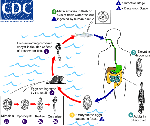
Opistorchiidae
Question 3: Opisthorchis tenuicollis (felineus)/Clonorchis sinensis (similar)
Answer: FH: fish-eating mammals. IH: 1st: water snail (Bithynia), 2nd: fish (bream, barbel). Location: liver, bile ducts. Infective: Metacercariae with fish tissue. Process: eggs shed with feces → snails ingest eggs → cercariae develops → fish infected → metacercariae are formed within fish in muscle → FH ingest fish, migrates to bile ducts, matures.
Question 4: Metagonimus yokogawai
Answer: FH: fish-eating mammals, humans. Location: Small intestine. IH: 1st water snails (semiliscospira) and 2nd: sweet fish (asia, balkans). Process: embryonated eggs each with miracidium is passed down with feces → snail ingest eggs → develops into sporocyst → redia → cercariae → released from snail → infects fish → encyst to metacercariae → FH infected by undercooked fish with metacercariae → excyst in SI.
H. heterophyes - similar, but it releases toxins in FH causing severe catarrhal inflammation (abd. pain, diarrhea).
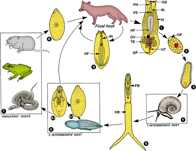
Diplostomidae
question 5: Alaria spp. (alata/canis/mercianae)
answer: FH: dogs, cats, red foxes, humans. Found in Small intestine. IH: 1st: Water snails (planorbis spp.), 2nd: frog, toads. Paratenic host: frogs, snakes, mice, birds. infective: Mesocercariae. Process: Adults live in SI → eggs passed in feces → hatch in water, releasing miracidia → 1st IH (snails), where it develops into cercariae (Furcocercariae) → released → 2nd IH (tadpole). The tadpole develops into frog - mesocercariae (special larval stage) → FH becomes infected after eating frog/snake. Mesocercariae migrate through lungs, trachea → adults in SI.
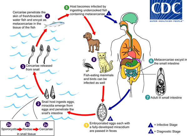
Paragonimidae
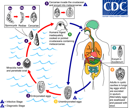
Question 6: Paragonimus westermani
Answer: Lung fluke. Indirect cycle. FH: dogs/cats/humans. IH: 1st: water snails (melania), 2nd: crabs, crayfish. Location: Lungs. infective: Metacercariae. Process:eggs released and hatch in water, releasing miracidia → infect snail → develops into cercariae → infects crabs/crayfish → metacercariae develops → FH infected by ingestion of raw or undercooked 2nd IH.
CESTODA (TAPEWORMS)
Pseudophyllidea
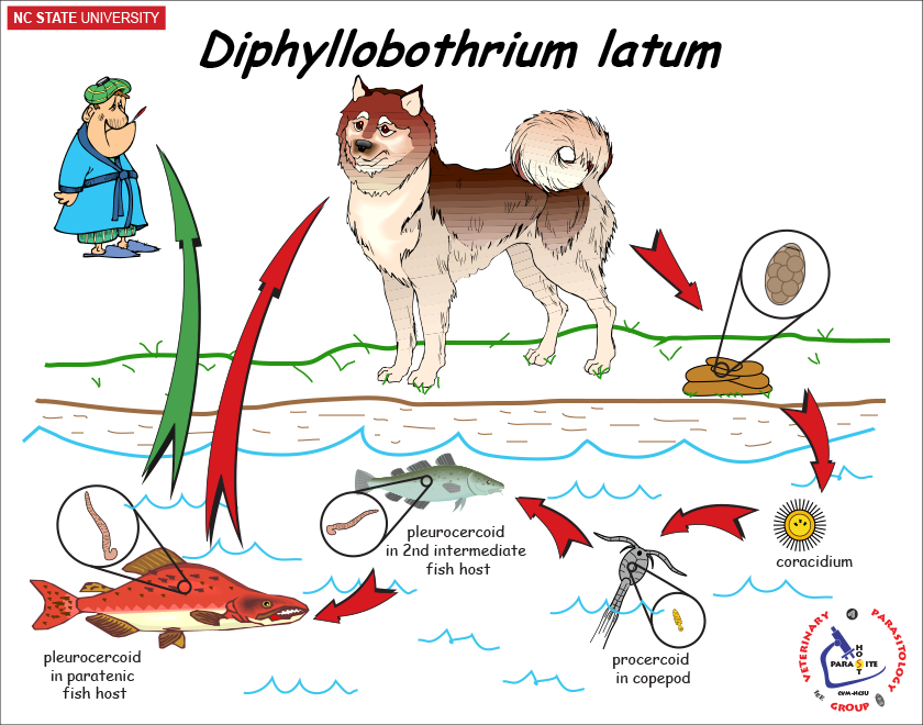
question 7: Diphyllobothrium latum
Answer: FH: fish-eating mammals. IH: 1st: water arthropod, 2nd: fish. Paratenic: carnivorous fish. Location: SI. Indirect LC. Infective stage: Plerocercoid for FH, procercoid for fish. (Pseudophyllidea). Process: Eggs shed in feces → embryonate in the water → coracidium hathces → taken up by 1st IH → procercoid develops → 2nd IH infected → plerocercoid developed in muscle → FH eats raw/undercooked fish → plerocercoid develops into immature worm → matures in SI.
Cyclophyllidea
Taeniidae
Question 8: Taenia solium
answer: FH: human, IH: pig, Location: muscle in IH, SI in man. Infective stage: cysticercus. Process: eggs/gravid proglottids are shed with feces → pigs ingest eggs → oncospheres hatch in their intestine → cysticercus develop in their muscle → humans get infected by eating undercooked pork. In the human intestine, cysticercus develops into adults.

Question 9: Taenia saginata
Answer: FH: human, IH: cattle. Location: muscle in IH, SI in human. Infective: cysticercus bovis. Process: same principle as t. solium.
Question 10: Taenia hydatigena
Answer: FH: dog/fox, IH: livestock, Location: abdominal cavity, liver. Infective stage: cysticercus tenuicollis. Process: Adults reside in SI of FH, gravid proglottids are shed with eggs in feces → ingested by IH → oncospheres hatch in the intestine of IH → cysticercus tenuicollis develop in the tissues (bladderworm with single invaginated scolex) → FH ingest the infected organs of IH → Tapeworm matures.


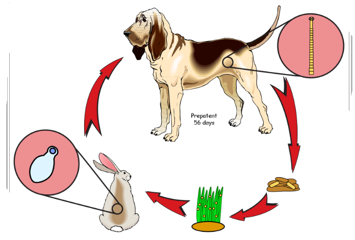
Question 11: Taenia pisiformis
Answer: FH: dogs/fox, IH: Lagomorphs (rabbits), location: serosa, abdominal cavity. Infective stage: Cysticercus pisiformis. Process: adults in SI of FH → releases gravid proglottids in feces → lagomorphs ingest the eggs → oncospheres hatch → serosa, abdominal cavity → cysticercus pisiformis develops → FH ingest the infected tissues → SI → matures.
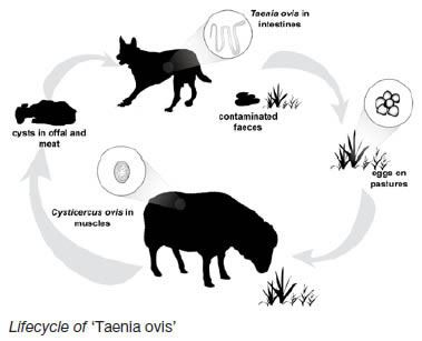
Question 12: Taenia ovis
Answer: FH: dog/fox, IH: sheep, Location: small muscles. infective stage: Cysticercus ovis. Process: adults in SI of FH → releases gravid proglottids in feces → sheep ingest eggs → oncosphere hatch in intestine → small muscles → cysticercus ovis develops → FH ingest infected tissues → tapeworm matures in SI.
Question 13: Taenia multiceps
Answer: FH: dog, fox, coyote, wolf. IH: sheep, cattle, man. Location: CNS, brain, spinal cord. Infective stage: Coenurus cerebralis (Zoonotic). Process: Adults tapeworms in SI of FH → gravid proglottids shed in feces → IH ingest the eggs → oncospheres hatch in intestine → CNS, brain, spinal cord → coenurus cerebralis develops → FH ingest the infected IH → matures in SI.

Question 13: Taenia serialis
Answer: FH: dog/fox, IH: lagomorphs (rabbits), location: tissue, infective stage: coenurus serialis. Process: adults in SI of FH → gravid proglottids are shed with feces → Lagomorphs ingest the eggs → oncospheres hatch in intestine → tissues → coenurus serialis develops → FH becomes infected → scolices evaginate + attach to SI, Matures.
Question 14: Taenia taeniaeformis
Answer: FH: CATS, IH: rodents/rabbits. Location: liver. Infective stage: Strobilocercus/cysticercus fasciolaris. Process: Adults in SI of FH → releases gravid proglottids in feces → IH eats eggs → oncospheres hatch → liver → cystercus fasciolaris develops → FH becomes infected, attaches to SI and matures.
Question 15: Echinococcus granulosus
Answer: FH: carnivores (dog, wolf, dingo), IH: ruminants, humans, camels, deer. Location: liver and lungs. SI of FH. Infective stage: hydatid cyst. Process: Eggs shed in feces → embryonated ingested by IH → releases oncospheres → gets to liver, lungs, tissues → develops into hydatid cyst → grows and releases brood capsules with scolices → FH ingests the tissues with cysts → SI -matures.

Question 16: Echinococcus multilocularis
Answer: FH: carnivores - fox, dog, wild canids, racoon, cat. IH: rodents/humans. Location: liver, lung but also brain, bone marrow, muscle, lymph nodes and heart. Infective: alveococcus. Process: Adults in SI of FH → releases eggs in feces → IH ingest eggs → oncosphere hatch in intestine → migrates → alveococcus (alveolar cyst) develops → FH becomes infected when ingesting the organs → SI and matures.

Dipylidiidae
Question 17: Dipylidium caninum
Answer: FH: dogs, cats, canids, fox, humans. IH: fleas, lice. Location: SI. Metacestode: cysticercoid. Process: adults in SI of FH → gravid proglottids → eggs shed in feces → IH eats eggs → cysticercoid develops → flea larvae matures → infective cysticercoid → FH eats flea → parasite matures.

Mesocestoididae
Question 18: Mesocestoides lineatus
Answer: FH: dogs, cats, wild canids. IH: 1st: arthropods - ant, beetle, oribatid mites (Cysticercoid here), 2nd: small mammals, reptiles, amphibians, birds (Tetrahyridium here). Location: SI. Process: adults matures in SI of FH → releases eggs in feces → 1st IH ingest eggs with oncosphere → cysticercoid develops → 2nd IH ingest this → tetrahyridium develops → FH ingest IH → matures in SI.

NEMATODA (ROUNDWORMS)
Strongyloididae
Strongyloides westeri - horse
Strongyloides ransomi - pig
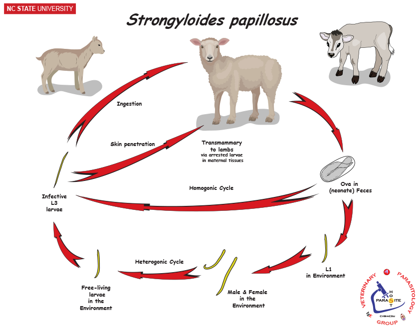
Strongyloides papillosus - ru
Strongyloides stercoralis - human
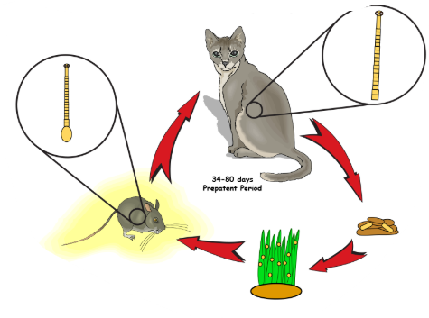
Question 19: Strongyloides spp.
Answer: FH: depends on species - S. westeri (horse - foals), stercoralis (man, dog), papillosus (ru), ransomi (pig). Location: SI of FH. Males and females reproduce sexually in the environment. Eggs - hatch in intestine of host. 2 forms of larvae - rhabditiform (L1, L2) and filariform (L3). Infection through skin and oral. Eggs released from FH → releases L1 → develops into L3 → autoinfection or passed into feces where it develops into L3 in the environment or free living adults - produce more.
Strongylidae
Question 20: Strongylus vulgaris
Answer: Direct LC, FH: horse, in Large intestine. Process: Eggs passed in feces → L3 development → host ingest these → larvae migrates to intestine → mucosa, moults to L4 → small arteries → cranial mesenteric artery + branches → L5 → returns to intestinal wall.

Ancylostomatidae
Question 21: Ancylostoma caninum/Tubaeforme
Answer: Direct LC. FH: caninum: car/man while tubaeforme: Cat. in SI. Infective stage: L3 eggs. Infection by: ingestion, per skin, transmammary + transplacental. Process: adults in SI of FH → eggs passed in feces → eggs develop → L1 into soil → L3 developed → infection of FH → if by skin, then it will enter blood, lungs then coughed up and swallowed → SI.
Ascarididae
Question 22: Ascaris suum
Answer: Direct LC, located in SI
Hepato-pulmonary passage of larvae after hatching
Host: pig
Process:
Unembryonated eggs in feces
moults to L3 larvae (infective stage)
ingestion of infective egg by host
hatched larvae → invades intestinal mucosa → hepato-pulmonary migration (to lungs)
further maturation in lungs → swallowing of larvae → SI, develops into adult worms

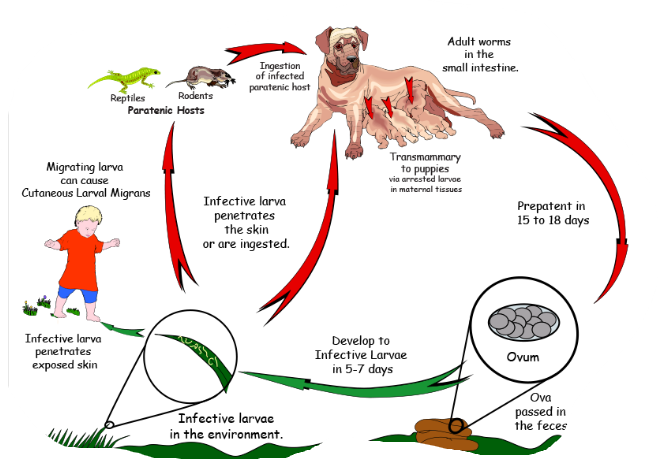
Question 23: Toxocara canis/cati
Answer: canis (canis as there is dog) or cati (cat)
direct LC, located in SI, diagnostic stage - umebryonated eggs in feces, infective - embryonated egg with L3
entero-hepato-pulmonal migration
FH: car (canis - dog, cat - cati, leonina - both), accidentally man
paratenic host: various small mammals, can be ingested by dogs (no development of parasite but remains infective)
Process:
eggs shed in feces of FH
eggs embryonate in environment with L3 larvae
Eggs ingested by host → can be FH (dog) or paratenic host (mouse, rabbit etc.)
In FH - larvae mature into adults in SI
vertical transmission: adults can be transmitted from mother to offspring
L3 larvae can get to other tissues → liver, lungs and brain

Metastrongylidae
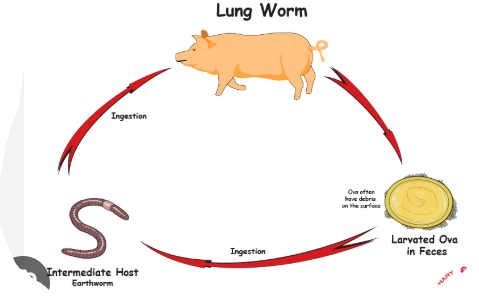
Question 24: Metastrongylus species
answer: FH: pigs (pdendotectus, apri, salmi, confusus), Location: in respiratory system of pig (lung worm). Indirect LC - IH: Earthworms. Infective: L3. Process: Adult in bronchi of pigs → eggs with L1 → coughed up → swallowed → feces → soil, ingested by IH → L3 develops here → Pigs eats eartworms → migrate to lungs via hepatic portal system → develop into L5.
Dictyocaulidae
Question 25: Dictyocaulus species
Answer: D. viviparus (cattle), filaria (small ru), arnfieldi (horse, donkeys). Location: respiratory system. Direct LC. Infective: L3. Process: Adults in bronchi + bronchioles of lungs → eggs with L1 in feces → develops to L3 in environment → host animal ingest these while grazing → matures in lungs.

Trichuridae
Question 26: Trichuris species
Answer: suis (pig), vulpis (dog), ovis (small ru), globulosa (ru). Location: large intestine. Direct LC, with infective stage: L1 eggs. Process: unembryonated eggs in feces → L1 larvae inside eggs → host ingest embryonated eggs → eggs hatch and L1 into SI → Large intestine → all 4 moults happens → matures.
Angiostrongylidae
Question 27: Angiostrongylus vasorum
Answer: Host: Dog/fox, location: Pulmonary arteries + right side of heart. IH: Snails/slugs. Process: adults in pulmonary arteries + right ventricle → eggs excreted → IH ingest L1 → inside, they develop into L3 (infective) → FH eats IH → intestinal wall → lymph nodes → liver → heart → pulmonary arteries and matures.
ACANTHOCEPHALA
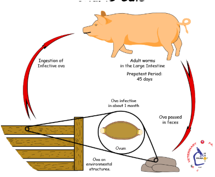
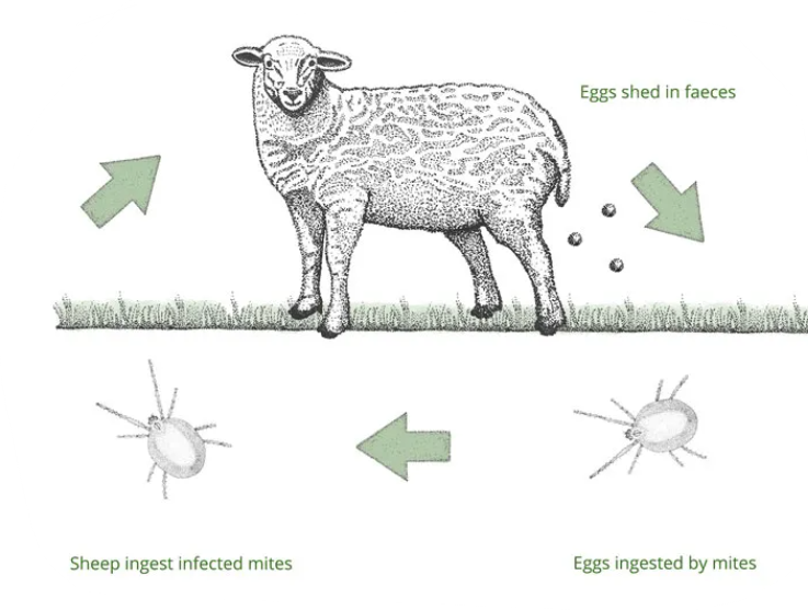
Question 28: Macracanthorhynchus hirudinaceus
Answer: FH: pigs, location: SI. IH: beetles/larvae. Infective stage: cystacanth larvae. Process: Adults in SI of pigs, releasing acanthor larvae eggs in feces → eggs ingestged by IH where they develop into acanthella larvae → cystacanth larvae (infective) → FH ingest beetles (IH) → matures.
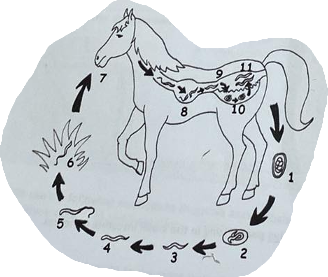
ANOPLOCEPHALIDAE - cestode
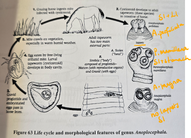
Question 29: Anoplocephala spp.
answer: Anoplocephala spp. (Perfoliata + magna) - also paranoplocephala mamillana.
Difference is that perfoliata is in SI and LI of horse, cysticercoid in oribatid mites. While magna is in SI of horses, scolex is small, no lappets.
indirect LC, IH: oribatid mites, FH: horse
infective stage: cysticercoid
Process:
grazing horse ingest mite infected with cysticercoid
Cysticercoid develops to adult in intestine of horse
gravid proglottids and embryonated eggs pass in horse feces
eggs eated by oribatid mites → cysticercoid develops here
mites crawl on vegetation → eaten by horse
Question 30: Prosthogonimus macrorchis
Answer: FH: birds, IH: 1st: snails, secondary: dragonflies. Location: oviduct and bursa. infective stage: Metacercariae (trematode typical). Process: Adults in bursa + oviduct in bird → eggs released with feces → snails ingest the eggs (miracidium → redia → cercariae) → 2nd IH eats snails (encyst as metacercariae) → FH eats dragonflies → excyst in bird intestine → bursa or oviduct - matures.

Question 31: Moniezia spp.
Answer: FH: ruminants (Expansa - young ruminants, benedeni - medium to large ru). IH: oribatid mites. Location: SI of ru. Infective stage: Cysticercoid (Cestode typical). Process: adults in SI of FH → eggs released with feces → mites eats the eggs → cysticercoid develops → ruminants eats the mites → mature in SI.
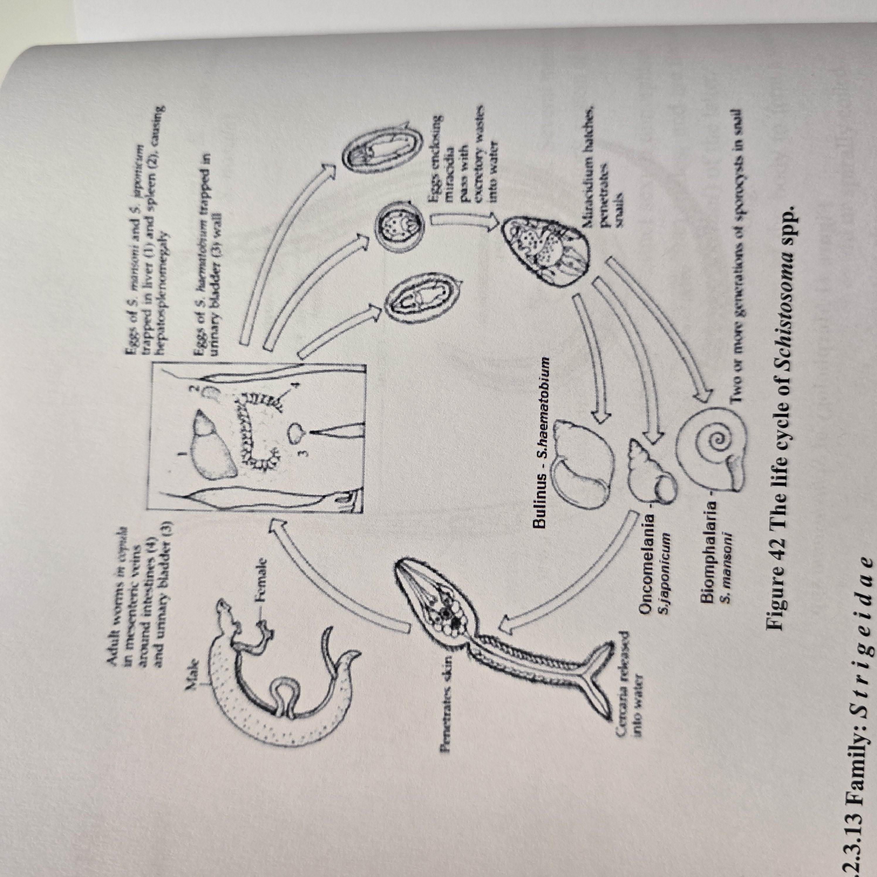
Question 32: Schistosoma spp.
answer: Schistosoma spp. (S. haematobium, Mansoni, japonicum) - trematode
Indirect LC, located in Blood vessels
IH: water snail
FH: man
process:
eggs hatch into miracidium → IH → pathogony → sporocysts → cercaria with forked tail (furcocercaria) → exits
FH gets infected by furcocercaria → looses their tail and become schistosomulae → circulation → matures.
Worms migrate to mesenteric or vesicular veins depending on species.
S. mansoni: mesenteric veins of LI
S. haematobium: veins of bladder
S. japonicum: mesenteric veins of SI
Embryonated eggs with miracidium released by urine/feces.
Question 35: cyathostomum genus
Answer: “small strongyles”, cycliocyclus
Nematode, from family Strongylidae.
Direct LC, located in Large intestine of horses
Process:
Adult cyathostomins live in LI → eggs pass in feces (unembryonated)
larva develops within each egg, then hatches
released larva develops → moults twice to infective L3 stage.
Infection of horse is by ingestion of these larvae → mucosal migration → moults to L4 in LI
Larvae migrates to lumen → moults to L5/adult and release eggs in feces