The Nervous System (March 12, 2025) (Presentation)
The Nervous System (NS)
Every conscious action that occurs in your body is governed by the nervous system (NS)
Functions include:
Communication from one end of the body to another.
Collection and integration of internal and external stimuli.
Formulation of appropriate response.
Most subconscious (or automatic) actions also are governed by the nervous system.
The immediacy of the nervous system response sets it apart from other control systems of the body.
It is responsible for maintaining homeostasis.
The nervous system works in concert with the endocrine system.
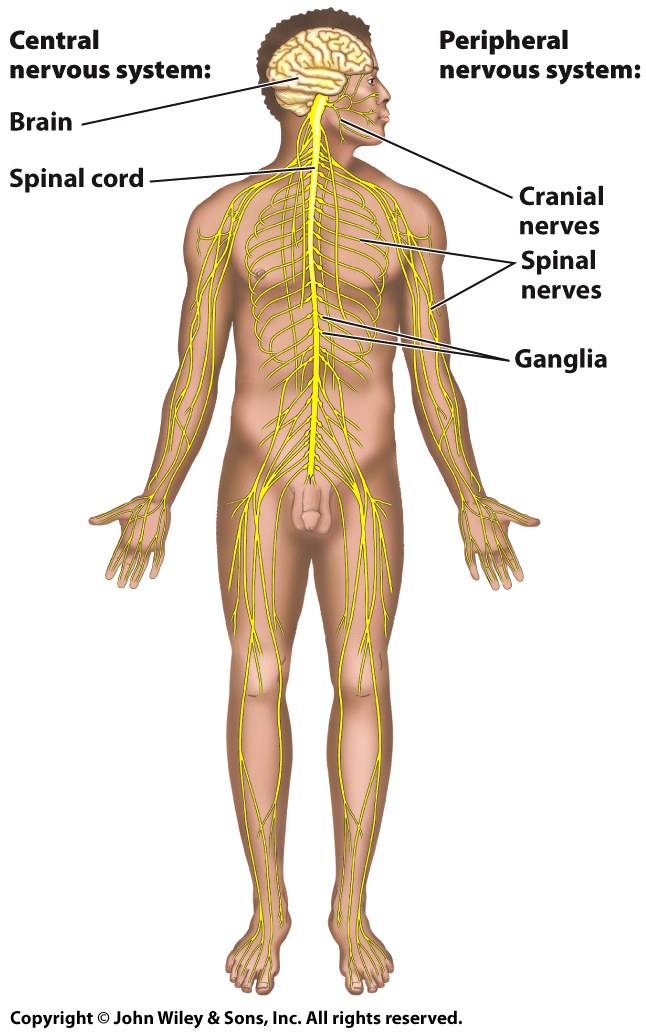
The Nervous System Has Two Components
Central Nervous System (CNS)
Composed of the brain and spinal cord.
Encased by the axial skeleton and covered by meninges.
Main integration system of the body.
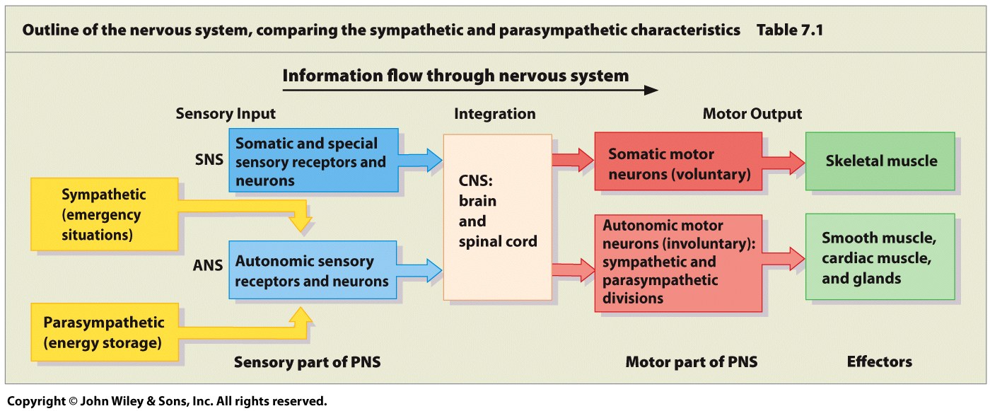
Peripheral Nervous System (PNS)
Composed of all neural tissue other than the brain and spinal cord.
Includes:
All the afferent and efferent neurons that extend from the CNS.
The autonomic, sensory, and somatic nerves of the body.
Central Nervous System (CNS) vs. Peripheral Nervous System (PNS)
Sensory information enters the CNS, which analyzes it and then sends a motor response through the PNS to muscular or glandular tissue.
Information reaches the CNS from the afferent division of the PNS.
The PNS picks up this information with one of three types of receptors.
Special senses (sight, hearing, taste, smell)
General sensory receptors (external temp., light, touch, pain)
Visceral receptors (proprioception, organ functions)
Motor responses are sent through the efferent division of the PNS.
Two Divisions of the PNS
The somatic division is involved in conscious movement.
It regulates voluntary movement of skeletal muscles.
The autonomic division governs the body’s responses to changes in homeostasis with involuntary, subconscious reactions.
It regulates functions such as blood vessel diameter and stomach activity.
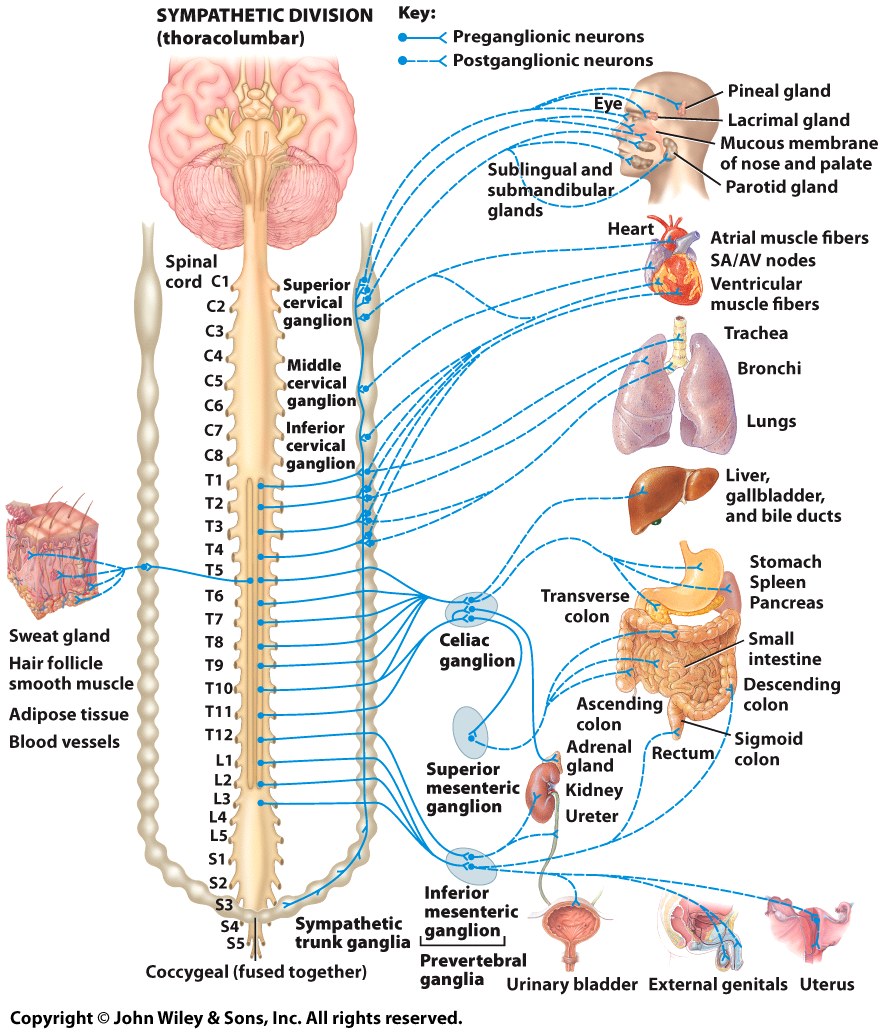
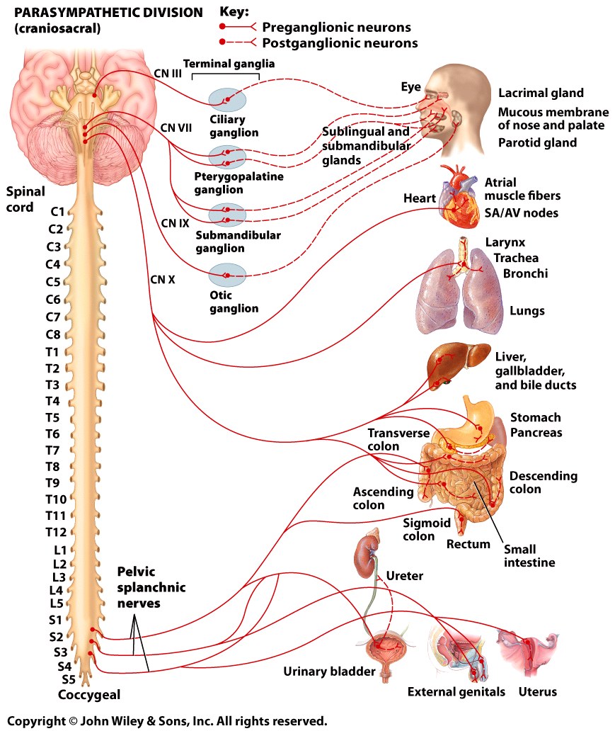
The autonomic nervous system has two subdivisions
Sympathetic NS
Parasympathetic NS
Autonomic NS
Sympathetic NS
Includes nerves that control the body when it is actively moving and burning energy.
Fight or flight responses.
Nerves in the thoracic and lumbar regions of the spinal cord.
Parasympathetic NS
Includes nerves responsible for digestion, energy storage and relaxation.
Rest and digest responses.
Nerves in the cranial and sacral regions of the spinal cord.
Most body organs are innervated by both the Sympathetic NS and Parasympathetic NS.
To maintain homeostasis.
SYMPATHETIC NS
PARASYMPATHETIC NS
Sympathetic vs. Parasympathetic
Neurons
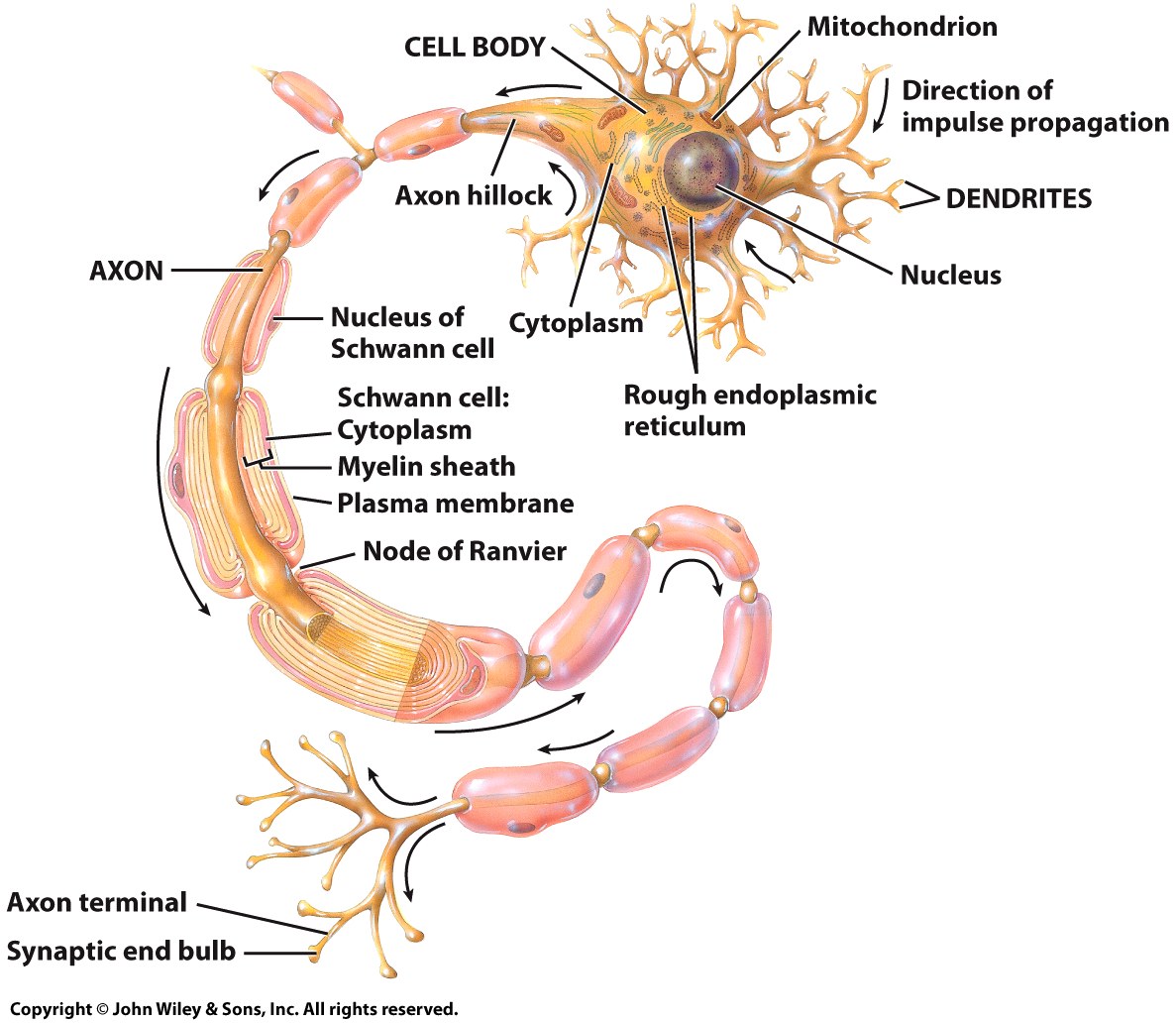
The NS contains neurons and neuroglial cells.
The neuron is the functional unit of the NS.
Functions include
Carrying sensory information to the brain.
Formulating responses.
Sending responses to organs.
Neurons arrange into bundles called nerves.
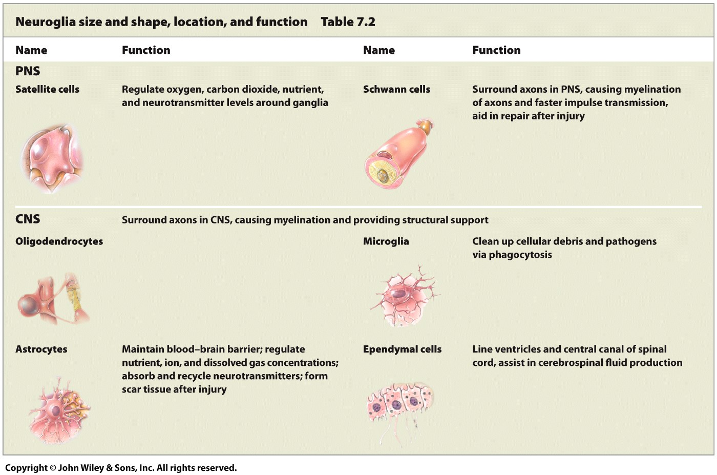
Neuroglia are the supporting cells of nervous tissue.
Neuroglia
PNS
Satellite cells
Regulate oxygen, carbon dioxide, nutrient, and neurotransmitter levels around ganglia.
Schwann cells
Surround axons in PNS, causing myelination of axons and faster impulse transmission, aid in repair after injury.
CNS
Oligodendrocytes
Surround axons in CNS, causing myelination and providing structural support.
Astrocytes
Maintain blood-brain barrier; regulate nutrient, ion, and dissolved gas concentrations; absorb and recycle neurotransmitters; form scar tissue after injury.
Microglia
Clean up cellular debris and pathogens via phagocytosis.
Ependymal cells
Line ventricles and central canal of spinal cord, assist in cerebrospinal fluid production.
Neurons
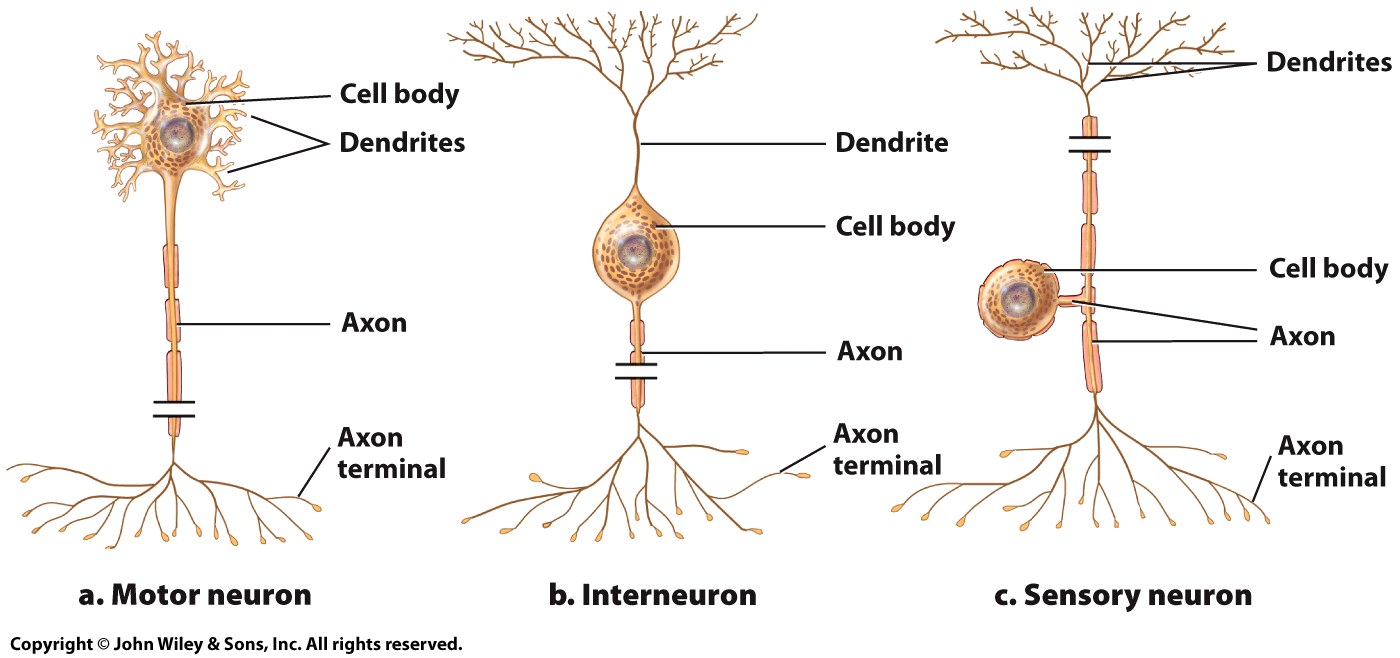
The three classes of neurons are based on function.
Sensory neurons detect conditions in the environment or body.
Motor neurons carry instruction to the body
Interneurons connect sensory and motor neurons.
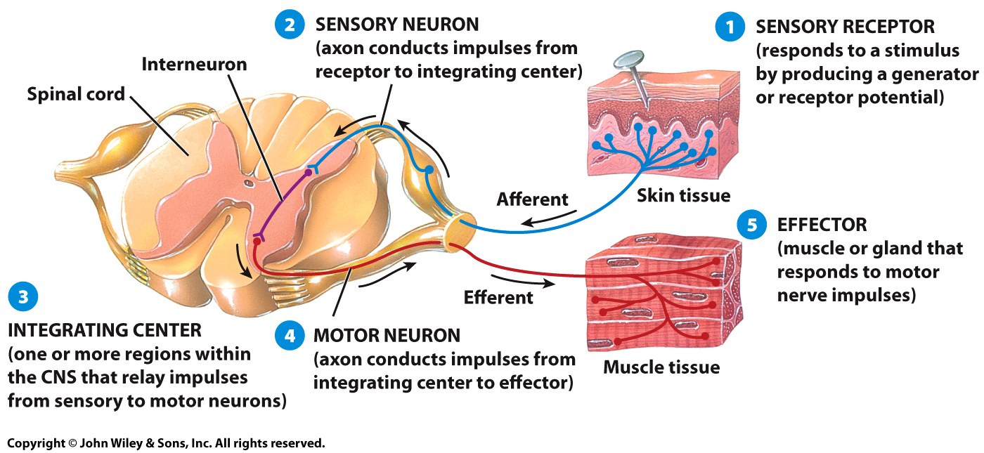
The Reflex Arc
Reflexes are rapid responses to sensory stimuli.
The stimulus from a sensory neuron bypasses the brain and instead runs through an interneuron within the spinal cord.
The motor neuron then transmits an immediate response to the effector organ.
INTEGRATING CENTER: (one or more regions within the CNS that relay impulses from sensory to motor neurons)
SENSORY NEURON: (axon conducts impulses from receptor to integrating center)
SENSORY RECEPTOR: (responds to a stimulus by producing a generator or receptor potential)
EFFECTOR: (muscle or gland the responds to motor nerve impulses)
MOTOR NEURON: (axon conducts impulses from integrating center to effector)
iNTEGRATING CENTER: (one or more regions within the CNS that relay impulses from sensory to motor neurons)
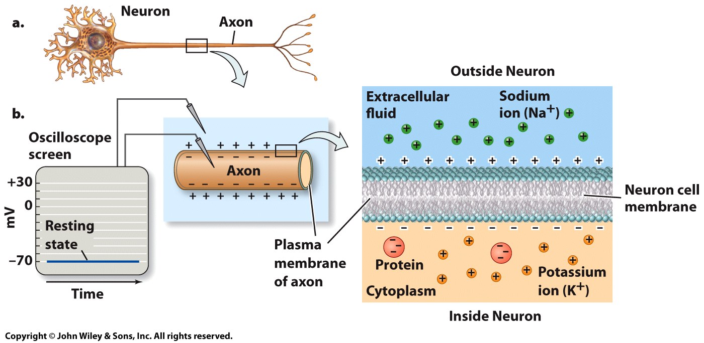
Membrane Potential
The membrane potential results from the difference in ion concentrations between the two sides of a cell membrane.
Measured as electrical differences.
The membrane potential of a neuron during a typical nerve impulse alternates between -70 mV and +30 mV
At rest, the inner side of a neuron’s membrane is negatively charged relative to it’s outer side and its membrane potential is -70 mV.
Variations in ion concentration across the cell membrane allow neurons to generate action potentials.
Gates and Channels Control the Flow of Ions
Ions cannot simply diffuse through the lipid bilayer of the cell membrane, they must ravel through channel proteins.
Voltage-gated channels - Respond to trans-membrane voltage changes.
Ligand-gated channels - Chemically regulated.
Mechanically-regulated channels - Respond to distortions of the membrane surface.
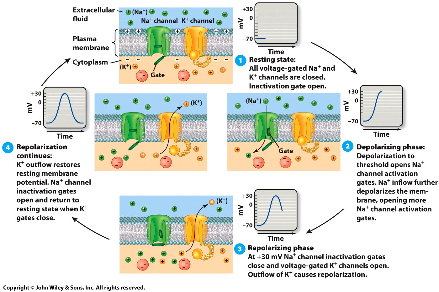
Neurons Work Through Action Potentials
An action potential is a brief change in electrical conditions at a neuron’s membrane.
When a neuron “fires”, the charge differential across the neuron’s membrane suddenly reverses polarity.
Action potentials are “all or nothing” events.
IPSPs and EPSPs also influence the generation of action potentials.
Resting state: All voltage-gated Na+ and K+ channels are closed. Inactivation gate open.
Depolarizing phase: Depolarization to threshold opens Na+ channel activation gates. Na+ inflow further depolarizes the membrane, opening more Na+ channel activation gates.
Repolarizing phase: At +30 mV Na+ channel inactivation gates close and voltage-gated K+ channels open. Outflow of K+ causes repolarization.
Repolarization continues: K+ outflow restores resting membrane potential. Na+ channel inactivation gates open and return to resting state when K+ gate close.
Graded Responses Created Fine Neural Control
Graded responses can be obtained by hyperpolarizing or depolarizing individual neural membranes.
Excitatory postsynaptic potentials (EPSPs)
Cause slight depolarization of the neuron.
Inhibitory postsynaptic potentials (IPSPs)
Hyperpolarize the neuron.
The membrane potential is further from the needed to generate an action potential, so a larger stimulus is required to begin an action potential.
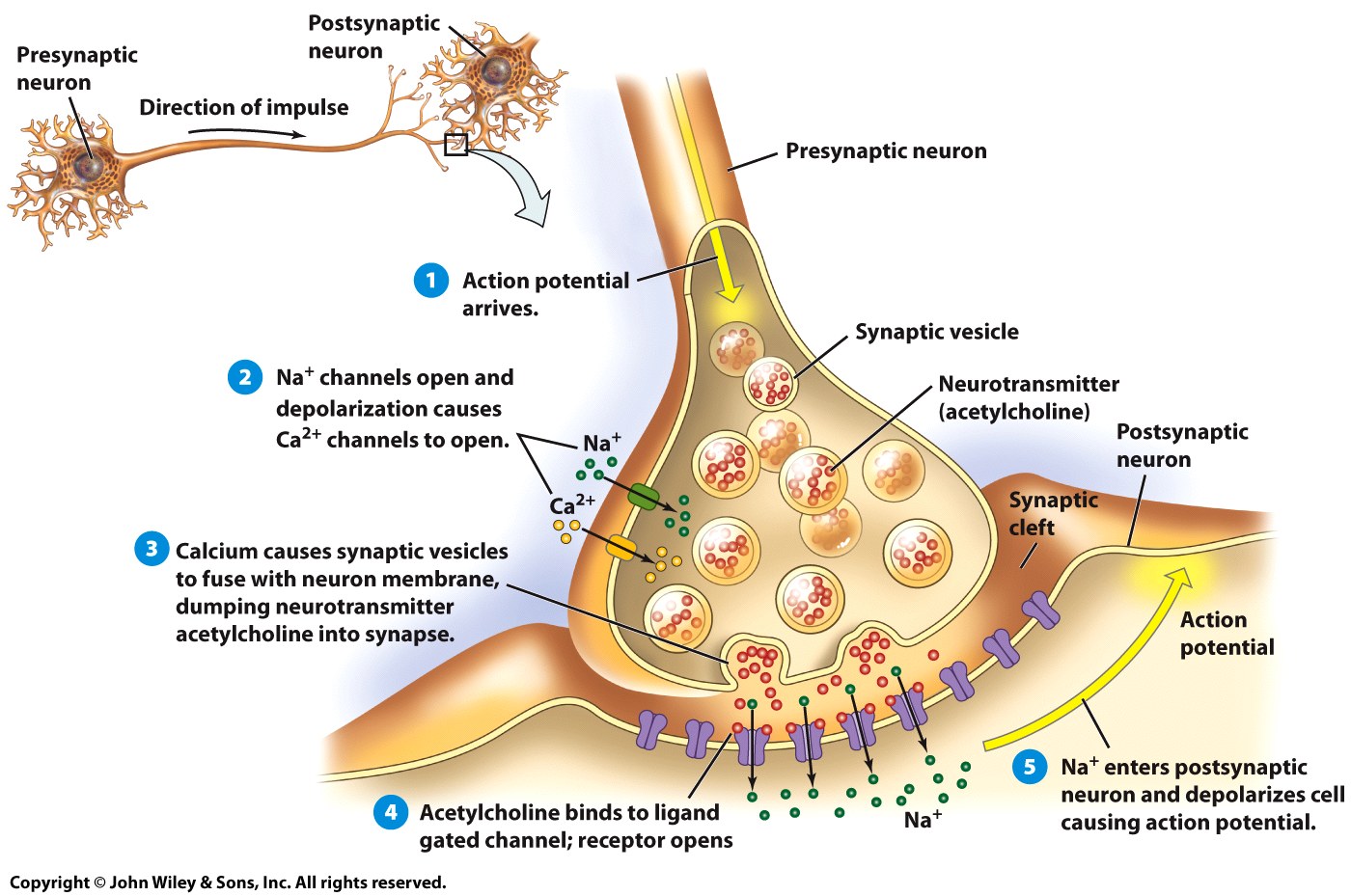
Neurotransmitters
Synapses separate one neuron from another.
Neurotransmitters are chemicals used to transmit the nerve impulse from one cell to the next.
Diffuse across the synaptic cleft.
Action potential arrives.
Na+ channels open and depolarization causes Ca2+ channels to open.
Calcium causes synaptic vesicles to fuse with neuron membrane, dumping neurotransmitter acetylcholine into synapse.
Acetylcholine binds to ligand gated channel; receptor opens.
Na+ enters postsynaptic neuron and depolarizes cell causing action potential.
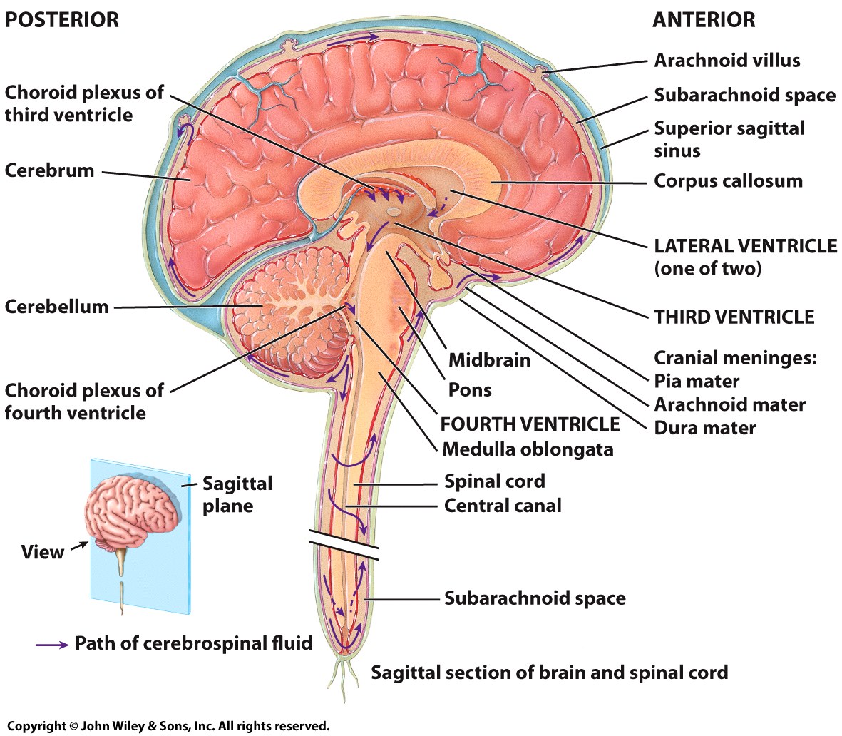
The Brain and Spinal Cord are Central to the NS
The volume of the human brain is about 1250 cm3
The human brain weights about 1,400 grams.
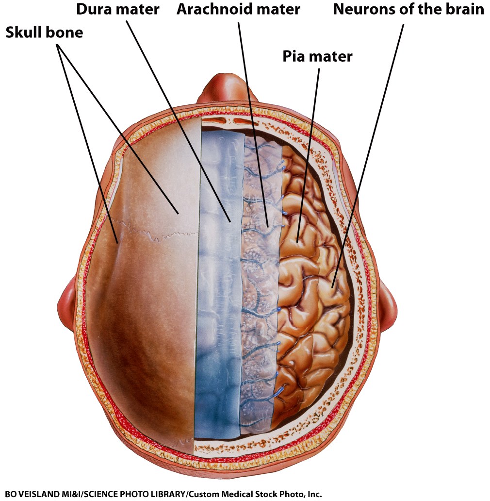
The cerebrospinal fluid and meninges protect and nourish the CNS.
The meninges are a series of three consecutive tissue coverings between the nervous tissue and the bone.
The CSF functions to maintain uniform pressure within the brain and spinal cord.
Layers of the Meninges
Dura mater
Tough connective tissue layer immediately beneath the skull.
Arachnoid mater
A thin, fragile layer which resembles a spider web.
Cerebrospinal fluid flows between the strands of the arachnoid mater.
Pia mater
The innermost layer, extremely thin and attached to neurons.
Meningitis is an inflammation of these three layers.
Cerebrospinal Fluid (CSF)
Provides a constant environment for the CNS.
Its is continuously produced and absorbed, creating a constant flow.
Ventricles make CSF.
CSF helps maintain the blood-brain barrier.
Permits passage of only certain ions and nutrients.
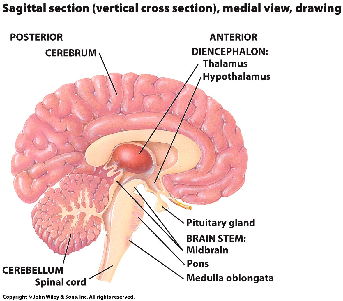
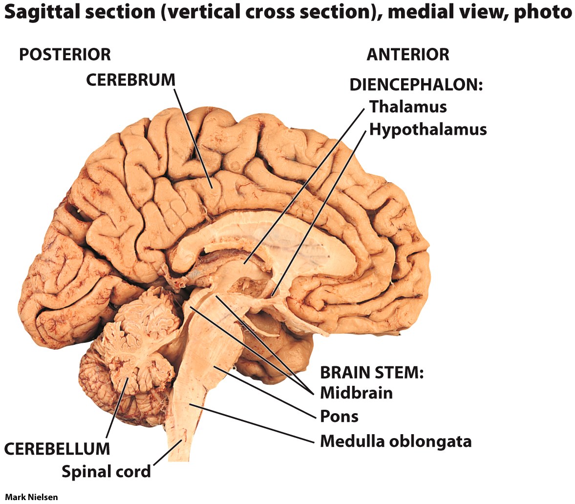
The Brain
The brain has 4 main parts.
The brain stem.
The cerebellum.
The diencephalon.
The cerebrum.
The Brain Stem
The oldest part of brain.
Made up of the
Mid brain, medulla oblongata, and pons.
Regulates
Heart rate, breathing, and blood pressure.
Has reflex centers for
sneezing, coughing, hiccupping and swallowing.
The Cerebellum
Focuses on muscles and movement.
Maintains muscle tone, posture and balance.
Fine tunes conscious and unconscious movements directed by the cerebrum.
Important in learning motor skills.
Riding a bike.
Taking notes in class.
Important in proprioception.
Knowing where the body is in space.
The Diencephalon
A relay center for sensory information and motor responses.
Center for visual and auditory startle reflexes are located here.
The thalamus and hypothalamus are also located here.
The hypothalamus
Secretes hormones that control the pituitary gland.
Also regulates circadian rhythm, body temperature, heart rate, blood pressure and controls smooth muscle contraction.
The thalamus is a relay station.
For incoming sensory information.
The limbic system
Communicates with the anterior portion of thalamus.
Is also responsible for emotions.
The Cerebrum
Largest portion of the brain.
Central processing center where learning, remembering, and activity planning takes place.
Information is processed and integrated and appropriate responses are generated.
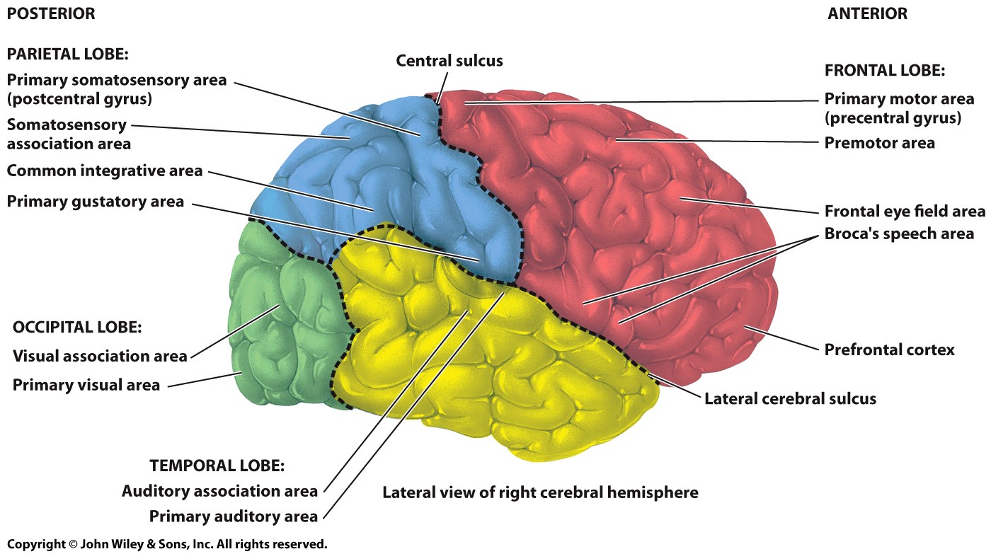
Organized into four lobes.
Frontal, parietal, occipital, temporal.
The surface of the cerebrum.
Covered in creases (sulci) and raised areas (gyri)
The outer layer of cerebrum, called the cerebral cortex. is composed of gray matter.
Folded to provide larger surface area.
Contains neuronal cell bodies, dendrites, and non-myelinated axons.
Responsible for sensations, voluntary movements, and thoughts.
The inner layer of the cerebrum consists of white matter that is composed of myelinated axons.
Myelinated axons are covered in lipids which allows for faster impulse transmission.
Information passed between areas of the brain via tracts of white matter.
The left and right cerebral hemispheres are distinct.
The right analyzes sensory input, recognizes faces, and functions in spatial relationships.
The left includes general language interpretation and speech, controls writing and speaking, categorizes items and makes logical decisions.
The cortex has motor areas, sensory areas, and association areas that integrate new information with stored information.
The primary motor area formulates voluntary motor commands.
Association areas of the cerebral cortex integrate and coordinate information.
The right and left sides of the brain connects through the transverse tracts of the corpous collosum.
Special senses are integrated in specific areas of the cerebral cortex.
Visual interpretation occurs in the occipital lobe.
Auditory interpretation occurs in the temporal lobe.
Taste interpretation occurs in the parietal lobes.
The Cerebral Lobes and their Functions
Posterior
Parietal Lobe:
Primary somatosensory area (postcentral gyrus)
Somatosensory association area
Common integrative area
Primary gustatory area.
Central sulcus
Occipital Lobe:
Visual association area.
Primary visual area.
Temporal Lobe:
Auditory association area.
Primary auditory area.
Anterior
Frontal Lobe:
Primary motor area (precentral gyrus)
Premotor area.
Frontal eye field area.
Broca’s speech area.
prefrontal cortex
Lateral cerebral sulcus.
Learning and Memory
Learning does not exist in a vacuum
The brain’s ability to learn is related to what else is going on.
Fight or flight conditions drastically reduce the ability to learn.
Learning is a type of memory.
Both play a critical role at both ends of life.
Memory occurs in three phases.
Immediate memory.
Short-term memory
Long-term memory.