Unit 2 - Cell Structure and Function
Cell Organelles
Eukaryotes - have (most) DNA stored in the nucleus, an organelle that is bounded by a double membrane.
Prokaryotes - characterized by having:
no nucleus
DNA in an unbound region called the nucleoid
No membrane-bound organelles
All cells have ribosomes. Both types of cells contain cytoplasm bound by the plasma membrane. Cells are the organization of life.
Cells →Tissues → Organs→ Organisms.
Cells have 3 main jobs:
make energy
need energy for all activities
need to clean up waste produced while making energy
make proteins
proteins do all the work in a cell, so we need lots of them
make more cells
for growth
to replace damaged or diseased cells
Organelles - functioning parts of a cell that do the work of keeping cells alive (tiny organs)
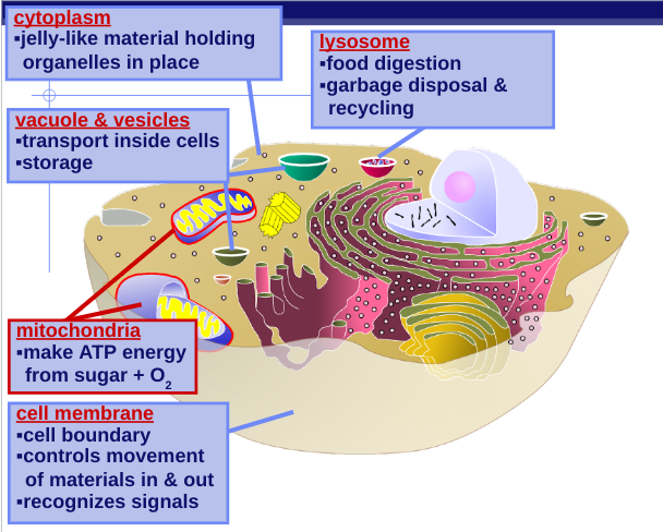
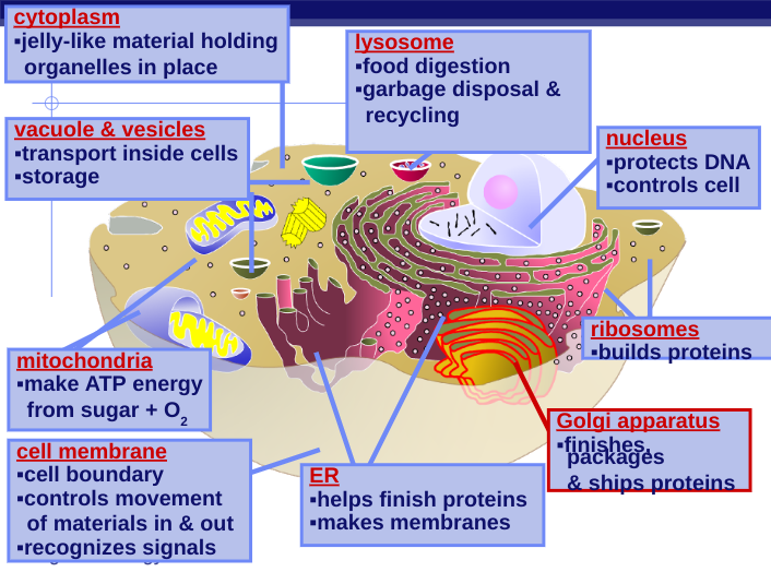
Cytoplasm - Semi-fluid substance that fills the interior of the cell and suspends organelles
How do cells power our bodies?
Cell membrane:
Function:
separates cell from outside
controls what enters or leaves cell
recognizes signals from other cells (communication between cells)
Structure
double layer of lipids
phospholipid bilayer
receptor molecules
proteins that receive signals
Vacuoles & Vesicles:
Function:
moving material around cell
storage
Structure:
membrane sac
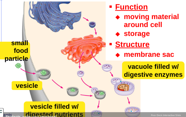
Lysosomes:
Function:
digest food
used to make energy
clean up and recycle
digest broken organelles
Structure:
membrane sac of digestive enzymes
Mitochondria
Function
make ATP energy from sugar via cellular respiration
sugar + O2 → ATP
fuels the work of life
Structure
double membrane
In both animal and plant cells
They have a smooth outer membrane and an inner membrane folded into cristae
The inner membrane creates two compartments: intermembrane space and mitochondrial matrix
Cristae presents a large surface area for enzymes that synthesize ATP
 Make Proteins
Nucleus:
Function:
control center of the cell
protects DNA
instructions for building proteins
Structure:
nuclear membrane
double membrane (lipid bilayer)
nucleolus (contained in the nucleus)
ribosome factory
chromosomes
condensed DNA
Nucleolus: located within the nucleus and is the site of ribosomal RNA (rRNA) synthesis makes ribosomes
Nuclear membrane: a double membrane; separating it from the cytoplasm — each membrane consists of a lipid bilayer
Nuclear pores: regulate the entry and exit of molecules (proteins)
Ribosomes:
Function:
protein synthesis
read instructions to build proteins from DNA
Structure:
complexes of rRNA and protein
some free in cytoplasm
some attached to ER
Endoplasmic Reticulum (ER):
Function:
site of protein synthesis (Rough ER)
helps complete the proteins after ribosome builds them
makes membrane lipids (Smooth ER)
Structure:
rough ER
ribosomes attached
smooth ER
no ribosomes
Golgi Apparatus:
Function:
finishes, sorts, labels, & ships proteins
like UPS headquarters
shipping and receiving department
ships proteins in vesicles
“UPS trucks”
Structure:
membrane sacs
 Cell Membrane Structure
Semipermeable: a material or membrane that allows some substances to pass through, but not all things
Phospholipids are amphipathic molecules, containing hydrophobic and hydrophilic regions.
Most of the lipids and some proteins in a membrane can shift about laterally. As temperatures cool, membranes switch from a fluid state to a solid state, this phase change is based on lipid composition.
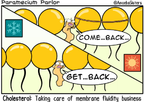
Cholesterol: regulates membrane fluidity - at warm temperatures (such as 37 C), restrains movement of phospholipids. At cool temperatures it maintains fluidity by preventing tight packing.
 Integral proteins: penetrate the hydrophobic interior of the lipid bilayer (sometimes called embedded proteins)
Integral proteins that span the membrane are called transmembrane proteins (membrane spanning proteins in the POGIL)
Peripheral (surface) proteins are loosely bound to the surface of the membrane
Six major functions of membrane proteins
Transport
Enzymatic activity
Signal transduction
Cell-cell recognition
Intercellular joining
Attachment to the cytoskeleton and extracellular matrix (ECM)
Cell to cell recognition is possible by membranes binding to each other, the carbohydrates serve as identification markers. Membrane carbohydrates may be covalently bonded to lipids (forming glycolipids) or, more commonly, to proteins (forming glycoproteins)
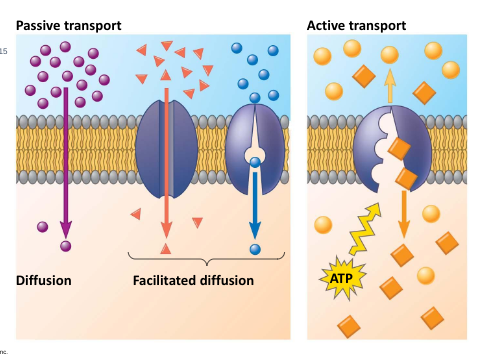
Passive transport: the movement of a substance across a membrane with no energy investment (ex. diffusion or osmosis)
Diffusion: the net movement of molecules from an area of high concentration to an area of low concentration (down the concentration gradient)
Individual molecules are moving randomly, and will continue to do so through dynamic equilibrium
Cell Membrane Transport (passive)
Osmosis: the diffusion of free water across a selectively permeable membrane.
Low concentration → High concentration
Isotonic solution: solute concentration is the same as inside the cell; no net water movement across the plasma membrane
Hypertonic solution: solute concentration is greater than that inside the cell; cell loses water
Hypotonic solution: solute concentration is less than that inside the cell; cell gains water
 Osmoregulation: the control of solute concentration and water balance, is a necessary adaptation for life in such environments.
Membrane transport using proteins (and other mechanisms)
Facilitated diffusion: Passive transport aided by proteins (no energy, still high to low)
in facilitated diffusion, transport proteins speed the passive movement of molecules across the plasma membrane
Channel proteins: provide corridors that allow a specific molecule or ion to cross the membrane
Channel proteins include
aquaporins, for facilitated diffusion of water
ion channels that open or close in response to a stimulus (gated channels)
needs another molecule to bond to the protein to change the shape to open up the channel
Carrier proteins: undergo a subtle change in shape that translocates the solute-binding site across the membrane
the shape change may be triggered by binding and release of the transported molecule
no net energy input is required
Active transport: moves substances against their concentration gradients (low concentration to high). This requires energy, usually in the form of ATP.
Sodium-potassium pump
 Membrane potential is the voltage across a membrane. Voltage is created by difference in the distribution of positive and negative ions across a membrane — this is needed to make things happen. This can create an electrochemical gradient - difference in concentration of both chemicals and charges across a membrane.
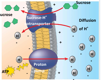
Electrogenic pump: a transport protein that generates voltage across a membrane. Ex: proton pump
Cotransport: occurs when active transport of a solute indirectly drives transport of other solutes
plant cells use the gradient of hydrogen ions generated by proton pumps to drive active transport of nutrients into the cell
 Exocytosis: transport vesicles migrate to the membrane, fuse with it, and release their contents.
Endocytosis: the cell takes in molecules and particulate matter by forming new vesicles from the plasma membrane
There are three types of endocytosis:
Phagocytosis (“cellular eating”)
Pinocytosis (“cellular drinking”)
Receptor-mediated endocytosis (need ‘keys’ to start endocytosis)
Scientific Theories of Cells
Hypothesis: a proposed explanation for a set of observations that can be tested.
Scientific Theory: an in-depth explanation of an observed phenomenon, that is supported with repeated testing and evidence
Cell Theory:
All living things are made of cells
Cells are the basic unit of structure and function to life
All new cells come from pre-existing cells
Endosymbiotic Theory: an explanation of the evolution of membrane bound organelles in eukaryotic cells. Some modern organelles such as the mitochondria were once prokaryotic microbes.
How did eukaryotic cells evolve?
infoldings of the cell membrane gave rise to the endomembrane system and the formation of the endoplasmic reticulum and nucleus
Ancestral eukaryotic cell engulfs (endocytosis) but does not consume aerobic bacteria that evolves into a mitochondria. A similar process occurred with photosynthetic bacteria that evolved into chloroplasts
What evidence supports endosymbiotic theory?
mitochondria and chloroplasts are the same size as prokaryotic cells and divide by binary fission
Mitochondria and chloroplasts have their own DNA which is circular, not linear
Evidence of prokaryotic life 3.5 billion years ago. Evidence of eukaryotic life 1.5 billion years ago