slides/AP videos 4.1-4.7
Cells most often communicate with each other via chemical signals
For example, the yeast, Saccharomyces cerevisiae, have two mating types, a and α
Cells of different mating types locate each other via secreted factors specific to each type
cells communicate with one another through direct contact with other cells
cells of multicellular organisms often maintain physical contact with other cells or make physical contact with other cells during certain activities
some unicellular organisms live in colonies and are in physical contact with other organisms in that colony
cells can send chemical signals directly to adjacent cells
cell membrane and cell wall modifications allow for communication to occur between adjacent cells
plant cells have plasmodesma
animal cells have gap junctions
cells can communicate with one another over short or long distance
cells use chemical signals to communicate over short and long distances
the cell receiving the signal is referred to as the target cell
short-distance communication
cell sends out local regulators (signals)
target cell is within a short-distance of the signal (local signaling)
often used to communicate with cells of the same type
long-distance communication
target cell is not in the same area as the cell emitting the signal
signal travels a long distance to reach target cell
often used to signal cells of another type
A signal transduction pathway is a series of steps by which a signal on a cell’s surface is converted into a specific cellular response
Cells in a multicellular organism communicate by chemical messengers
Animal and plant cells have cell junctions that directly connect the cytoplasm of adjacent cells
In local signaling, animal cells may communicate by direct contact, or cell-cell recognition
In many other cases, animal cells communicate using local regulators, messenger molecules that travel only short distances
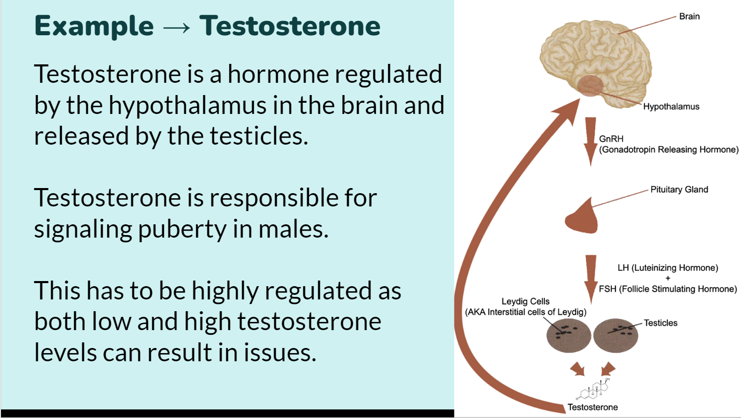
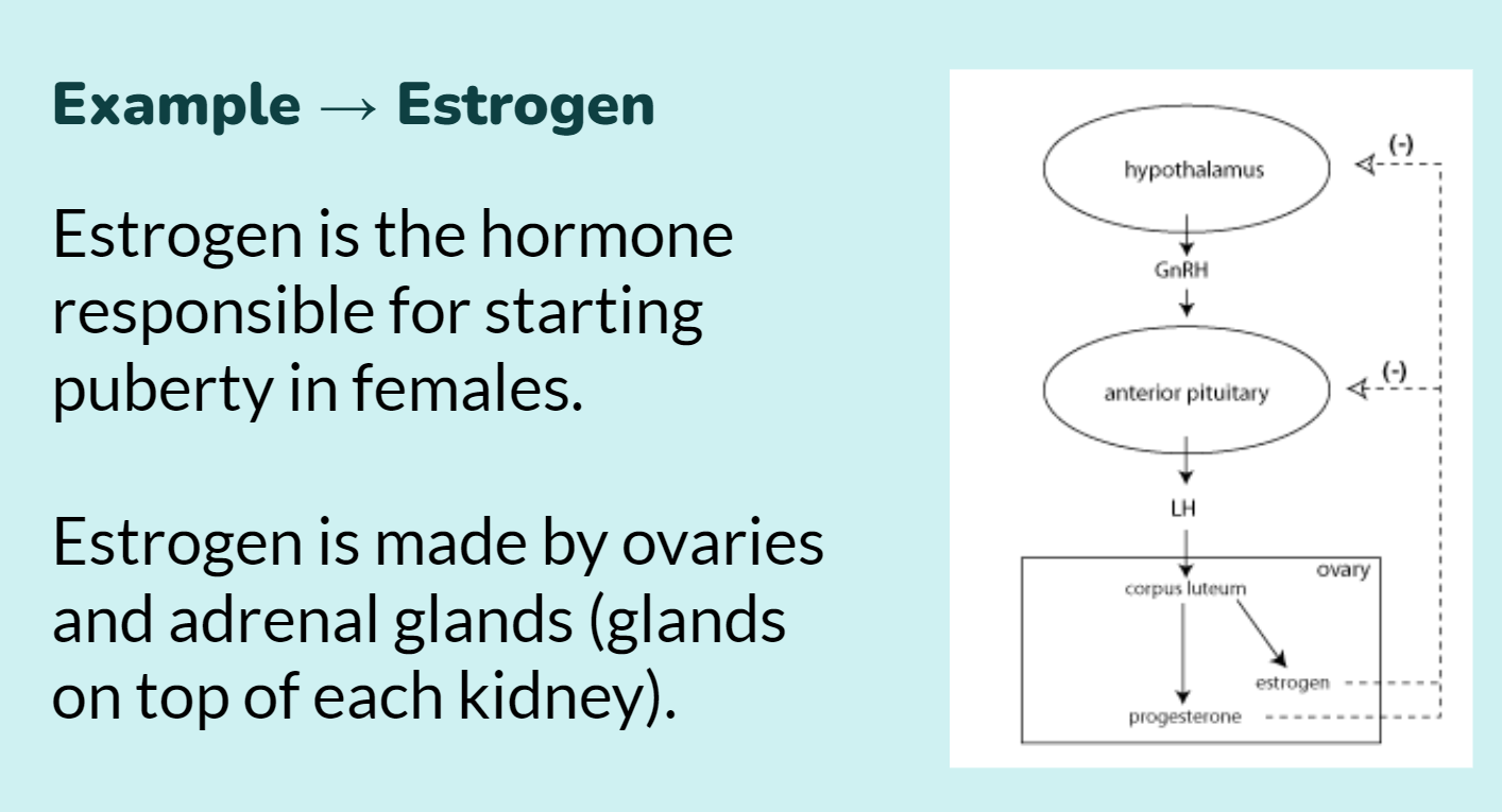
In long-distance signaling, plants and animals use chemicals called hormones
The ability of a cell to respond to a signal depends on whether or not it has a receptor specific to that signal
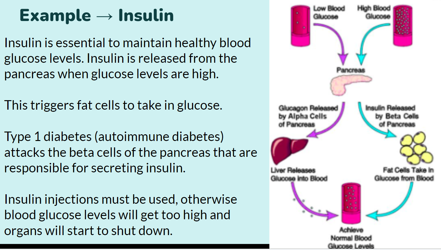
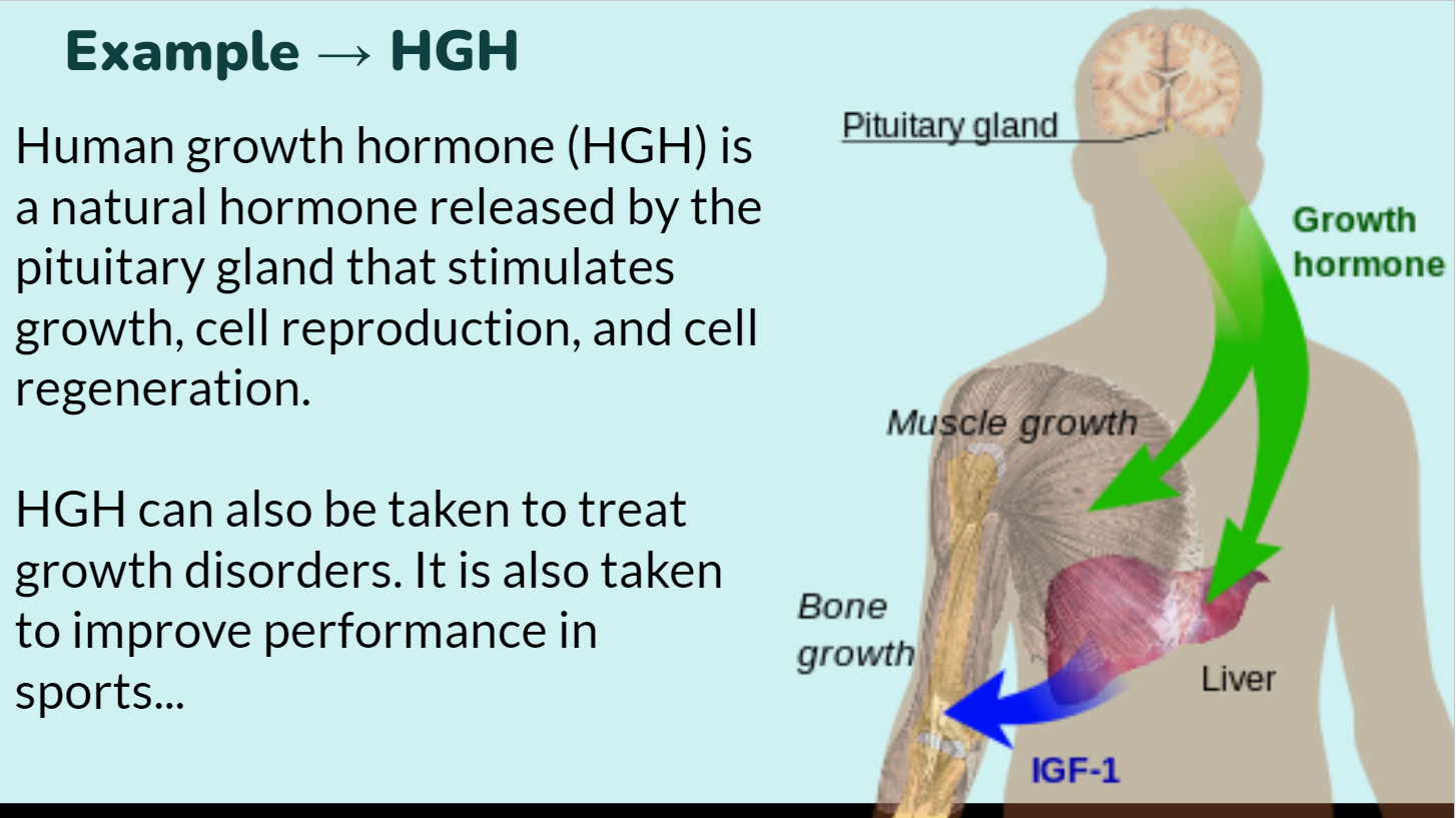
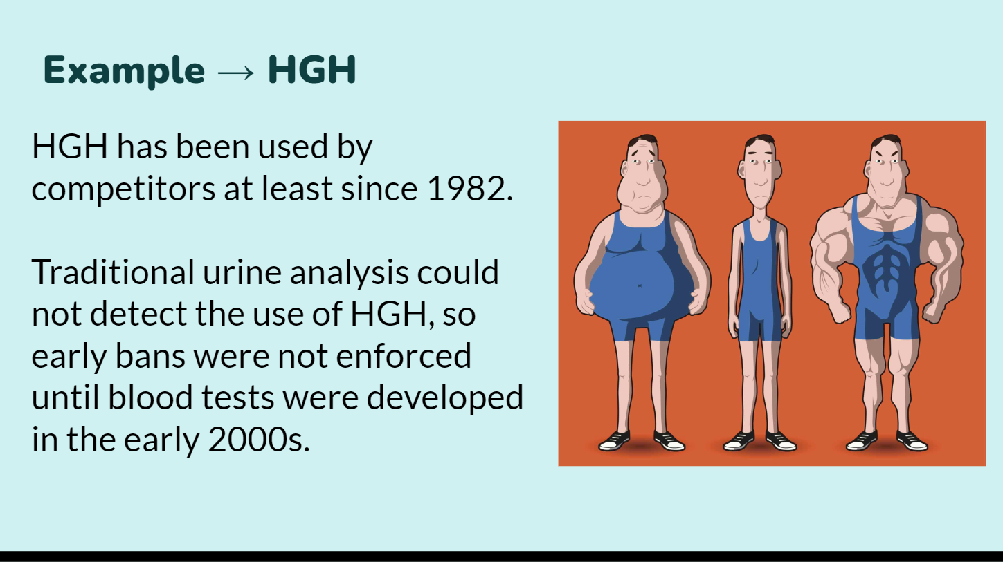

Earl Sutherland first discovered signal transduction by studying the effects of epinephrine on cells
Epinephrine is known as the fight-or-flight hormone
signal transduction pathways link signal reception with cellular responses
Sutherland identified three stages of signal transduction:
Reception - detection of a signal molecule coming from outside the cell
Transduction - convert signal to a form that can bring about a cell response
Response - specific cellular response to the signal molecule
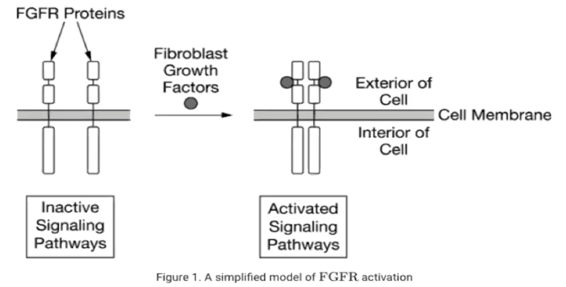
Reception occurs when a signal molecule, or ligand, binds a receptor protein, altering the receptor’s shape
Ligands are highly specific to particular receptors
Receptors are either on the cell surface (membrane receptors) or inside of the cell (cytosolic or intracellular receptors)
Membrane receptors have polar ligands, while cytosolic receptors have nonpolar ligands
A G-proteincoupled receptor (GPCR) is an example of a membrane receptor
The ligand binds the GPCR extracellular domain, slightly altering the receptor’s shape
G-protein is activated by the GPCR and released, as it displaces its GDP with a GTP molecule
The active G-protein may begin transduction
One GPCR can activate dozens of G-proteins
G-protein dephosphorylates its own GTP, forming GDP and inactivating itself
When ligand concentrations drop, the ligand dissociates from the receptor, deactivating the GPCR
Intracellular receptor proteins are found in the cytosol or nucleus of target cells
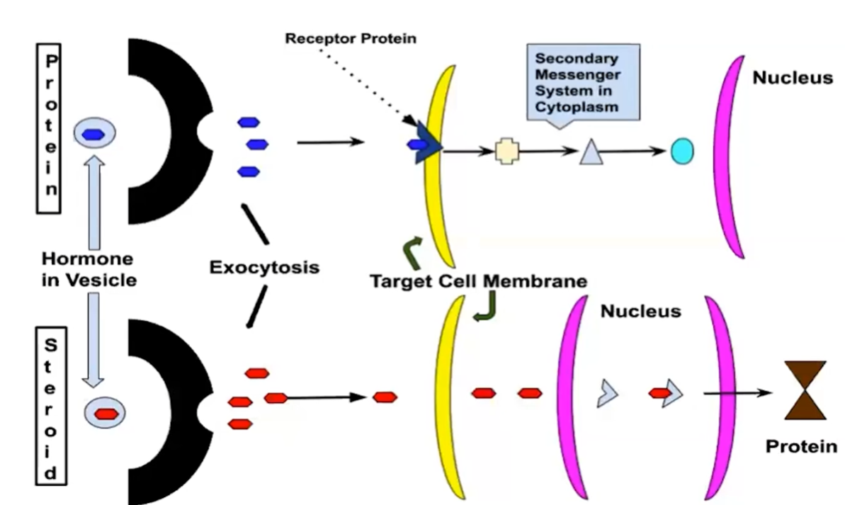
Small or hydrophobic chemical messengers can readily cross the membrane and activate receptors
Examples of hydrophobic messengers are the steroid and thyroid hormones of animals
A ligand-gated ion channel receptor acts as a gate when the receptor changes shape
When a signal molecule binds as a ligand to the receptor, the gate allows specific ions, such as Na+ or Ca2+, through a channel in the receptor
The conformational change in the receptor precipitates a step-wise molecular relay in the cell, indirectly causing a change in the cellular part performing the response
Multistep pathways can
Amplify a signal: A few molecules can produce a large cellular response
Provide more opportunities for coordination and regulation of the cellular response
In many pathways, the signal is transmitted by a cascade of protein phosphorylations
many signal transduction pathways include protein modification and phosphorylation cascades
regulate protein synthesis by turning on/off genes in nucleus
regulate activity of proteins in cytoplasm
cascades of molecular interactions relay signals from receptors to target molecules
phosphorylation cascade: enhance and amplify signal
Protein kinases transfer phosphates from ATP to protein, a process called phosphorylation
The molecules that relay a signal from receptor to response are mostly proteins
Like falling dominoes, the receptor activates another protein, which activates another, and so on, until the protein producing the response is activated
At each step, the signal is transduced into a different form, usually a shape change in a protein
signaling begins with the recognition of a chemical messenger - a ligand - by a receptor protein in a target cell
the ligand-binding domain of a receptor recognizes a specific chemical messenger, which can be a peptide, a small chemical, or a protein, in a specific one-to-one relationship
g protein-coupled receptors are an example of a receptor protein in eukaryotes
The extracellular signal molecule (ligand) that binds to the receptor is a pathway’s “first messenger”
signaling cascades relay signals from receptors to cell targets, often amplifying the incoming signals, resulting in the appropriate responses by the cell, which could include cell growth, secretion of molecules, or gene expression
after the ligand binds, the intracellular domain of a receptor protein changes shape, initiating transduction of the signal
binding of ligand-to-ligand channels can cause the channel to open or close
Second messengers are small, nonprotein, water-soluble molecules or ions that spread throughout a cell by diffusion (molecules that relay and amplify the intracellular signal)
Second messengers participate in pathways initiated by GPCRs
Cyclic AMP is a common second messenger
A cell’s response occurs when cell signaling leads to regulation of transcription or cytoplasmic activities
A cell’s response can be fine-tuned in the following ways:
Amplifying the signal (and thus the response)
Specificity of the response
Overall efficiency of response, enhanced by scaffolding proteins
Termination of the signal
signal transduction pathways influence how the cell responds to its environment
the environment is not static, and organisms need to regulate pathways to respond to changes in the environment
the ability to respond to stimuli is a characteristic of life and necessary for survival

signal transduction may result in changes in gene expression and cell function
signaling pathways can target gene expression and alter the amount and/or type of a particular protein produced in a cell
changes in protein type and/or amount can result in a phenotype change
Signal transduction can alter phenotype or cause apoptosis
An example of signal transduction altering phenotype is observed in Y-chromosomalSRY gene activation
Phenotype is manifest in an organism’s appearance
Apoptosis is programmed cell death and occurs during embryonic and fetal development or if a cell is damaged
Components of the cell are chopped up and packaged into vesicles that are digested by scavenger cells
Apoptosis prevents enzymes from leaking out of a dying cell and damaging neighboring cells
Caspases are the main proteases (enzymes that cut up proteins) that carry out apoptosis
Apoptosis can be triggered by
An extracellular death-signaling ligand
can be the response of a signal transduction
DNA damage in the nucleus
Protein misfolding in the endoplasmic reticulum
Alterations to any component of a signal transduction pathway will affect response
Alterations can include mutation or chemical interactions
Altering any domain in the receptor can affect transduction
Ras, a proto-oncogene, can cause cancer if mutated
changes in signal transduction pathways can alter cellular response
mutations in any domain of the receptor protein or in any component of the signaling pathway may effect the downstream components by altering the subsequent transduction of the signal
changes in protein structure can result in change in function
one disruption in a pathway can affect the downstream reactions
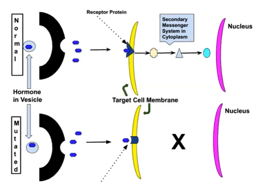
chemicals that interfere with any component of the signaling pathway may activate or inhibit the pathway
Organisms must respond to environmental change in order to maintain homeostasis
Homeostasis is the constant set of internal conditions of an organism
the maintenance of a stable internal environment
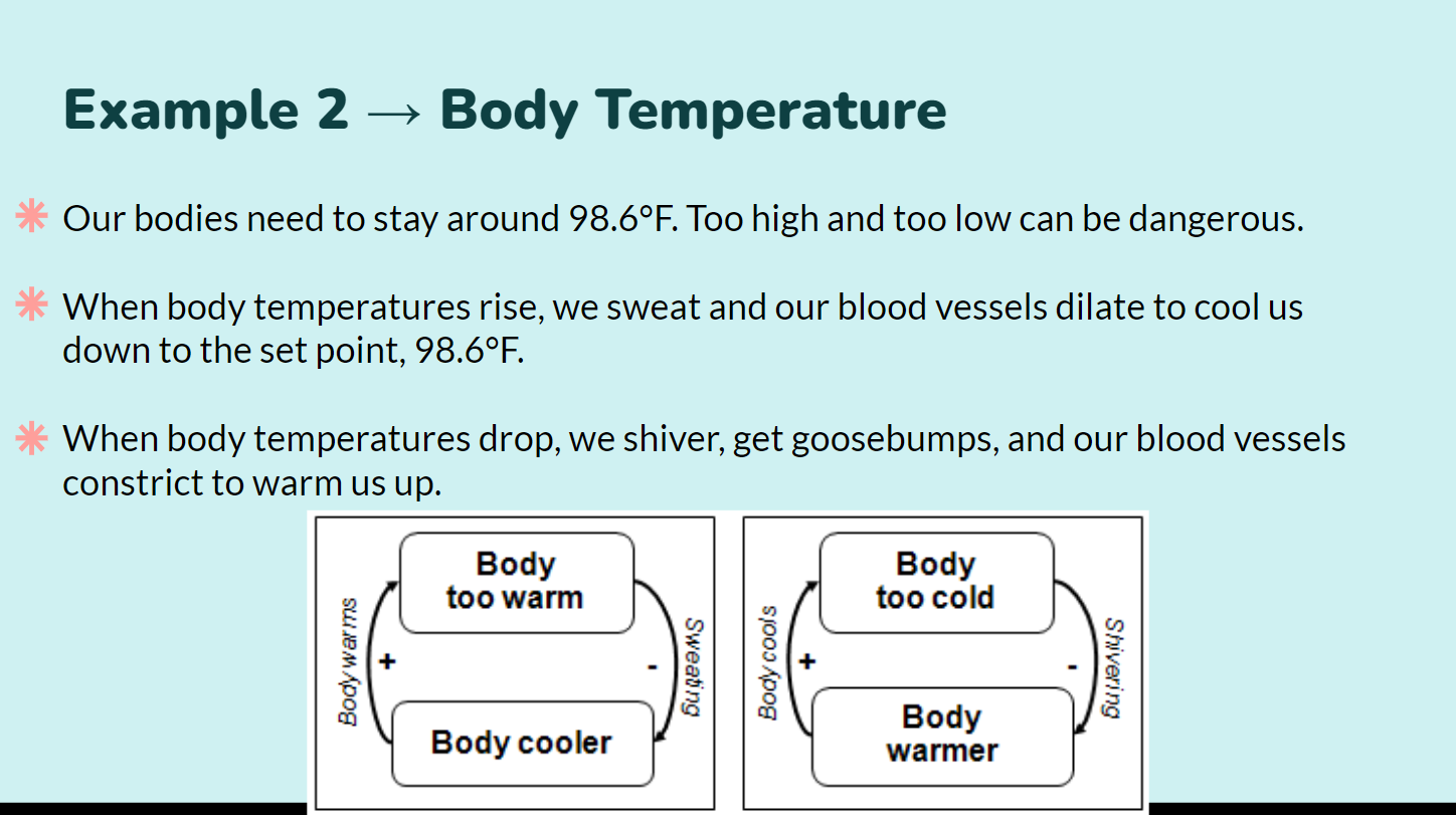
organisms use feedback mechanisms to maintain their internal environments and respond to environmental changes (both internal and external)
feedback mechanisms are processes used to maintain homeostasis by increasing or decreasing a cellular response to an event
If a stimulus causes an organism’s to migrate away from its homeostatic level, negative feedback mechanisms will restore homeostasis
“More gets you less”
negative feedback mechanisms maintain homeostasis for a particular cell condition
negative feedback mechanisms maintain homeostasis for a particular condition by regulating physiological processes
if a system is disrupted, negative feedback mechanisms return the system back to its target set point
these processes operate at the molecular and cellular levels

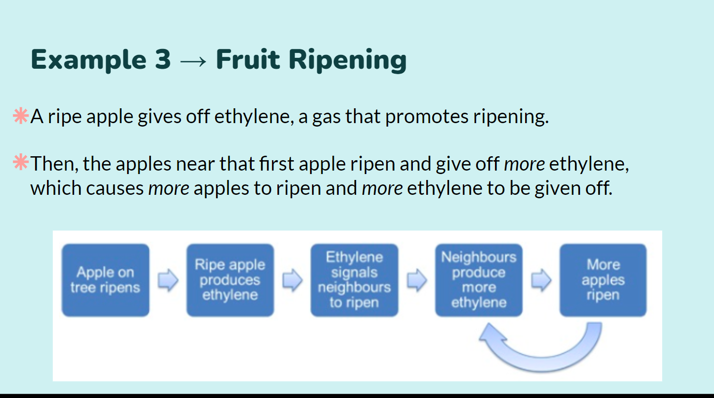
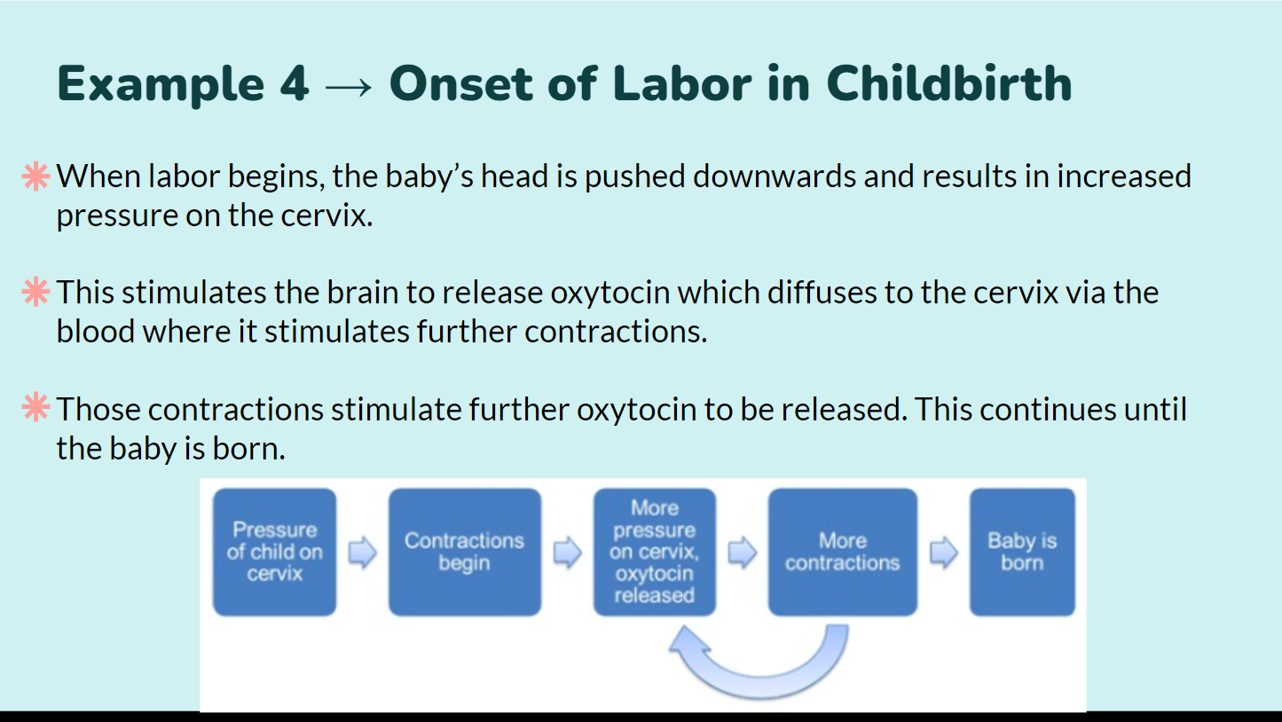
 Rarely, a stimulus causes an organism’s to migrate away from its homeostatic level, and the organism will amplify its response using positive feedback
“More gets you more”
positive feedback mechanisms amplify responses and processes in biological organisms
the variable initiating the response is moved farther away from the initial set point, disrupting homeostasis
amplification occurs when the stimulus is further activated, which, in turn, initiates an additional response that produces the system change
 Cell cycle is the life of a cell from formation to its own division (a highly regulated series of events for the growth and reproduction of cells)
the cell cycle consists of two highly regulated processes
interphase
growth and preparation
interphase involves 3 sequential stages
G1 - cell growth
S - copies of DNA are made
G2 - the cytoplasmic components are doubled in preparation for division
m-phase
mitosis - division of the nucleus
cytokinesis - division of the cytoplasm
in eukaryotes, cells transfer genetic information via a highly regulated process
mitosis…
plays a role in cell growth, tissue repair, and asexual reproduction
ensures the transfer of a complete genome from a parent cell to two genetically identical daughter cells
alternates with interphase in the cell cycle
occurs in a sequential series of steps
prophase
metaphase
anaphase
telophase
is followed by cytokinesis
cytokinesis ensures equal distribution of cytoplasm to both daughter cells
cells can enter G0 where cell division no longer occurs, a cell can reenter division with appropriate signals
nondividing cells may exit the cell cycle or be held at a particular stage
Cell division is an important component of cell cycle
In unicellular organisms, division of one cell reproduces the entire organism
Multicellular organisms depend on cell division for embryonic development, growth and repair
For cells capable of division, the genome must be replicated to ensure the next generation of cells receives the entire complement of DNA
All the DNA in a cell constitutes the cell’s genome
A genome can consist of a single DNA molecule (common in prokaryotic cells) or a number of DNA molecules (common in eukaryotic cells)
DNA molecules in a cell are packaged into chromosomes
Eukaryotic chromosomes consist of chromatin, a complex of DNA and protein that condenses during cell division
Every eukaryotic species has a characteristic number of chromosomes in each cell nucleus
Somatic cells (non-reproductive cells) have two sets of chromosomes and are diploid (2n)
Gametes (reproductive cells: sperm and eggs) have half as many chromosomes as somatic cells and are haploid (n)
In preparation for cell division, DNA is replicated and the chromosomes condense
Each duplicated chromosome has two sister chromatids (joined copies of the original chromosome), which separate during cell division
The centromere is the narrow “waist” of the duplicated chromosome, where the two chromatids are most closely attached
During cell division, the two sister chromatids of each duplicated chromosome separate and move into two nuclei
Once separate, the chromatids are called chromosomes
Eukaryotic cell division consists of
Mitosis, the division of the genetic material in the nucleus
a process that ensures the transfer of a complete genome from a parent cell to daughter cells
daughter cells carry genomes identical to the parent cell genome
Cytokinesis, the division of the cytoplasm
The cell cycle consists of
Mitotic (M) phase: mitosis and cytokinesis
Interphase: cell growth and copying of chromosomes in preparation for cell division)Interphase (about 90% of the cell cycle) can be divided into subphases
G1 phase (“first gap”)
S phase (“synthesis”)
G2 phase (“second gap”)
The cell grows during all three phases, but chromosomes are duplicated only during the S phase
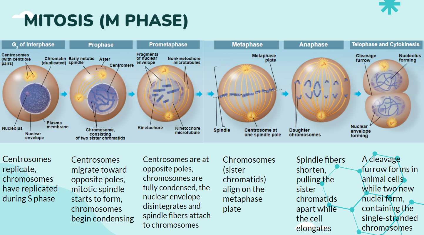
Mitosis is conventionally divided into five phases
Prophase
nuclear envelope begins to disappear
DNA coils into visible chromosomes
fibers begin to move double chromosomes toward the center of the cell
Prometaphase
fibers begin to move double chromosomes toward the center of the cell (sometime considered its own phase, sometimes grouped with prophase)
Metaphase
fibers align double chromosomes across the center of the cell
Anaphase
fibers separate double chromosomes into single chromosomes (chromatids)
chromosomes separate at the centromere
single chromosomes (chromatids) migrate to opposite sides of the cell
Telophase
nuclear envelope reappears and establishes two separate nuclei
each nucleus contains a complete genome
chromosomes will begin to uncoil
Cytokinesis overlaps the latter stages of mitosis
cytokinesis will separate the cell into two daughter cells, each containing identical genomes
The sequential events of the cell cycle are directed by a distinct cell cycle control system, which is similar to a clock
The cell cycle control system is regulated by both internal and external controls
The clock has specific checkpoints where the cell cycle stops until a go-ahead signal is received
For many cells, the G1 checkpoint seems to be the most important
If a cell receives a go-ahead signal at the G1 checkpoint, it will usually complete the S, G2, and M phases and divide
If the cell does not receive the go-ahead signal, it will exit the cycle, switching into a nondividing state called the G0 phase
internal controls or checkpoints regulate progression through the cycle
G1 checkpoint
at the end of G1 phase
cell size check
nutrient check
growth factor check
DNA damage check
G2 checkpoint
at the end of G2
DNA replication check
DNA damage check
m-spindle checkpoint
fiber attachment to chromosome check
Two types of regulatory proteins are involved in cell cycle control: cyclins and cyclin-dependent kinases (Cdks)
interactions between cyclins and cyclin-dependent kinases control the cell cycle
cyclins
a group of related proteins associated with specific phases of the cell cycle
different cyclins are involved in different stages of the cell cycle
concentrations can fluctuate depending on cell activity
produced to promote cell cycle progression
degraded to inhibit cell cycle progression
used to activate cyclin-dependent kinases (CDKs)
cyclins are specific to the CDK they activate
cyclin-dependent kinases (CDKs)
group of enzymes involved in cell regulation
requires cyclin binding for activation
phosphorylate substrates, promotes certain cell cycle activities
Cdks activity fluctuates during the cell cycle because it is controlled by cyclins, so named because their concentrations vary with the cell cycle
MPF (maturation-promoting factor) is a cyclin-Cdk complex that triggers a cell’s passage past the G2 checkpoint into the M phase

Cell cycle can be influenced by external factors
Growth factors are ligands that initiate cell division
Anchorage dependence typically requires cells to be bound to a substratum in order to divide
Density-dependent inhibition causes cells to cease dividing once they fill a space
Cancer cells do not respond normally to the body’s control mechanisms
cancer is the result of an unregulated cell cycle with uncontrolled cell division (if there is a change in the DNA in a region coding for one of the proteins needed to regulate the cell cycle, the cell cycle could go unregulated)
Cancer cells may not need growth factors to grow and divide
They may make their own growth factor
They may convey a growth factor’s signal without the presence of the growth factor
They may have an abnormal cell cycle control system
Cancer cells that are not eliminated by the immune system form tumors, masses of abnormal cells within otherwise normal tissue
If abnormal cells remain only at the original site, the lump is called a benign tumor
Malignant tumors invade surrounding tissues and can metastasize, exporting cancer cells to other parts of the body, where they may form additional tumors
