4.3 Thoracic Wall: Vessels & Nerves
Thoracic Wall: Vessels & Nerves
Overview of the Thoracic Wall
Vessels and Nerves: Essential components for understanding blood supply, drainage, and nerve distribution in the thoracic wall.
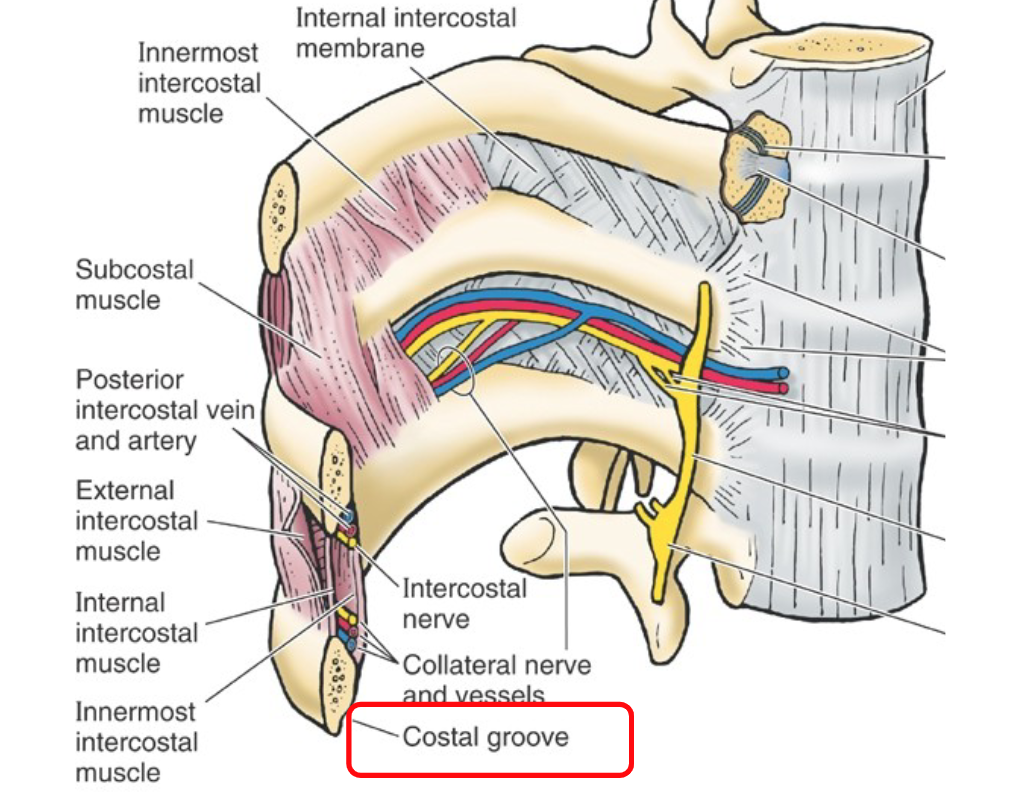
Mnemonic to Remember: "VAN" (Vein, Artery, Nerve) is used to describe the typical arrangement of vessels in the intercostal spaces.
Anatomy of the Thoracic Wall
Blood Supply
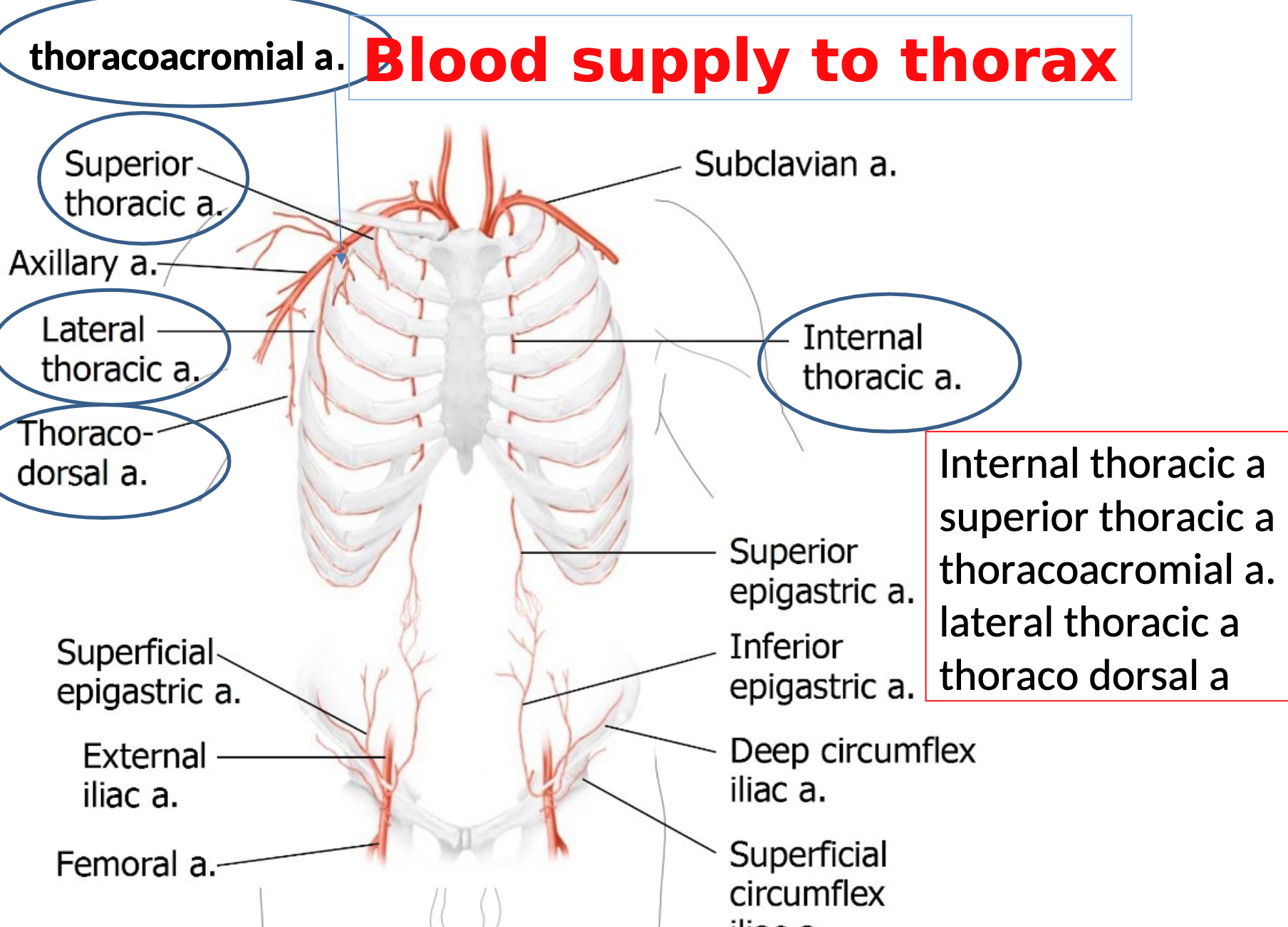
Key Arteries:
Subclavian Artery: Supplies blood to the upper limb, neck, and parts of the thorax.
Axillary Artery: Continuation of the subclavian artery, supplying the thoracic and pectoral regions.
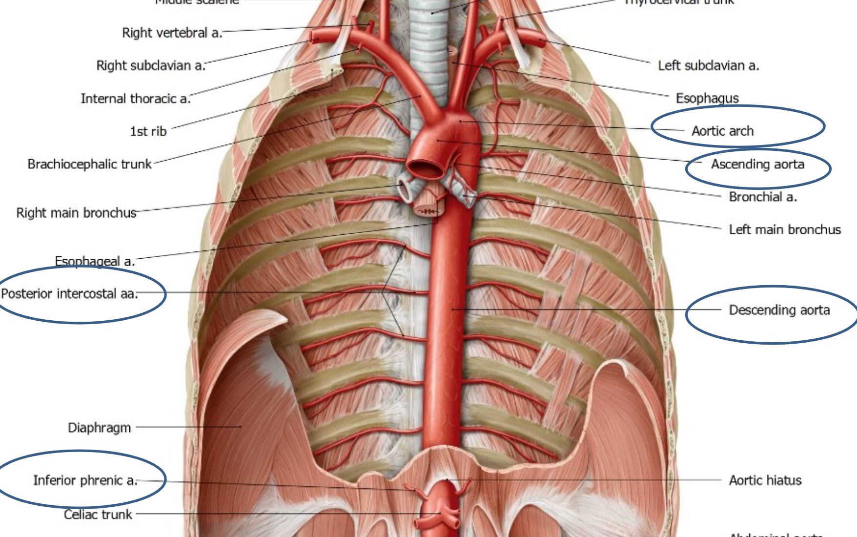
Thoracic Aorta: The main arterial supply from the heart to the thorax.
INTERCOSTAL VESSELS & NERVES
Intercostal space containing intercostal muscles & intercostal
• vein, artery, nerve – VAN - Near angle of rib, they travel along costal
grooves
• Follow same path (arteries travel with veins and nerves) – also – collateral
Branches of the Thoracic Aorta:
Posterior Intercostal Arteries: Supply blood to intercostal spaces 3-11 and are branches directly from the thoracic aorta.
Anterior Intercostal Arteries: Branch from the internal thoracic artery, which is a branch of the subclavian artery, supplying the first two intercostal spaces.
Diaphragm Blood Supply:
Supplied by Musculophrenic Artery (from the internal thoracic artery) and Inferior Phrenic Arteries (branches of the abdominal aorta).
Venous Drainage
Key Veins:
Azygos Vein (right): Drains posterior thoracic wall.
Hemiazygos Vein (left inferior): Drains lower left regions.
Accessory Hemiazygos Vein (left superior): Drains upper left regions.
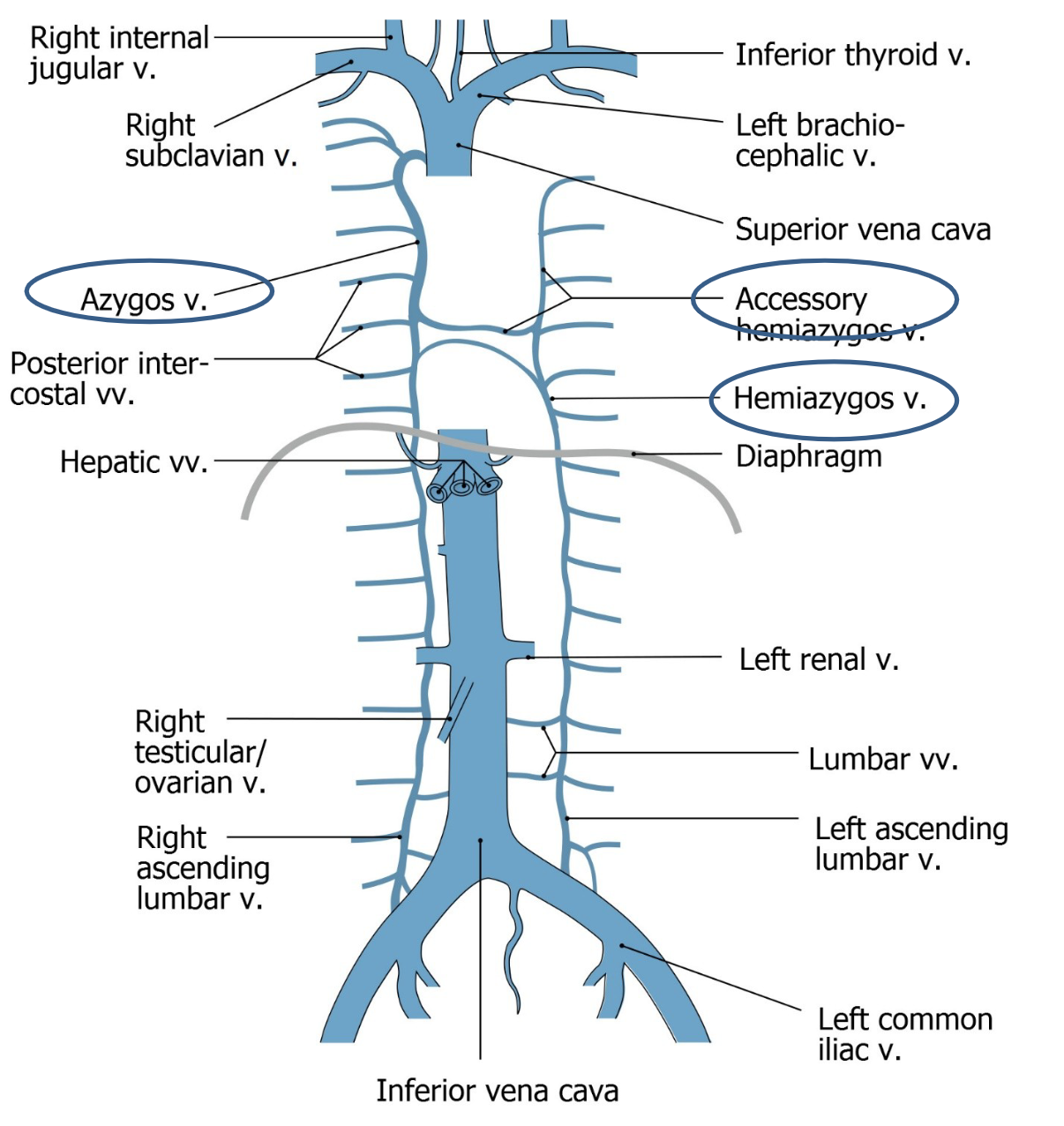
Venous Pathway:
Posterior intercostal veins drain into the azygos system, while anterior intercostal veins drain into the Internal Thoracic Vein, which then leads to the Subclavian Vein.
Major Lymphatic Vessel: Thoracic Duct - Drains lymph from the lower body and left side of the upper body into the venous system.
Nerve Supply
Posterior ramus supplies skin (sensory) and muscles (motor) of the back
Anterior ramus (intercostal nerves) innervate the skin and muscles of the anterior and lateral thorax and abdomen (intercostal
muscles, rectus abdominus and abdominal obliques
Intercostal Nerves (T1-T12): These are branches of the anterior rami of thoracic spinal nerves.
T1-11 = intercostal nerves, T12 = subcostal nerve
T1: Joins the brachial plexus.
T2: Supplies the floor of the axilla and parts of the arm.
T3-T6: Provide sensory and motor innervation to the intercostal muscles in their respective spaces.
T7-T11: Supply the anterior abdominal wall.
T12: Known as the subcostal nerve supplying the lower anterior abdominal wall.
Cutaneous Branches:
Anterior and lateral branches provide sensory innervation to the skin of the thorax, while collateral branches accompany the intercostal vessel.
Summary of Key Structures
Blood Supply:
Thoracic aorta, subclavian artery, axillary artery.
Venous Drainage:
Azygos vein, hemiazygos vein, accessory hemiazygos vein, thoracic duct for lymphatic drainage.
Nerve Innervation:
Intercostal nerves from T1-T12 supply the thoracic wall and parts of the abdomen.
Additional Notes
The Intercostal Muscles play a crucial role in respiration, with blood supply from both the intercostal arteries and the internal thoracic artery.
There are complex anastomoses between anterior and posterior intercostal arteries, ensuring adequate blood supply throughout the thoracic wall.
Autonomic fibers from intercostal nerves can innervate multiple body areas, illustrating the complex neural control present in thoracic innervation.
Further Reading
Explore anatomy books or resources on thoracic vascular anatomy for more detailed illustrations and comprehensive understanding of thoracic structures.