Chapter 1 Human Physiology
1.1 The Scope of Human Physiology
Physiology: The study of the functions of the body parts.
Pathophysiology: The study of disease states (physiological disfunction)
Review Question
Distinguish between anatomy, physiology, and pathophysiology and describe how they are related: Anatomy is the study of the structures of the parts of the body, Physiology is the study of the function of those structures, and pathophysiology is the study of the function og body parts in disease.
1.2 How is the Body Organized
The simplest structural unit of a multicellular organism is a cell
Cell Differentiation: The process by which an unspecialized cell takes un a particular function.
Four Major Categories of Cells: Muscle, neurons, epithelial, connective-tissue
Differentiated cells with similar properties aggregate to form tissues.
Four Major Types of Tissues: Muscle, nervous, epithelial, connective
Muscle Cells and Tissue
Three Types of Muscle Cells: Skeletal, cardiac, smooth
Skeletal muscle cells are attached to bones or skin through other structures and produces movements.
Cardiac muscle is only found in the heart
Smooth muscle cells make up part of the walls of many tubes in the body (blood vessels, tubes of the stomach, the esophagus)
Cardiac and smooth mucles are both involuntary (cannot be consciously altered)
Neurons and Nervous Tissue
A neuron is a cell of the nervous system that is specialized to initiate, integrate, and conduct electrical signals to other cells
Cellular extentions from many neurons can be packaged together with connective tissue to form a nerve, which carries the signals from many neurons between the nervous system and other parts of the body
Epithelial Cells and Epithelial Tissue
Functions of Epithelial Tissues
Barriers that cover internal and external surfaces, allows for compartmentalization
Structures, such as the basement membrane, basolateral side, luminal side, and tight junctions between epithelium
Basolateral side is across from the luminal side, which is the side of a tube
Selective secretion and absorbtion of ions & organic molecules
Protection
Epithelial cells are named by their shape
Epithelial cell types include cuboidal (cube-shaped), columnar (elongated), squamous (sqashed) and ciliated
Epithelial tissue may form any type of epithelial cell
Simple epithelium: single cell thick tissue of epithelial cells
Stratified epithelium: thicker epithelial tissue with many layers of cells (e.g. skin)
Epithelial tissue is called epithelium
Basement membrane: where the epithelium rests, it’s an extracellular protein layer which anchors the tissue
Connective-Tissue Cells and Connective Tissue
Connective-tissue cells connect, anchor, and support the structures of the body
Epithelial: Examples of these cells include adipose, blood, fibrous connective tissue (tendons), cartilage, and bone
Connective tissue also forms the extracellular matrix for cell scaffolding and cell signaling (collagen, elastin, protein fibers, polysaccs)
Organs
Organ: A discrete structure that porforms a specific functions
Organs are comprised of all cell and tissue types
Functional unit: The “working unit” of an organ (e.g. nephrons in kidneys)
Organ system: A collection of different organs working together to perform an overall function
1.3 Body Fluid Compartments
Typical Values
Body fluids are localized into 2 compartments - intracellular fluid (ICF) and extracellular fluid compartments (ECF)
ICF accounts for 2/3 of body water
Water inside cells
ECF is about 1/3 of body water
Water outside cells
Interstitial fluid (outside of capillaries and cells) & plasma
Average body weight of a human is 70kg
Plasma volume is 3 to 3.5 litres, about 8lbs of body weight
ECF parts are compartmentalized by capillaries
Electrolyte Composition
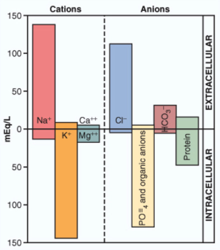
The main cation of extracellular fluid is ________. : Sodium
The main cation of intracellular fluid is __________. : Potassium
The main anion of extracellular fluid is _________. : Chloride
The main anion of intracellular fluid is __________. : Phosphate
ICF and ECF both have more cations than anions.
Ion & Water Movement
Steady state implies the normal amouns of water and ions
Equilibrium implies that the values are the same
1.4 Homeostasis
Homeostasis: The process by which physiological variables are kept relatively “stable” despite imposed challenges.
Set point: The regulated physiological range at which a certain variable will vary
Steady state: The active maitenence of a a variable within its set point
Integrated physiological function: Each variable can be regulated independently, but regulation of one variable may influence regulation of another
E.g. sweating reduces body temperature, but decreases fluid volume
1.5 Homeostatic Control Systems
Negative Feedback: The homeostatic control system where a change in a variable away from its set point intitiates a response bringing it back towards its set point
E.g. high room temperature will lead the body to sweat
Positive Feedback: The homeostatic control system where a change in a variable away from the set point causes a chain of events that further increases that change.
E.g. childbirth, where the uterus stretches more as the baby grows more
Typically exist for variables that are normal at low-level conditions
Feedforward Regulation: The homeostatic control system where the body adjusts a variable prior to any actual change, in anticipation of future needs.
E.g. when you start running, you start breathing heavier to anticipate your oxygen needs; or salivation prior to a meal
1.6 General Characteristics of Homeostatic Control Systems
Local vs Reflexes
Homeostic control systems regulate variables via reflexes and/or local responses, both of which require cell communication
Homeostatic Reflexes: Unlearned control systems linking stimuli with one/more responses, mediated by a reflex arc
Components of Reflex Arc: Stimulus, receptor, afferent pathway, integrating center, efferent pathway, effector, response
Works in a global sense (whole body)
The integrating center compares the stimulus to a set point
Homeostasis is integration (multiple stimuli, multiple effectors)
Local Homeostatic Responses: Links a stimulus to a response, but occurs within a local area (no pathways or integrating center)
Cell Communication
Both reflexes and local responses require intercellular chemical messenges for cellular communication
Chemical messengers
Categorized by which cells release them and where they go
The same messenger can play multiple roles
Classes of Intercellular Messengers
Hormones are associated with homeostatic reflexes
Secreted by an endocrine cell into the bloodstream
Carried to target cells, can go a long way
Neurotransmitters are associated with homeostatic reflexes
Released by a nerve cell into a synapse
Close contact only
Paracrine agents are associated with local homeostatic responses
Released by a cell and targets cells nearby
E.g. epithelial cells in blood vessels might release a paracrine agents to tell the muscles nearby to contract and move the blood along
Autocrine agents are associated with local homeostatic responses
The substance comes back to the same cell and signals it
E.g. building up a muscle during exercise