Chapter 6 Human Physiology
Structures & Functions of Neural Tissue
Overview
- Central Nervous System: Brain and spinal cord
- Peripheral Nervous System: Nervous structures that aren’t the brain and spinal cord
- The functional unit of the nervous system is the neuron
The Neuron
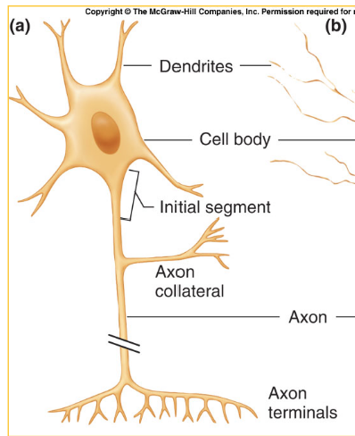
- The neuron is made up of the cell body, debdrites, and axon
- Soma: The cell body of the neuron that includes the nucleus and organelles
- Dendrites: The spikes of a neuron that increase receiving surface
- Axon: The main stem of a neuron that carries output “message” to target cells
- Initial segment: The beginning of the stem of the neuron, part of the axon
- Axon collateral branches: The extra branching on the axon of a neuron
- Axon terminals: The ending “base” of the axon of the neuron; also valled varicosities
- Some axons are myelinated
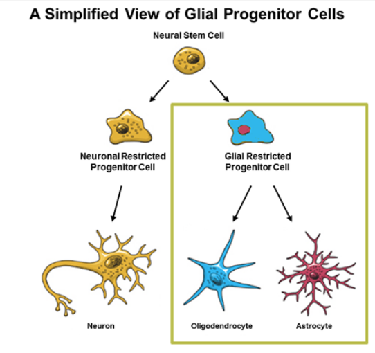
- Muelination: 20 to 200 layers of modified plasma membrane, formed by glial cells
- Glial cells: Non-neuronal cells (i.e. not nerves) of the brain and nervous system that provide support (physical and metabolic) with a capacity for cell division
- Oligodendrocytes: The glial cells of the CNS
- Schwann Cells: The glial cells of the PNS
- Nodes of Ranvier: specialized axonal segments that lack myelin, allowing the saltatory conduction of action potentials
- Myelination occurs to insulate the axons (prevent ion leakage) and speed up conduction velocity
Functional Classes
- Afferent neurons: Neurons that relay sensory information to the CNS
- Efferent neurons: Neurons that relay commands away fron the CNS
- Interneurons: Neurons that connect afferent and efferent neurons
- Interneurons are all part of the CNS.
- 99% of all neurons are interneurons
The Synapse
- Synapse: The junction between two neurons
- The synapse releases neurotransmitters
- Presynaptic neuron: The neuron that releases a neurotransmitter
- Postynaptic neuron: The neuron that receives a neurotransmitter
Neural Support & Maintenance
- Axon damage & regeneration
- In the PNS, Schwann cells can help with axon regeneration
- In the CNS, there is no significant regeneration.
Membrane Potentials
Overview
- Membrane potential: Charge inside the cell relative to the extracellular fluid
Resting Membrane Potential
- A chemical gradient exists for certain diffusible ions, which causes charge to move across the cell membrane
- Sodium and Potassium (& also chloride and calcium)
- Some ions can move more than other ions
- Ion permeabilities can differ, allowing some ions to have bigger effects on membrane potential than others
- The [Na+] gradient tends to create membrane potential of +60 mV
- The [K+] gradient tends to create membrane potential of -90 mV
- The actual resting membrane potential is about -70mV
- Sodium
- Sodium is in much higher concentrations outside of the cell because of ion pumps
- Equilium is reached when the concentration gradient pushing sodium in is equal to the electrical gradient that pushes sodium out
- +60 mV equilibrium potential for sodium
- Potassium
- Potassium is in much higher concentrations inside of the cell because of ion pumps
- Equilium is reached when the concentration gradient pushing out potassium is equal to the electrical gradient that pushes sodium in
- -90 mV equilibrium potential for potassium
- More leak channels for potassium than sodium, so overall charge is more influenced by potassium
Graded Potentials
- At rest, a membrane potential is polarized (is negative at resting)
- Graded potential: Local change in membrane potential due to the opening/closing of ion channels, takes place in the dendrites and the cell body (recieving parts)
- A graded potential can cause either…
- Depolarization, or excitatory post-synaptic potential, is when the graded potential moves the overall potential closer toward 0 from the initial -90
- Sodium and calcium are likely depolarizing ions
- Hyperpolarization, or inhibitory post-synaptic potential, is when the graded potential moves the overall potential further away from zero (more negative)
- Potassium and chloride are likely hyperpolarizing ions
- The size of the potential change is proportional to the size of the stimulus
- Bigger stimulus = opens/closes more ion channels
- Spatial summation: many graded potentials located close together on a neuron
- Temporal summation: Multiple, repeated graded potentials coming from the same source over a short period of time
Action Potentials
- The action potential is the long-distance communication mechanism of the body, and is a large, rapid change in membrane potential
- The purpose of most graded potentials is to influence action potentials
- If the sum of the graded potentials reaches a threshold potential at the initial segment, axons will generate axon potentials
- Action potentials are always the same magnitude regardless of the size of initiating stumuli (it’s all or none!)
- Action potentials can propagate along an excitable membrane wihtout decreasing in size
- An action potential depolarizes the membranes around it to the threshold, which sets off new action potentials
- So it’s not a local response that moves along, it’s more of a flame travelling across gunpowder
- Backwards propagation is prevented by the refractory period
- Velocity of action potential propogation depends on axon diameter (larger = faster) and myelination (more = less conduction)
- Voltage-gated ion channels are required for an excitable membrane (aka a membrane that can do an action potential)
- Refactory periods: Times during which it is difficult or impossible to fire a second action potential at a point on the neuron
- Absolute refactory period: The period from the firing of the action potential to repolarization
- Relative refractory period: The period from repolarization to the end of after-hyperpolarization