chapter 7 axial skeleton
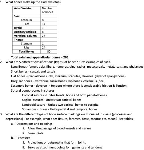
1. What bones make up the axial skeleton?
Total axial and appendicular bones = 206
2. What are 5 different classifications (types) of bones? Give examples of each.
Long Bones- femur, tibia, fibula, humerus, ulna, radius, metacarpals, metatarsals, and phalanges
Short bones - carpals and tarsals
Flat bones – cranial bones, ribs, sternum, scapulae, clavicles. (layer of spongy bone)
Irregular bones – vertebrae, facial bones, hip bones, calcaneus (heel)
Sesamoid bones - develop in tendons where there is considerable friction & Tension
Sutural bones- bones in sutures
Coronal sutures - Unites frontal bone and both parietal bones
Sagittal sutures - Unites two parietal bones
Lambdoid suture - Unites two parietal bones to occipital
Squamous sutures - Unite parietal and temporal bones
3. What are the different types of bone surface markings we discussed in class? (processes and depressions) For example, what does fissure, foramen, fossa, meatus etc. mean? See tables.
a. Depressions and openings
i. Allow the passage of blood vessels and nerves
ii. Form joints
b. Processes
i. Projections or outgrowths that form joints
ii. Serve as attachment points for ligaments and tendons

4. How many bones make up the cranium? How many bones make up the facial bones? Name them.
• 8 Cranial Bones
(Bones of the Braincase)
◦ Frontal bone (1)
◦ Parietal bone (2)
◦ Temporal (2)
◦ Occipital bone (1)
◦ Sphenoid bone (1)
◦ Ethmoid bone (1)
} 14 Facial Bones
◦ Mandible (1)
◦ Maxilla (2)
◦ Zygomatic bone (2)
◦ Nasal bones (2)
◦ Lacrimal Bones(2)
◦ Palatine bones (2)
◦ Inferior Nasal conchae (2)
◦ Vomer (1)
5. Know that different structures you can find on each part of the skull. HINT: Stick to the slides. For example, foramen magnum is located on the occipital bone.
26
· Frontal – supra-orbital margin, supra- orbital foramen, frontal sinuses
· Parietal Bones (2) form greater portion of the sides and roof of the cranial cavity
· Temporal Bones- Temporal squama, zygomatic process
i. Mandibular fossa and articular tubercle articulate with mandible (condyle) to form temporomandibular joint (TMJ) (jaw joint)
ii. Mastoid portion, mastoid process, internal auditory meatus, styloid process, petrous portionk
· Occipital Bone – foramen magnum, occipital condyles (Articulates with depressions of atlas (C1) – allows for dorso-ventral movement of head (up and down), external occipital protuberance (above foramen magnum)
· Sphenoid bone – (articulates with every bone) body, sella turcica, greater wings, lesser wings, pterygoid processes
· Ethmoid bone - cribriform plate, crista galli, perpendicular plate, superior nasal concha, middle nasal concha
6. What bones make up the zygomatic arch? What are the names of the processes that make up the arch?
Zygomatic process of the temporal Bone and the temporal process of zygomatic bone
7. Which bone serves as a bridge uniting the cranial and facial bones?
Sphenoid bone
8. What are the different parts of the sphenoid and ethmoid bones? Where on the skull are each located? sphenoid bone- Joins anteriorly with frontal and ethmoid bones, laterally with temporal bones, and posteriorly with occipital bone
} Ethmoid bone - Located in anterior part of cranial floor medial to orbits
9. What are the functions of the nasal conchae?
greatly increase vascular and mucous membrane surface area in nasal cavity
a. which warms and moistens (humidifies) inhaled air before it passes to lungs
b. Conchae cause inhaled air to swirl, inhaled particles becoming trapped in mucus that lines nasal cavity. Keeps air clean before entering lungs
10. Describe the different facial bones.
11. 14 Facial Bones:
a. Mandible (1)
b. Maxilla(2)-forms upper jawbone
c. Vomer (1)-component of nasal septum
d. Nasal bones (2)
e. Zygomatic bones (2)-forms cheek bones
f. Lacrimal bones(2)-form part of medial wall of each orbit
g. Palatine bones (2)-form posterior portion of hard palate
h. Inferior Nasal Conchae (turbinates) (2)-form wall of nasal cavity
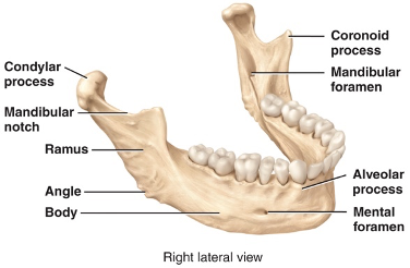
12. What are the different parts of the mandible? Hint: we covered this in lab too.
13. What 3 components make up the nasal septum?
Vomer, perpendicular plate of ethmoid bone, septal nasal cartilage
14. What 7 bones make up the eye orbit? (3 cranial and 4 facial)
3 cranial bones – frontal, sphenoid, ethmoid bone
4 facial bones- palatine, zygomatic, lacrimal, maxilla (cheek bones)
15. What are fontanels? At what age does each close?
• Fontanels- (soft spots) - areas where unossified connective tissue develops into dense connective tissues of skull
Anterior fontanelle – closes 18-24 months of age
Posterior fontanelle – closes 2-3 months after birth
Posterolateral fontanel (mastoid) closes 6-8 months after birth
Anterolateral fontanel (sphenoidal) – 6 months after birth
16. What are sutural bones? What are suture lines and name the different ones (what bones are they found between, each of them)? Slide 7
a. small extra bone plates located within the sutures of cranial bones
i. Classified based on location not by shape
17. What is the function of the hyoid bone?
18. In which bones can you find paranasal sinuses? What are the functions of these sinuses?
Frontal bone, ethmoid bone, sphenoid bone, maxillary bone
Function of paranasal sinuses
1. increase surface area of nasal mucosa, thus increasing production of mucus to help moisten and cleanse inhaled air
2. Serve as resonating chamber within skull that intensify and prolong sound (sound funny when have cold)
· Paranasal sinuses become blocked by excess mucus production, changing quality of your voice
19. Know the different parts of the vertebral column. How many vertebrae make up each segment? (26 bones)
· 7 cervical vertebrae in the neck region labeled C1-C7
· 12 Thoracic vertebrae that articulate with the ribs (T1-T12)
· 5 lumbar vertebrae that support the lower back labeled L1-L5
· sacrum and coccyx are single bones that result from the fusion of several vertebrae
20. What are the 4 curves of the adult spinal column? Which are primary and which are secondary?
Cervical and lumbar are convex (bulging out) (primary curvature) hold you up
Thoracic and sacral curves are concave (cupping in) (secondary curvature)
21. Thoracic vertebrae can be distinguished from other vertebrae by presence of what structure? We discussed this in lab.
Costal facets
22. What is another name for C1 and C2? Which type of movement of the head does each permit (dorsoventral or lateral)?
C1 atlas up and down
C2 axis move head from side to side
23. What structure on the atlas and dens makes it different than any other cervical vertebrae? We discussed this is lab as well.
24. At what age do the bones of the coccyx vertebrae fuse? Sacrum?
Sacrum- Fuse between 16-18; complete fusion at age 30
Coccyx - fuse around age 20-30 yo
25. Which bones make up the thoracic cage (thorax)?
• 12 pairs of ribs
• 7 pairs true ribs. 5 pairs of false ribs (8-12)
• Sternum
• costal cartilages
• 12 thoracic vertebrae