Applied Anatomy and Physiology
1.1 The structure and functions of the musculoskeletal system
Bones
Are made of connective tissue reinforced with calcium and specialized bone cells.
Most bones contain bone marrow (where blood cells are made).
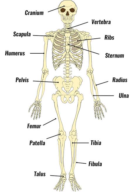
Location of the following bones;
Cranium and vertebrae: Head and Neck
Scapula and Humerus: Shoulders
Ribs and Sternum: Chest
Humerus, Radius, and Ulna: Elbow
Pelvis and Femur: Hip
Femur and Tibia: Knee
Patella: In front of knee joint
Tibia, Fibula, and Talus: Ankle
Functions and Structure of the Skeleton
Skeleton: a supportive and protective structure of an organism.
Skeletal System: helps the body hold organs and gives them its shape.
Also called the musculoskeletal system.
It consists of all of the bones, cartilage, tendons, and ligaments in the body
Has connective tissue that helps support joint movement and safety.
It includes muscles that help to move and create new blood cells that keep you healthy.
It provides an attachment point for muscles as when muscles contract they pull the bone.
The shape and type of the bones determine the amount of movement.
Short bones allow finer controlled movements
Long bones allow gross movement
Flat bones protect vital organs
The different joint types allow different types of movement.
Functions of the Skeleton
Significant functions of the skeleton in physical activity involve;
Support and Structural Shape: It provides a framework to give support and shape to the body.
Protection: Bones are crucial in protecting organs during physical activities that involve potential impact or injury.
Movement: The framework along with the muscle system works together to perform and facilitate movements.
Mineral storage: Bones serve as a reservoir for essential minerals (especially calcium and phosphorus) because, during physical activity, the body can utilize the stored minerals to maintain physiological functions.
Production of blood cells: The bone marrow handles the production of blood cells. Its function is crucial for oxygen transport, immune response, and blood clotting.
Muscles of the Body
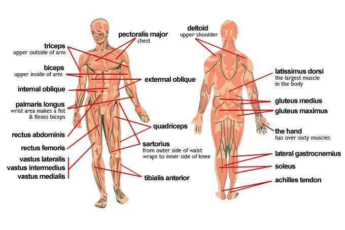
Latissimus Dorsi: A broad, flat muscle that occupies the majority of the lower posterior thorax (lower back side of the chest).
Function: To stabilize your back while producing movements on the shoulder joint (internal rotation, extension, and adduction).
Deltoid: The ball-and-socket joint that connects your arm to the trunk of your body that is located in your shoulders.
Functions: Arm abductions, flexions, extensions, compensation for lost arm strength if you have an injury, and stabilization of your shoulder joint to prevent dislocations when carrying or lifting.
Rotator Cuffs: A group of muscles and tendons that hold the shoulder joint in place and allow you to move your arm and shoulder.
Functions: To stabilize the glenohumeral joint (a ball and socket joint) by compressing the humeral head against the glenoid.
Pectoralis: It is a thick, fan-shaped muscle that lies underneath the breast tissue and forms the anterior wall of the axilla.
Functions: Acts as a strong adductor and internal rotator of the humerus at the shoulder joint.
Biceps: It is a large muscle situated on the front of the upper arm between the shoulder and the elbow.
Functions: Flexion and Supination (outward rotation) of the forearm.
Triceps: A large, thick muscle on the dorsal part of the upper arm.
Functions: Extension of the elbow joint.
Abdominals: These are the muscles forming the abdominal walls, the abdomen being the portion of the trunk connecting the thorax and pelvis.
Functions: Supports the trunk, allows movement, and holds organs in place by regulating internal abdominal pressure.
Hip flexors: A group of muscles toward the front of the hip.
Functions: To flex the hip, bringing the knee closer to the chest.
Gluteals: A group of muscles that make up the buttock area.
Functions: To help stabilize the upper body and pelvis, aid in locomotion, and extend the hip.
Hamstring Group: They are muscles in the posterior compartment of the thigh that consist of biceps femoris, semitendinosus, and semimembranosus.
Functions: To extend the hip and flex the knee.
Quadriceps Group: Group of muscles in front of the thighs.
Functions: To perform various movements such as kicking, running, jumping, and walking.
Gastrocnemius: A complex muscle that is fundamental for walking and posture, forming the major bulk at the back of the lower leg.
Functions: Plantarflexor of ankle joint and knee flexor.
Tibialis Anterior: An anterior leg muscle that acts as the main foot dorsiflexor on the ankle joint.
Functions: Inversion and adduction of the foot.
Tendons: A cord of strong and flexible tissue (similar to a rope) that connects the muscles to your bones.
Functions: To transfer muscle-generated force to the bony skeleton, facilitating movement around a joint.
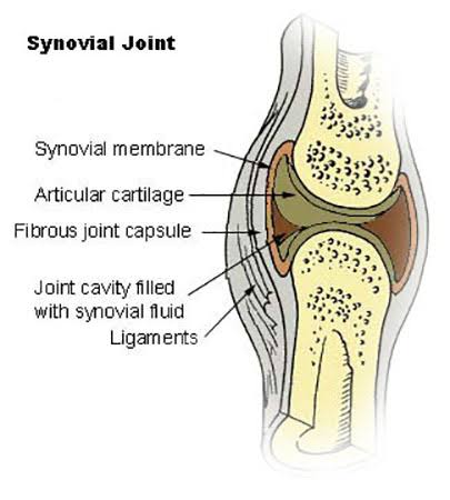
Structures of a Synovial Joint
How the following help to prevent injury:
Synovial Membrane: Helps to protect the joints they surround.
Synovial Fluid: Helps to reduce friction between the articular cartilages of synovial joints during movement.
Joint Capsule: It seals the joint space, and provides passive stability by limiting movements and active stability via its proprioceptive nerve endings.
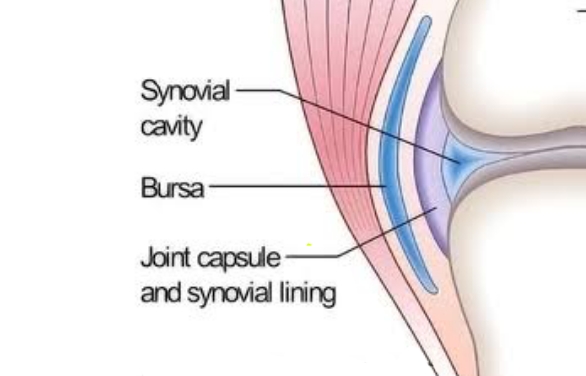
Bursae: These are small fluid-filled sacs that reduce friction between moving parts in your body's joints.
Cartilage: Reduces friction and prevents them from rubbing together when you use your joints.
Ligaments: They stabilize the joint or hold the ends of two bones together.
Types Of Freely Moving Joints
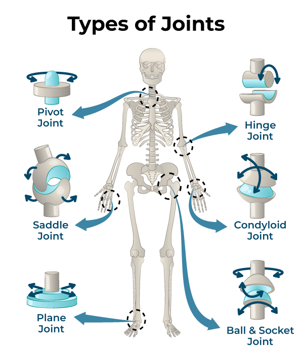
Hinge Joint: Located in the elbow, knee, and ankle. These are two bones that open and close in one direction only (along one plane)
Ball and Socket Joint: Located in the hip and shoulder. These are the rounded heads of one bone that sits within the cup of another, allowing movement in all directions.
Saddle Joint: The joint at the base of the thumb that permits back-and-forth and side-to-side movement, but does not allow rotation.
Candyloid Joint: Located at the jaw or finger joints, allowing movement without rotation.
Pivot Joint: The joint between the first and second vertebrae of the neck where one bone swivels around the ring formed by another bone.
Gliding Joint: Like in wrist joints, the smooth surfaces slip over one another, allowing limited movement.
Differences in Joint Design
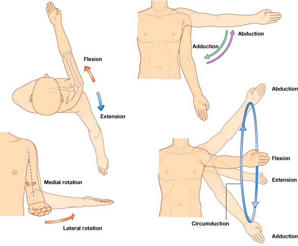

Flexion/extension at the shoulder, elbow, hip, and knee
The elbow and knee joint is a hinge joint that allows bending and straightening movements in one plane.
The shoulder and hip is a ball-and-socket joint that provides a wide range of motion, allowing flexion, extension, abduction, adduction, and rotation.
Abduction/Adduction at the shoulder
The ball-and-socket joint type of shoulder allows the humerus to move outward and upward (abduction), and inward and downward (adduction) allowing the arms to be raised laterally and be brought back to the body’s midline.
Rotation of the shoulder
The shoulder’s ball-and-socket joint structure facilitates and allows internal and external rotation due to a high degree of mobility.
Circumduction of the shoulder
Due to the shoulder’s joint structure, it allows it to move in a circular path by combining flexion, extension, abduction, and adduction accordingly.
Plantar flexion/Dorsiflexion at the ankle
The ankle’s hinge joint type allows movement in one plane. Therefore, the ankle allows the foot to pivot around its axis while the top of the foot moves away from the leg (plantar flexion).
Ankle’s joint type also allows the foot to pivot in the opposite direction by bringing the top of the foot closer to the leg (dorsiflexion)
Major muscles and muscle groups that work and affect movement in physical activity
Major muscle groups operating at these joints:
Shoulder: rotator cuffs muscle, deltoid, trapezius, and rhomboids.
Elbow: brachialis (lateral portion), the anconeus, the supinator muscle, brachioradialis, and triceps brachii.
Hip: gluteal, adductor, iliopsoas, and lateral rotator.
Knee: the quadriceps (on the anterior side of the knee and femur), and the hamstrings (on the posterior side).
Ankle: the tibialis anterior, the extensor digitorum longus, the extensor hallucis longus, and the peroneus tertius
Prime Movers (Agonist): a muscle that provides the primary force of driving action.
Antagonist Muscle: a muscle that provides resistance or reverses a given movement.
Bones located at the joint:
Shoulder: the scapula (shoulder blade), clavicle (collarbone) and humerus (upper arm bone)
Elbow: humerus, ulna, and radius
Hip: the femur (thighbone), and the pelvis (made up of three bones called ilium, ischium and pubis)
Knee: femur (thighbone), tibia (lower leg bone), and patella (kneecap)
Ankle: talus (small ankle bone), tibia, and fibula
Types of Muscle Contractions
Isometric Contractions: is a muscle contraction doesn't noticeably change the length and the affected joint also doesn't move.
Isotonic Contractions: a contraction where the tension in the muscle remains unchanged despite a change in muscle length.
Concentric Contractions: shortening of the muscle with the requisite movement of the origin or insertion and limb translation.
Eccentric Contractions: a lengthening contraction that occurs when a force applied to the muscle exceeds the momentary force produced by the muscle itself.
1.2 The structure and functions of the cardio-respiratory system
The Pathway of Air
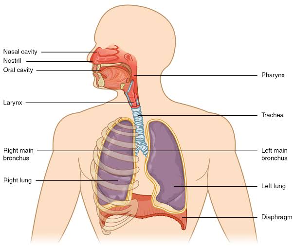
Air is inhaled through the nose and/or mouth. It then moves through the pharynx, larynx, and trachea to the bronchi, bronchioles, and alveoli, into the lungs.
Pharynx: a muscular tube in the middle of your neck.
Larynx: a hollow tube in the middle of your neck containing the vocal chords.
Trachea: a long, U-shaped tube that connects your larynx (voice box) to your lungs.
Bronchi: are large tubes that connect to your trachea and direct the air you breathe to your right and left lungs.
Bronchioles: the smallest airways.
Alveoli: are very small air sacs where the exchange of oxygen and carbon dioxide takes place.
Gaseous Exchange
Gas exchange: occurs in the respiratory zone of the lung in which the alveoli are present.
Features that assist in gaseous exchange:
large surface area of alveoli
moist thin walls (one cell thick)
short distance for diffusion (short diffusion pathway)
lots of capillaries
large blood supply
movement of gas from high concentration to low concentration.
Oxygen combines with haemoglobin in the red blood cells to form oxyhaemoglobin.
Haemoglobin: contains iron that allows it to pick up oxygen from the air we breathe and deliver it everywhere in the body. (It can also carry carbon dioxide)
Oxyhaemoglobin: a compound of haemoglobin with oxygen that is the chief means of transportation of oxygen from the air in the lungs, by way of the blood to the tissues.
Blood Vessels
Are the channels that carry blood throughout your body.
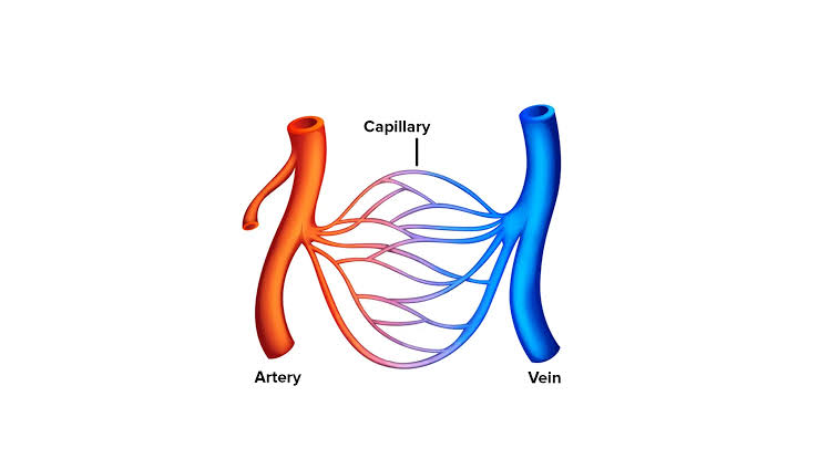
Structure of arteries, capillaries and veins
Arteries: are blood vessels that bring oxygen-rich blood from your heart to all of your body's cells.
Size/Diameter: 3 mm to 5 mm (µm)
Wall Thickness: 1.50 mm in young patients, 1.69 mm in older patients, and 2.01 mm in those with symptomatic claudication.
Capillaries: are delicate blood vessels that transport blood, nutrients, and oxygen to cells in organs and body systems.
Size/Diameter: 8 to 10 mm
Wall Thickness: approximately 0.5 mm
Veins: are blood vessels located throughout your body that collect oxygen-poor blood and return it to your heart
Size/Diameter: normally between 7 to 15 mm
Large Veins:
Wall thickness: Around 1-2 millimetres (mm) or more.
Medium-Sized Veins:
Wall thickness: Varies, but typically in the range of a few hundred micrometres (µm).
Small Veins (Venules):
Wall thickness: Around 50-200 µm.
Functions of Blood Vessel Structure
Arteries
Arteries’ three-layered structure allows them to withstand high pressure generated by the heart during systole. It carries oxygenated blood away from the heart to supply tissues and organs with oxygen and nutrients.
Arteries do not directly participate in gas exchange.
Elastic fibres allow arteries to stretch during systole and recoil during diastole. It helps maintain continuous blood flow by smoothing out pulsatile surges generated by the heart's contraction, making the contraction and relaxation of smooth muscle contribute to blood pressure regulation.
The smooth muscles in the tunica media can constrict or relax to adjust the diameter of the artery. As exercise increases the demand for oxygen and nutrients for active muscles, arteries that supply the muscles undergo vasodilation that increases blood flow. Conversely, arteries in less active areas may experience vasoconstriction to redirect blood flow to where it is needed most.
Vasoconstriction: the narrowing (constriction) of blood vessels by small muscles in their walls.
Vasodilation: the widening of blood vessels as a result of the relaxation of the blood vessel's muscular walls.
Capillaries
Capillaries connect arteries and veins, allowing for the exchange of oxygen and nutrients from the bloodstream to the tissues (oxygenated blood) and the removal of waste products from the tissues to the bloodstream (deoxygenated blood).
The thinness of capillary walls ensures a short diffusion distance, facilitating efficient gas exchange between the bloodstream and tissues.
Capillaries’ small diameter and high total cross-sectional area create resistance to blood flow, leading to a decrease in blood pressure as blood moves from arteries to capillaries.
Some capillaries may constrict to redirect blood flow away from less active tissues and toward active muscles (vasoconstriction). Capillaries in active muscles may dilate to increase blood flow, allowing for enhanced oxygen and nutrient delivery (vasolidation).
Veins
Veins are generally responsible for carrying deoxygenated blood from the body's tissues back to the heart except for pulmonary veins that carry oxygenated blood from the lungs to the left atrium of heart.
Veins do not directly participate in gas exchange.
Veins contribute to blood pressure regulation by facilitating venous return, which is the flow of blood back to the heart. The muscular walls of veins and the presence of venous valves help prevent the backflow of blood, ensuring efficient return to the heart.
Veins can also undergo vasoconstriction and vasolidation during exercise.
Arteries And Veins Associated With Blood Flow
 Arteries
Aorta: the largest artery in the body and originates from the left ventricle of the heart.
Function: It carries oxygenated blood away from the left ventricle to various parts of the body.
Right and Left Pulmonary Arteries: These are arteries that arise from the right ventricle of the heart.
Function: Carries the deoxygenated blood from the right ventricle to the lungs for oxygenation.
Veins
Superior Vena Cava: Collects deoxygenated blood from the upper part of the body.
Function: It returns deoxygenated blood to the right atrium of the heart.
Inferior Vena Cava: Collects deoxygenated blood from the lower part of the body.
Function: It returns deoxygenated blood to the right atrium of the heart.
Pulmonary Veins: The four pulmonary veins (two from each lung) carry oxygenated blood from the lungs to the left atrium of the heart.
Function: Returns the oxygenated blood to the left atrium, initiating systemic circulation.
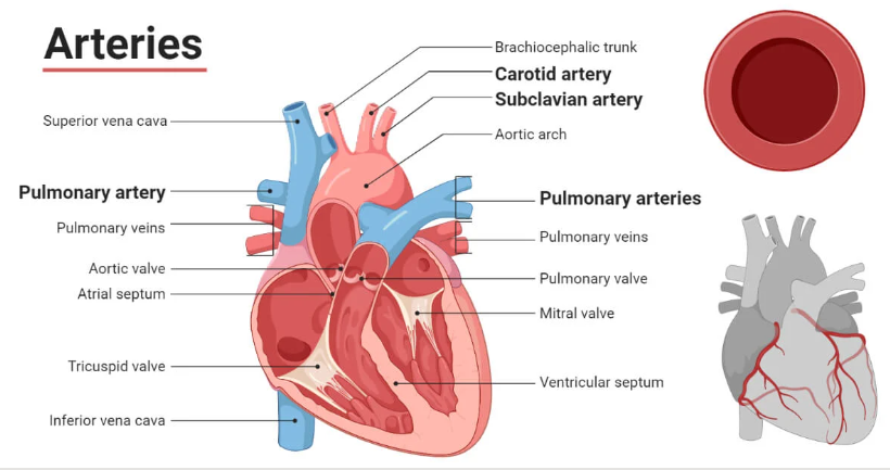
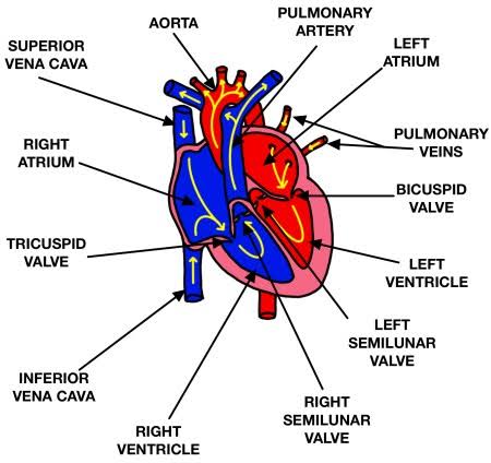
Structure of the Heart
The heart is divided into four chambers namely left and right atria (located on the top) and left and right ventricles (located at the bottom.
Heart Chambers
Atria (Upper Chambers)
Left Atrium: It receives oxygenated blood from the lungs through the four pulmonary veins.
Right Atrium: It receives deoxygenated blood from the body through the superior and inferior vena cava.
Ventricles (Lower Chambers)
Left Ventricle: Pumps oxygenated blood to the body through the aorta.
Right Ventricle: Pumps deoxygenated blood to the lungs through the pulmonary arteries.
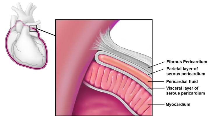
Heart Wall
Endocardium: The innermost layer lining the heart chambers.
Myocardium: The thick, muscular middle layer responsible for pumping blood.
Epicardium: The outermost layer, also known as the visceral pericardium, which is a protective layer covering the heart.
Heart Valves
Atrioventricular (AV) Valves
Tricuspid Valves: Are valves located between the right atrium and right ventricle.
Bicuspid or Mitral Valve: Are valves located between the left atrium and left ventricle.
Semilunar Valves
Pulmonary Valve: It guards the entrance to the pulmonary artery from the right ventricle.
Aortic Valve: It guards the entrance to the aorta from the left ventricle.
Blood Vessels (connected to the Heart)
Aorta: The largest artery that carries oxygenated blood from the left ventricle to the systemic circulation.
Pulmonary Artery: Artery that carries deoxygenated blood from the right ventricle to the lungs for oxygenation.
Superior and Inferior Vena Cava: These are veins that bring deoxygenated blood from the body to the right atrium.
Pericardium
Fibrous Pericardium: The tough outer sac that encloses and protects the heart.
Serous Pericardium: A double-layered membrane consisting of the parietal and visceral layers (epicardium).
Coronary Arteries and Veins
Coronary Arteries: Supply the heart muscle (myocardium) with oxygen and nutrients.
Coronary Veins: It collects deoxygenated blood from the myocardium and return it to the right atrium.
The Cardiac Cycle and the Pathway of the Blood
The Cardiac Cycle
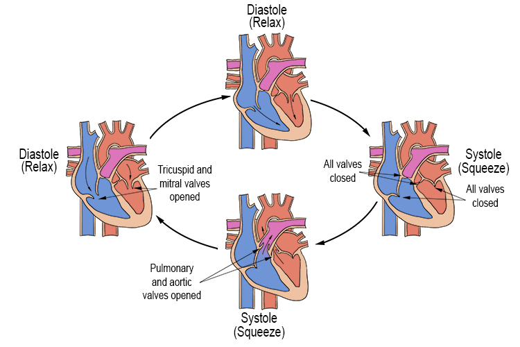
Atrial Systole (Contraction): The contraction of the atria forces blood into the ventricles, completing the filling of the ventricles.
Ventricular Systole (Isovolumetric Contraction): The ventricles contract in response to electrical signals from the atrioventricular (AV) node.
As the ventricular pressure increases, the semilunar valves (pulmonary and aortic valves) are still closed, causing the volume of blood in the ventricles to remain constant (isovolumetric contraction).
Ventricular Ejection: Once ventricular pressure exceeds the pressure in the pulmonary artery and aorta, the pulmonary and aortic valves open and the blood is ejected, initiating the flow of blood to the lungs and the systemic circulation.
Atrial Diastole (Relaxation): After contraction, the atria relax (diastole), allowing blood from the veins to flow into the atria.
Ventricular Filling: As the atria relax, the ventricles also begin to fill with blood. The tricuspid and mitral valves (atrioventricular valves) are open during this phase.
Ventricular Diastole (Isovolumetric Relaxation): The ventricles relax (diastole), causing a decrease in ventricular pressure. During this phase, all four heart valves are closed, and the volume of blood in the ventricles remains constant.
Closure of Semilunar Valves: When the ventricular pressure drops below the pressures in the pulmonary artery and aorta, the pulmonary and aortic valves close.
Pathway of the Blood
Deoxygenated blood from the body enters the right atrium.
The right atrium contracts then pumps deoxygenated blood into the right ventricle.
From the right ventricle, the pulmonary artery then transports deoxygenated blood to the lungs.
Gas exchange (blood is oxygenated) occurs in the lungs.
The pulmonary vein then transports oxygenated blood back to the left atrium.
Cardiac Output, Stroke Volume, and Heart Rate
Cardiac Output: Refers to the key physiological parameter that represents the volume of blood pumped by the heart in one minute.
It is influenced by various factors such as heart rate, stroke volume, and the body's demand for oxygen and nutrients.
Formula: Cardiac Output (CO) = Heart Rate (HR) × Stroke Volume (SV)
Heart Rate (HR): The number of heartbeats per minute.
Stroke Volume (SV): The volume of blood ejected by the left ventricle with each contraction.
Mechanics of Breathing
Lungs can expand more during exercise (inspiration) due to the use of pectorals and sternocleidomastoid. During exercise (expiration), the rib cage is pulled down quicker to force air out quicker due to the use of the abdominal muscles.
Changes in air pressure cause the inhalation and exhalation.
Inhaling at rest has the coordinated action of intercostal muscles, rib cage, and diaphragm to increase the volume of the thoracic cavity, allowing air to flow into the lungs.
Intercostal Muscles: Helps to expand and shrink the size of the chest cavity.
Rib Cage: Assists and expands through intercostal muscle contractions with upward and downward movement, increasing the lateral dimensions of the chest.
Diaphragm: Contracts and moves downward, increasing the space in your chest cavity, and your lungs expand into it.
Exhaling at rest involves the relaxation of the diaphragm and external intercostal muscles.
External Intercostal Muscles: Relaxes during exhalation.
Rib Cage: Relaxation of the external intercostal muscles allows the ribcage to move downward and inward.
Diaphragm: Relaxes and moves upward to its resting position, reducing the volume of the chest cavity.
Interpretation of a spirometer trace
Spirometer Trace: A graphical representation of the volume of air inspired or expired by a person as a function of time during respiratory manoeuvres.
Components
Tidal Volume: The volume of air inspired or expired during normal, quiet breathing.
Appears as regular, rhythmic waves during normal breathing (on the trace).
Expiratory Reserve Volume: The additional volume of air that can be expired beyond the tidal volume during a forced exhalation.
Is seen as the increased vertical distance from the baseline during a forced exhalation.
Inspiratory Reserve Volume: The additional volume of air that can be inspired beyond the tidal volume during a deep inhalation.
Is seen as the increased vertical distance from the baseline during a deep inhalation.
Residual Volume: The volume of air that remains in the lungs after a maximal exhalation.
Not directly measured on the trace. However, it can be observed through baseline level, if the spirometer does not return to zero, and through the volume above baseline.
1.3 Anaerobic and aerobic exercise
Aerobic exercise and anaerobic exercise
Aerobic Exercise: a rhythmic and repetitive physical activity that uses your body’s large muscle groups, increasing the heart rate and the oxygen that the body uses.
Aerobic means “with oxygen”.
(glucose + oxygen → energy + carbon dioxide + water)
Benefits:
Builds stronger bones
Improves muscle strength, endurance, and flexibility
Improves balance
Increases mental function
Assists weight management and/or weight loss.
Examples: Walking or jogging, cycling, cardio equipment, and swimming.
Anaerobic Exercise: Involves short, fast, high-intensity exercises that don’t make your body use oxygen like it does for cardio (or aerobic) activities.
Anaerobic means “without oxygen”
(glucose → energy + lactic acid)
Benefits:
Strengthen bones
Burn fat
Boost muscle development
Helps keep muscle mass as you age
Examples: High-intensity interval training (HIIT), strength training and weight lifting that challenges your body, jump squats, box jumps, and plyometrics.
The use of aerobic and anaerobic exercise in practical examples of differing intensities
Aerobic Exercise
Low Intensity: Walking at a moderate pace.
A low-intensity aerobic exercise sustainable for an extended period enhances cardiovascular health and endurance and is suitable for beginners.
Moderate Intensity: Jogging and/or Running at a moderate pace.
Increases heart rate and breathing that improves cardiovascular health.
High Intensity: High-Intensity Interval Training (HIIT).
There are alternating short bursts of intense exercises and periods of rest or low-intensity tasks.
Anaerobic Exercise
Low Intensity: Lifting heavy weights with a low number of repetitions.
Low-repetition and high-weight anaerobic exercise focuses on building strength and muscle mass where there is longer rest intervals between sets.
Moderate Intensity: Bodyweight exercises with moderate repetitions.
Moderate intensity- anaerobic involves weight resistance, helping to build strength, muscular endurance, and flexibility.
High Intensity: Short, intense sprints over a short distance.
Sprinting is a high-intensity anaerobic which engages fast-twitch muscle fibres, improving speed, power, and anaerobic capacity.
Excess post-exercise oxygen consumption (EPOC)
Commonly referred to as oxygen debt.
A physiological phenomenon where the body continues to consume oxygen at an elevated rate after the cessation of exercise.
It is caused by anaerobic exercise and lactic acid production, as the body relies on energy systems that do not require oxygen that leads to the production of lactic acids.
During EPOC, the performer must maintain an increased breathing rate to repay oxygen debt after exercise (oxygen debt repayment).
The recovery process from vigorous exercise
Methods to Recover from Exercise
Cool Down – It is to maintain or gradually recover breathing rate or heart rate (blood flow), stretching, removal of lactic acid.
Promotes the circulation of blood, helping to clear byproducts from the muscles that contribute to reducing muscle soreness (faster recovery).
Manipulation of diet – Refers to rehydration and taking up carbohydrates for energy.
Water, carbohydrates, and protein intake are crucial for muscle repair and growth, endurance, body composition, and energy availability to prevent fatigue.
Ice Baths or Massage – To prevent the delayed onset muscle soreness (DOMS).
Helps to reduce swelling and modulate muscle contractions to recover from muscle fatigue.
1.4 The short and long-term effects of exercise
Immediate effects of exercise (during exercise)
Hot/Sweaty/Red skin
Increase in depth and frequency of breathing
Increased heart rate
Short-term effects of exercise (up to 36 hours after exercise)
Tiredness or Fatigue
Lightheadedness
Nausea
Aching or Delayed onset muscle soreness (DOMS)
Long-term effects of exercise (months and years of exercising)
Change in body shape
Improvements in specific components of fitness
Builds and improves muscle strength and endurance
Improve stamina
Increase in the size of the heart (hypertrophy)
Lower resting heart rate (bradycardia)
Components of Fitness
Flexibility
Reaction Time
Balance
Muscle Strength and Endurance
Cardiorespiratory Endurance
Accuracy
Coordination
Agility
Power
Speed
Body Composition