Week 7: Nervous System
finished
Neurological Examination 🧠
A neurological examination assesses the central nervous system (CNS), consisting of the brain and spinal cord, and peripheral nerves. The exam includes:
Examination of the skull and spine
Assessment of consciousness
Evaluation of sensitivity
Superficial sensitivity
Deep sensitivity (e.g., proprioceptive)
Evaluation of senses (sight, hearing, smell)
Assessment of reflexes
Cranial nerve reflexes
Spinal reflexes
Assessment of motor function
Ataxia
Paralysis
Examination of cerebrospinal fluid (CSF)
Nervous System Functions and Classifications ⚙
The nervous system has multiple key functions:
Sensory-motor system: Responsible for posture and gait.
Autonomic nervous system: Regulates smooth muscle function and endocrine glands.
Overall function: Controls the relationship between an animal and its environment and maintains endogenous homeostasis.
Primary vs. Secondary Disorders 🦠
Neurological disorders can be classified as:
Primary: infective agents, nutritional deficiency, degenerative changes
pregnancy toxemia, enzootic ataxia
Secondary: infective processes elsewhere in the body (overcoming of the blood-brain barrier), functional diseases like decrease in Mg, liver coma, heart failure. And reflex disturbances - pain (colic)
grass tetany, hepatic encephalopathy
🩺Examination of Nervous system:
Examination of skull and spine:
Skull → inspection (size, shape, symmetry) and palpation (tenderness, consistency)
symmetry - symmetrical, normal size and shape
palpation: hard consistency, not painful
spine - inspection: curvature - lordosis, kyfosis, scoliosis
position of head and neck - opistotonus, pleurotonous, emprostotonous, torticolis.
1. Consciousness:
Measurement of reaction to stimuli (excitation and depression). Done by applying various stimuli to get the animalˋs reaction.
Excitation Responses: Cringing, frenzy, mania (e.g., in rabies).
swishing with tail
Depression Responses:
1st Degree: Slower reactions,apathy, reduced reflexes. (Daze, somnolence).
2nd Degree: Loss of consciousness, autonomic functions remain. (coma).
examine corneal reflexes → touching cornea to see if the animal blinks.
Learn classification according to the book here!
2. Perception/Sensitivity:
Senses: Touch, sight, smell, taste, hearing.
first examining → by hopping etc. (the tests in video).
False Insensibility: Misinterpretation of sensory inputs.
blindness, deafness (clapping to check), anosmia (distemper in dod).
3. Reflexes:
Types of Reflexes: Autonomic (internal organs) and Somatic (muscles).
Reflex Functions: Can be absent, diminished, or exaggerated.
Principal Reflexes to examine:
Corneal Reflex: Touching the cornea causes blinking.
Pupillary Reflex: Light exposure leads to pupil constriction.
miosis: poisoning (morphine), increased intercranial tension, diseases of cervical part of spinal cord
mydriasis: coma, epilepsis, retina diseases, poisoning (atropine)
Spinal cutaneous: touching (pricking) with a needle
panniculus reflex - irritating the skin with sharp object along spine and hips → bilateral contraction
pedal reflex → stretching the limb at max. extension
positive: flexion of joints
Withdrawal - touching/pinching
dog & cat: interdigital skin
horse & cattle: coronet skin
positive result: movement with the leg
Anal or perineal reflex: anal skin
positive: jerky contractions of the anal sphincter
negative: recumbent cows and horses
Spinal Limb Reflexes:
Spinal limb reflex: | Description | Positive reaction |
Patellar reflex  | tapping (striking) on the patellar ligament | forward extension of leg as quadriceps femoris muscle contracts |
Extensor carpi radialis 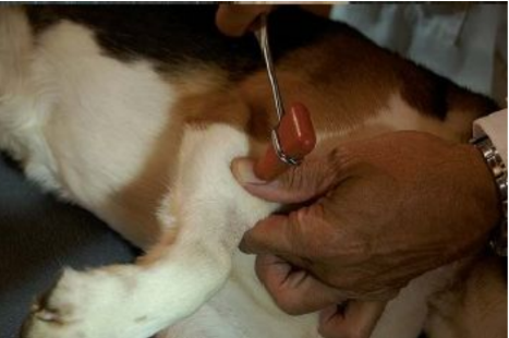 | Tapping (striking) on extensor muscle group distal to the elbow | forward extension of the leg, due to contraction of the muscle |
Postural:
Postural - tests: | How: |
Proprioceptive positioning reaction (paw replacement) 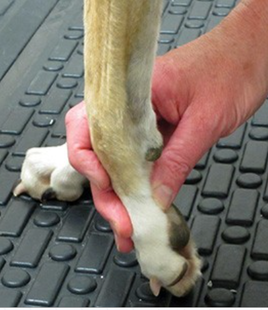 | flex the paw so dorsum of paw is on the floor, do not let patient put weight on it. Patient should return the paw to normal position. |
Wheel-barrowing reaction 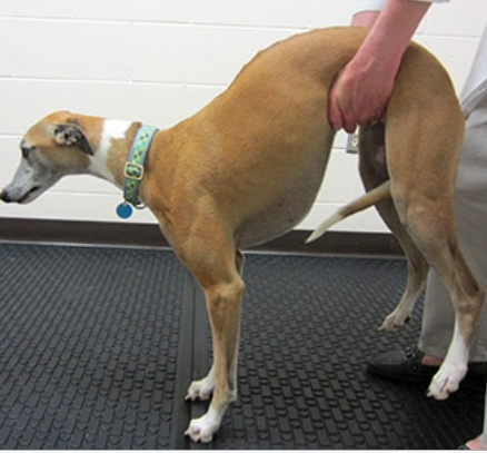 | Lifting pelvic limbs from ground and move the patient forward. |
Hopping reaction 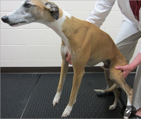 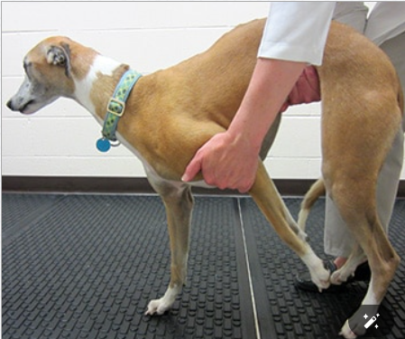 | placing one hand under abdomen to lift pelvic limb from ground or by lifting thoracic limb back along chest, while pushing animal toward the standing limb. |
Extensor postural thrust reaction 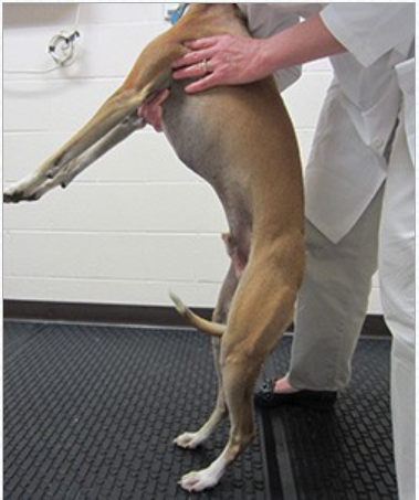 | Patient is lifted straight up, then lowered to the ground. As the pelvic limb paws touch the ground, patient extend the hocks and takes a few steps backwards to find its balance. |
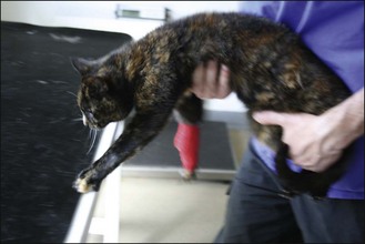
Placing:
With patient in your arms, go to table/other surface and let dorsum of paw touch the table, the paw “away” from your body is tested.
In visual placing - animal can see the table
In tactile placing - animal`s eyes are covered
 |  |
Motor disturbances:
Paralysis: Can be central (brain/spinal) or peripheral (nerves)
can be complete/incomplete (paresis)
flaccid/spastic
Spasms: Muscle contractions
tonic (continuous), clonic (alternating of contractions and relaxations), epileptiform attacks, trismus - continuous spasm of jaw muscles, convulsions - shaken
causes: decrease of Mg, transit tetany
Tremors: Rapid clonic contractions, often due to low chloride levels or dehydration.
Nystagmus: Jerking eye movements; can be horizontal or vertical.
Ataxia: Coordination issues among muscle groups.
Forced Movements: from conditions like focal encephalitis, parasites, repeated stimulation of motor centres
circling, compulsive walking
clonic and tonic - basic classification
How is cerebrospinal fluid (CSF) analyzed?
CSF Puncture Methods: Postoccipital and lumbar punctures.
Normal CSF Properties:
Sensory Analysis
Color: Normally colorless. Blood contamination results in a pink color.
Transparency: Normally clear. Turbidity indicates the presence of cells or fibrin.
Specific gravity: Normal range is 1.005 - 1.001.
Biochemical Analysis
pH: Normal range is 7.0 - 7.5.
Concentration of glucose, proteins, macronutrients, and trace elements.