Exam 3
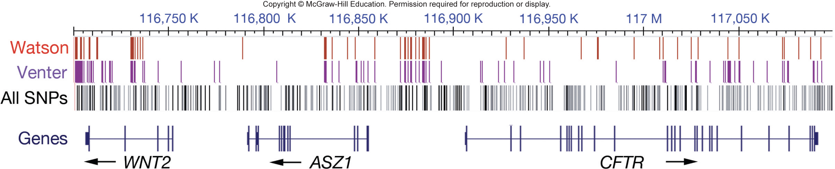
A wild-type human genome sequence does not exist
- The genome sequences of only 3 people reveal over 5 million DNA polymorphisms
- DNA polymorphisms: sequence differences
Most polymorphisms do not influence phenotype
- codons make up less than 2% of the human genome
- many mutations in codons don’t change the amino acid
- many deleterious mutations disappear from the population through natural selection
- \
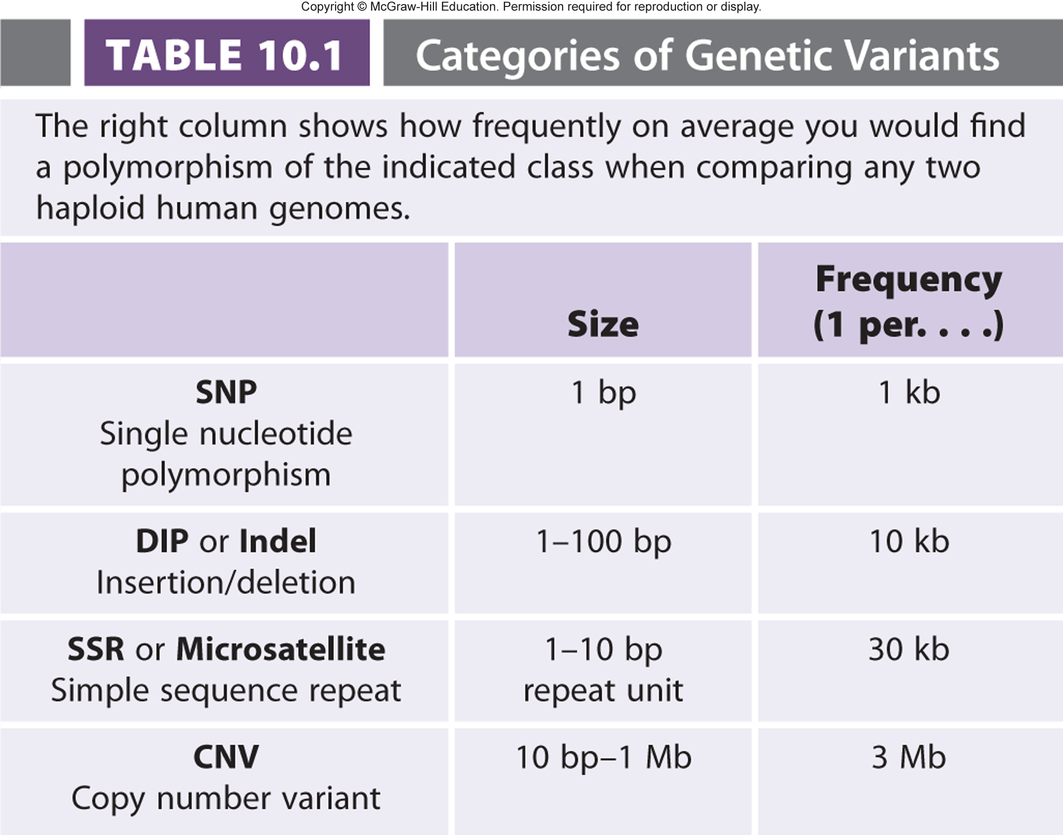
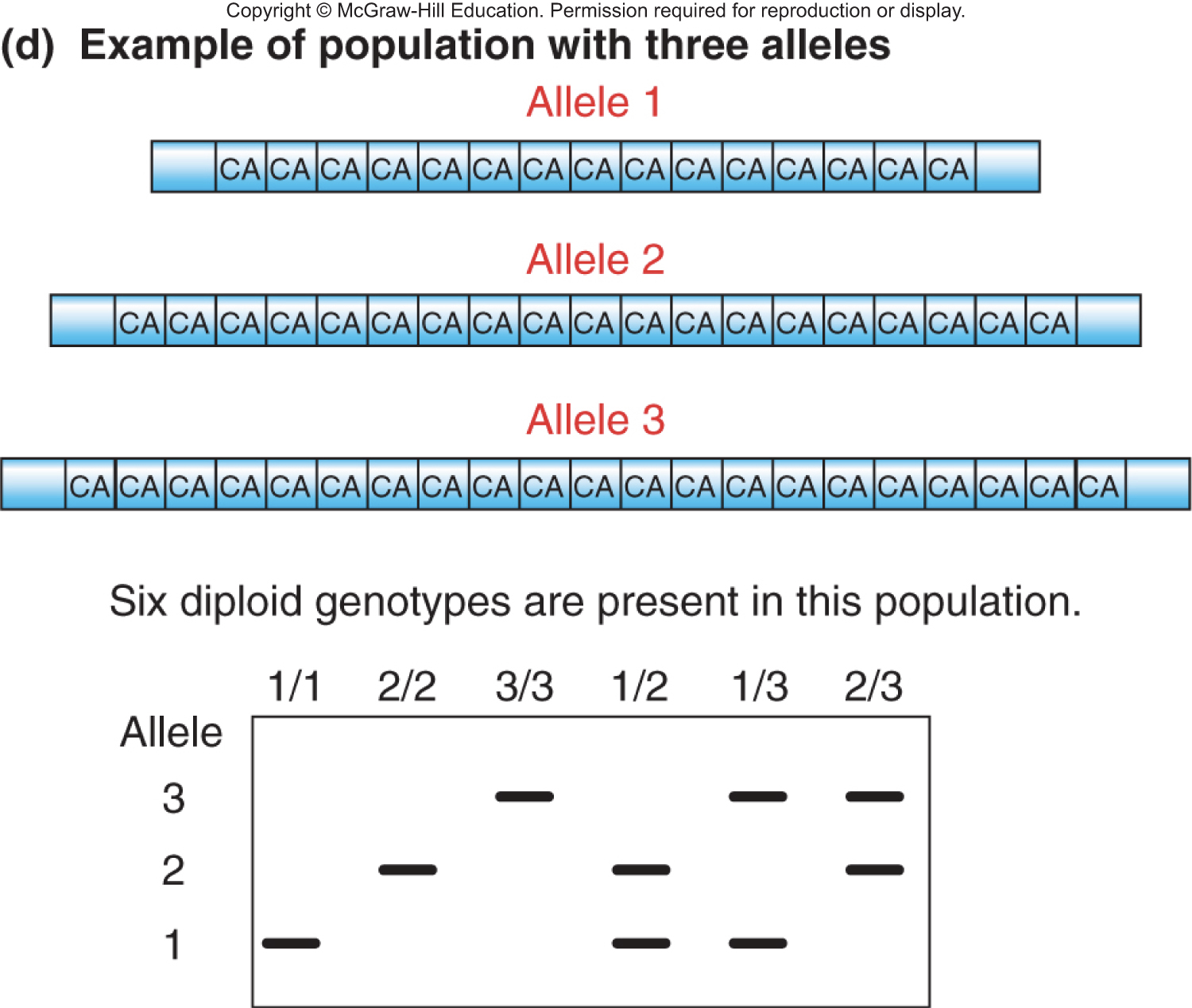
Four categories of genetic variation
- single nucleotide polymorphisms: only 1 base pair changes
- deletion-insertion polymorphisms (DIPs): short insertions or deletions of a single or a few base pairs
- simple sequence repeats (SSRs or microsatellite): 1-10 base sequence repeated 15-100 times in tandem
- copy number variants (CNVs): large bocks of duplication or deletion with population frequency of <1%
- \
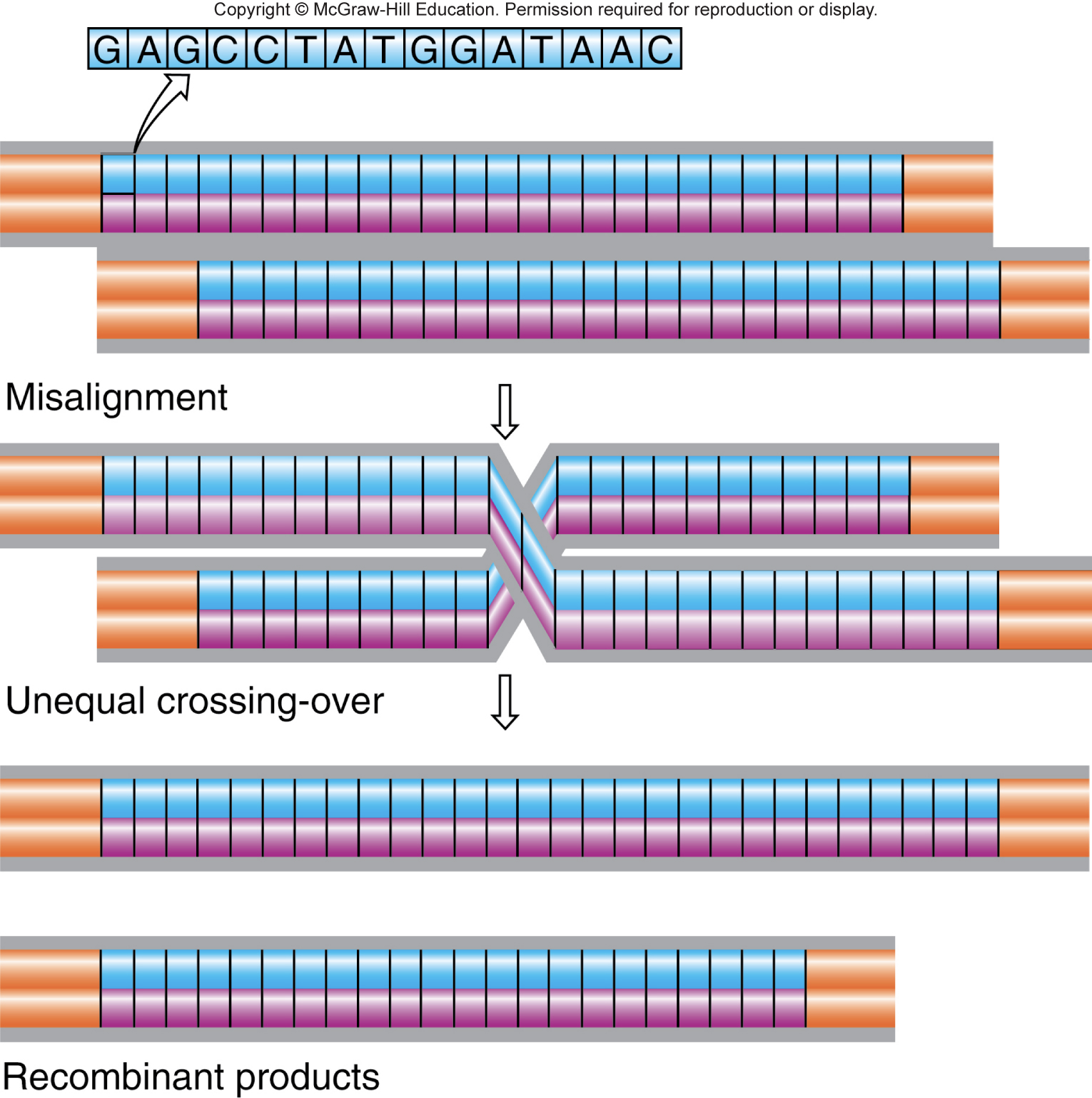
Unequal crossing-over produces new alleles of copy number variants (CNVs)
- CNVs are tandem sequence repeats more than 10 bp long
- Misalignment during meiosis leads to unequal crossing over
- not a common event, so most CNVs are inherited, rather than being a new mutation
- \
Determining genotype depends on isolating a gene and analyzing the alleles
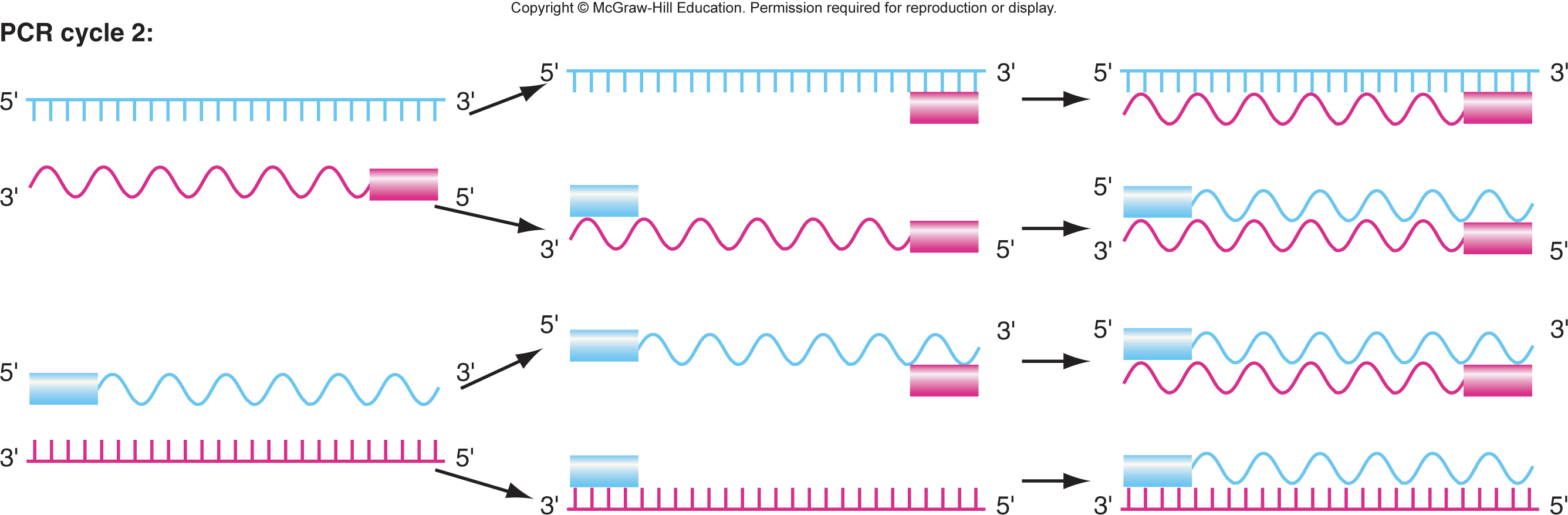
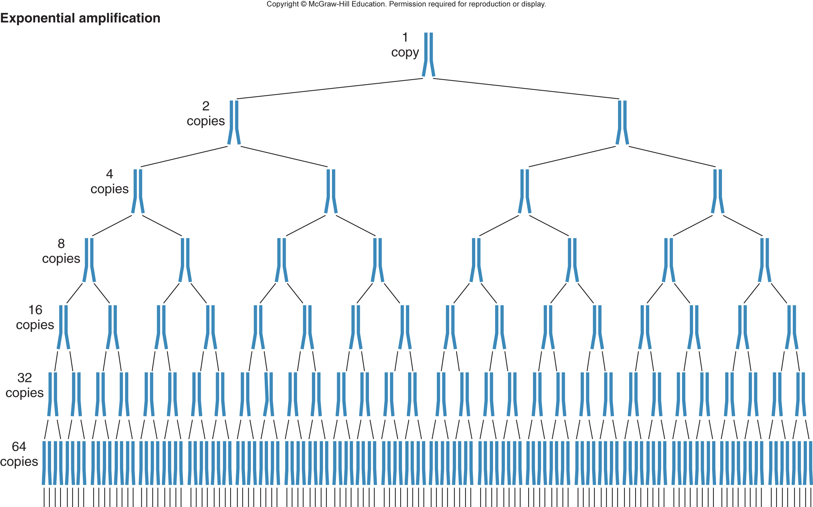
- Polymerase chain reaction (PCR): method of making many copies of a target region of DNA
- PCR was first developed in 1985 by Kary Mullis
- PCR is a faster, less expensive, and more flexible way to amplify specific fragments of DNA (compared to molecular cloning)
- PCR is extremely efficient- can amplify DNA from a single cell or from some archaeological samples
Two oligonucleotide primers define the target region
- one oligonucleotide primer is complementary to one strand of DNA at one end of the target region
- the other oligonucleotide primer is complementary to the other strand of DNA at the other end of the target region
- \
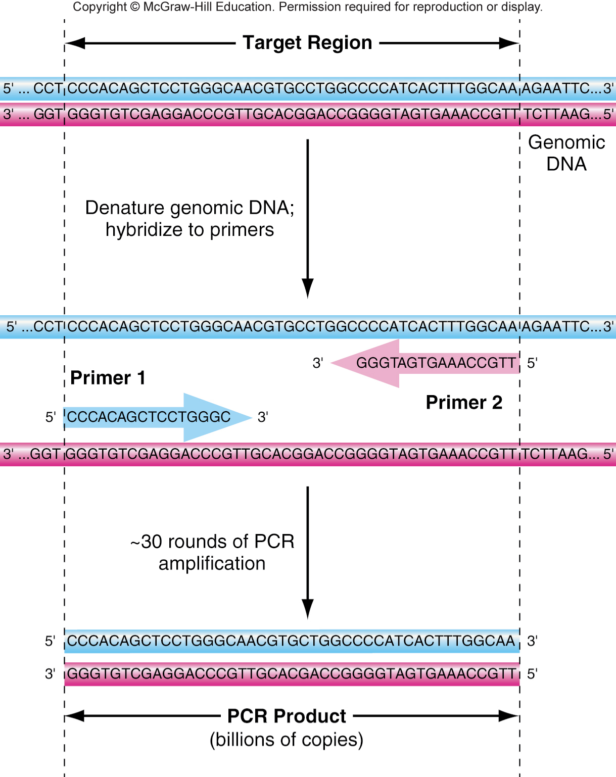
Three steps of PCR: Denature strands, Base pairing of primers, Polymerization from primers along the templates. Repeated multiple times.
\
\
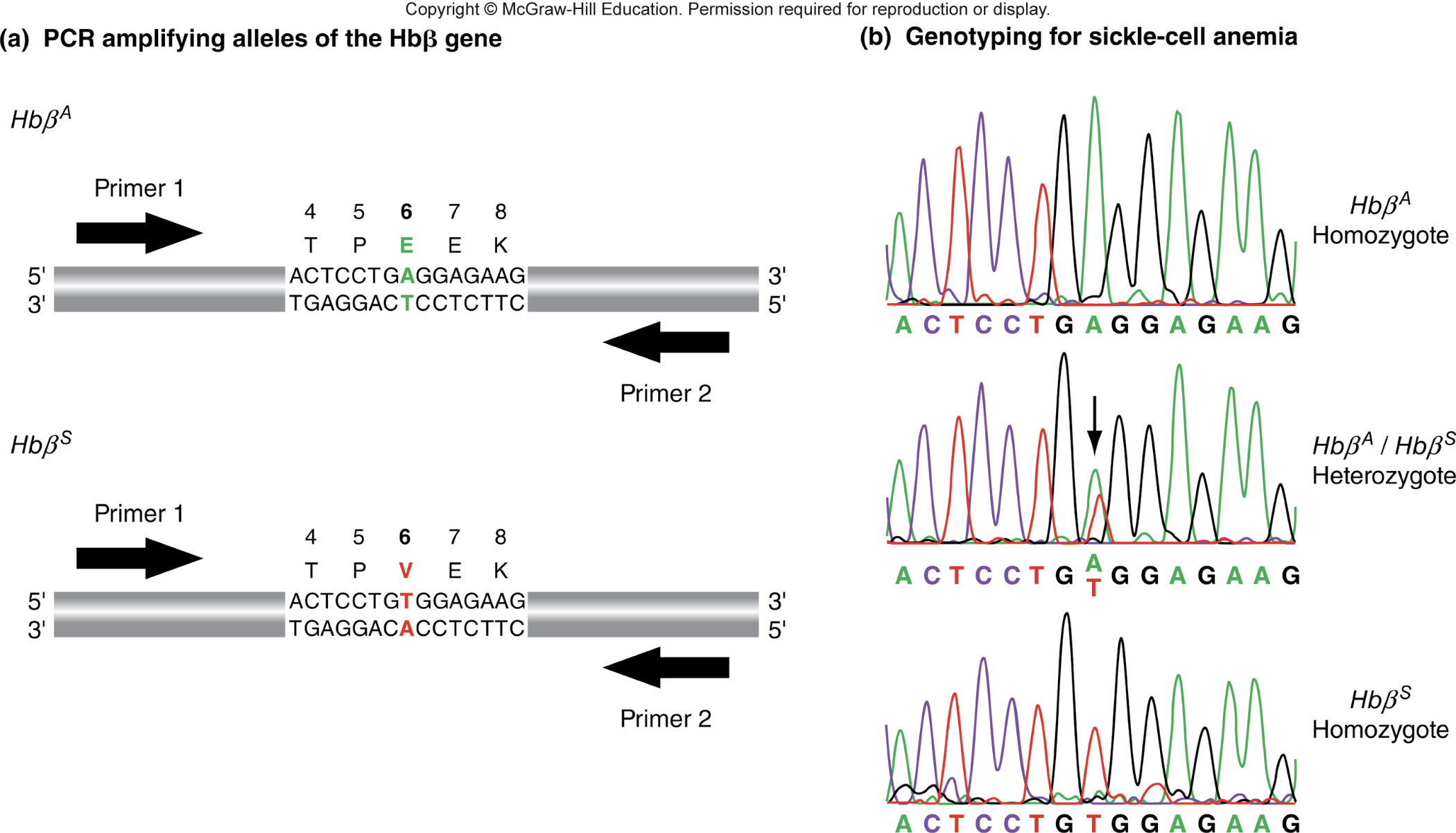
Determining genotype by sequencing PCR products
- sickle cell anemia is caused by a SNA in the HbB gene, chromosome 11 (glutamic acid - GAG, valine - GTG
- genotyping can identify carriers and homozygous individuals
- \
Determining genotype by PCR product size
target regions containing SSRs or DIPs can be amplified by PCR
PCR products vary in size
\
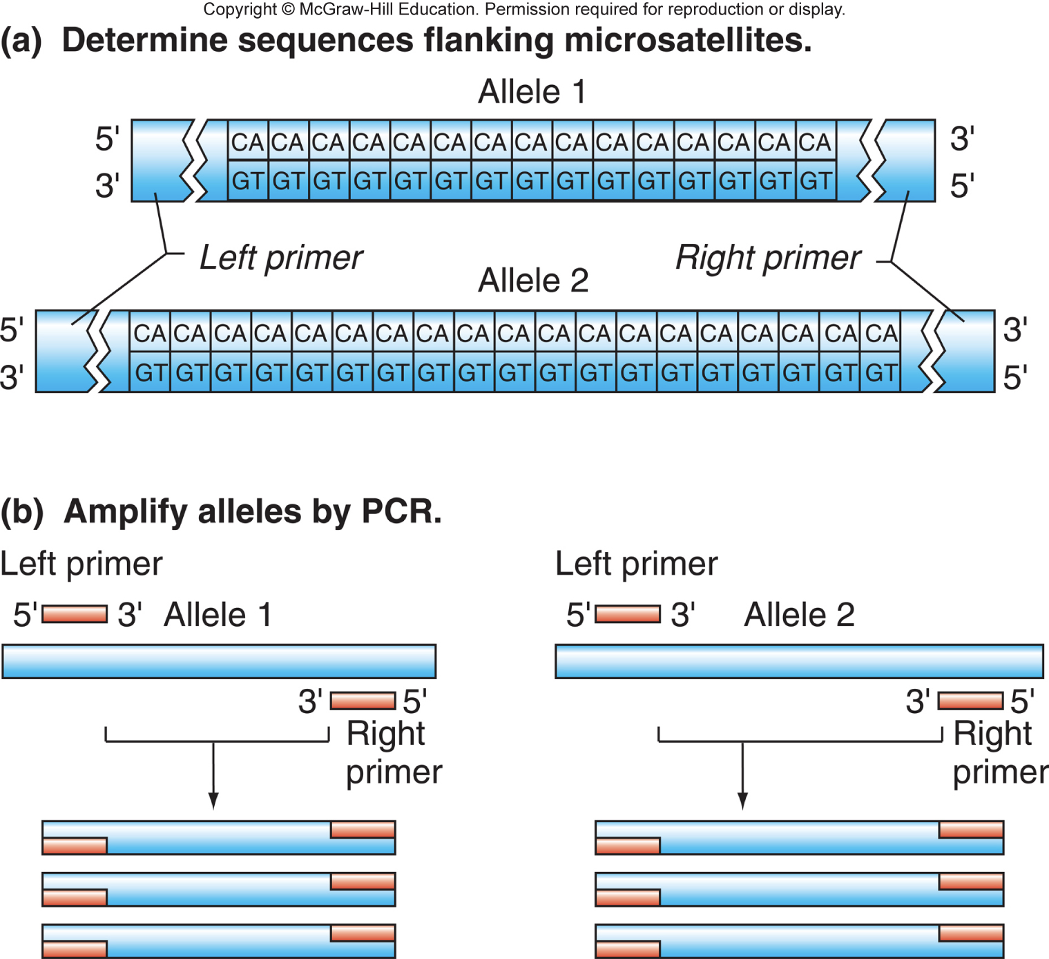
size variations from PCR can be detected by gel electrophoresis
\
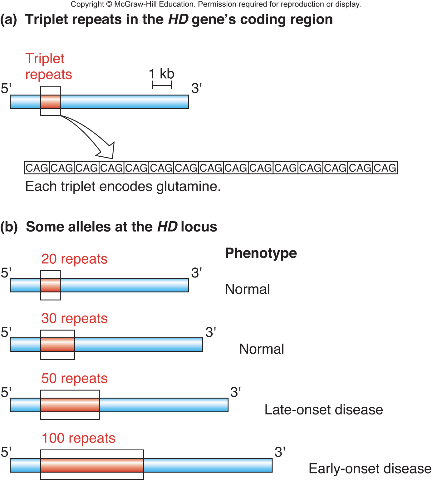
Analysis of the Huntington’s disease locus by PCR
- Huntington’s disease is an autosomal dominant disorder
- In Huntington’s, normal allele has <34 CAG repeats
- disease-causing alleles have 42 or more CAG repeats in Huntington’s
- \
- \
Fetal and embryonic cells can be genotyped using PCR
- Prenatal genetic diagnosis: genotypic fetal cells isolated by amniocentesis (getal cells in amniotic fluid extracted using a needle)
- Preimplantation embryo diagnosis: utilizes in vitro fertilization and PCR to genotype embryos before placing in womb
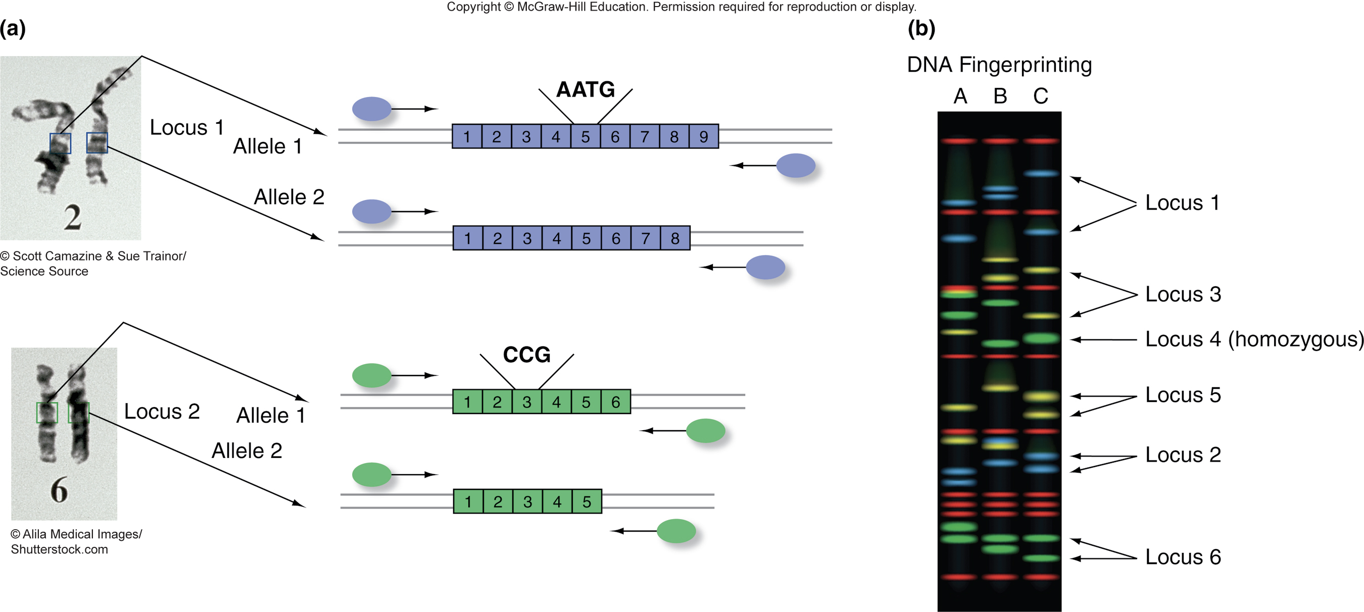
Forensic DNA fingerprinting
- STR loci are highly polymorphic: many alleles exist in the population and an individual person carries only 2 alleles at a given locus
- Genotype is discovered through PCR at many STR loci: 20 pairs of PCR primers are labeled with fluorescent dyes, the probability that 2 people have the same alleles at 20 STR loci is very remote
- CODIS database is maintained by the FBI: Data from all 20 STR loci can match DNA from crime scene to a person, or can establish innocence
Multiplex FCR is used fro DNA fingerprinting
- \
- \
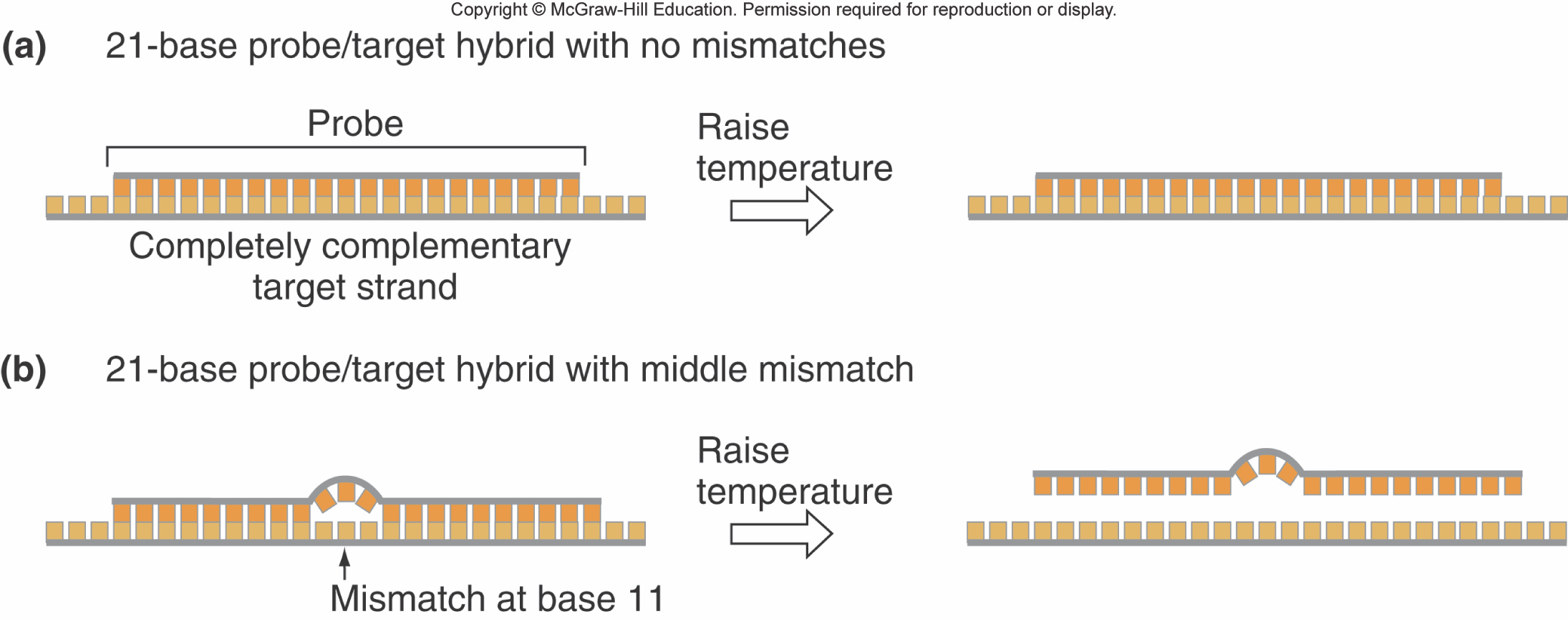
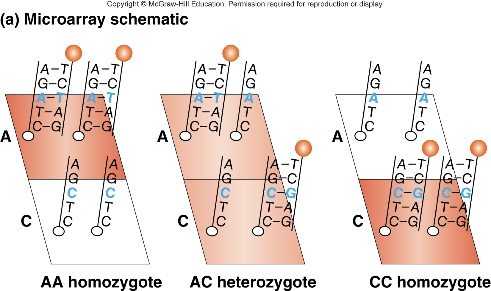
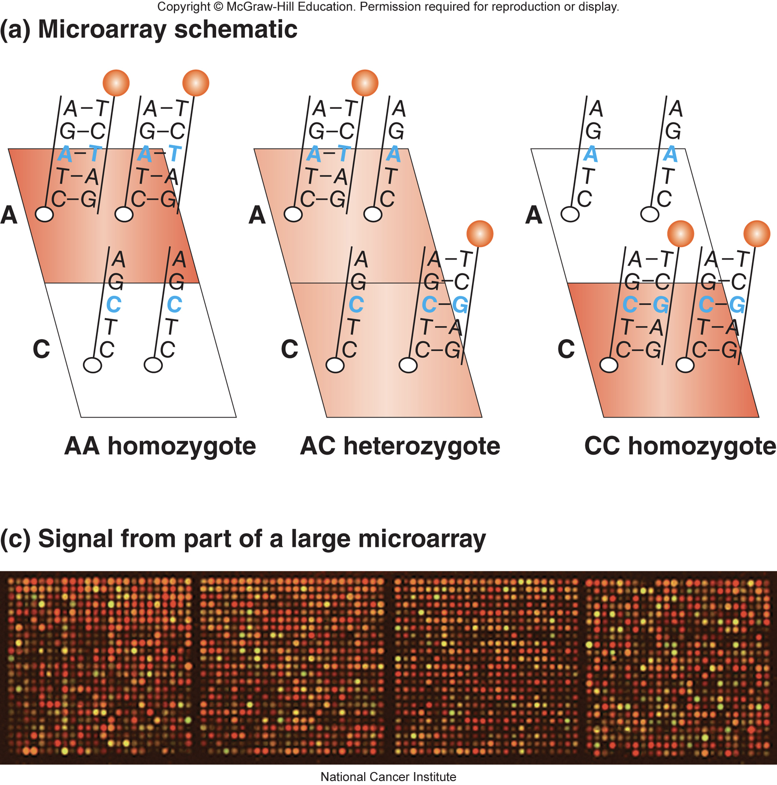
Short hybridization probes can distinguish single-base mismatches
- Hybridization of short (<40) oligonucleotides to sample (target) DNAs (allele-specific hybridization)
- In hybridization, if no mismatch between probe and target, hybrid will be stable at high temp
- In hybridization, if there is mismatch between probe and target, hybrid will not be stable at high temp
- \
Hybridization probes are used on microarrays for genotyping
- allele-specific oligonucleotides(ASO) are attached to a solid support (like a silicon chip)
- two oligonucleotides are shown here, but many are put on one array
- \
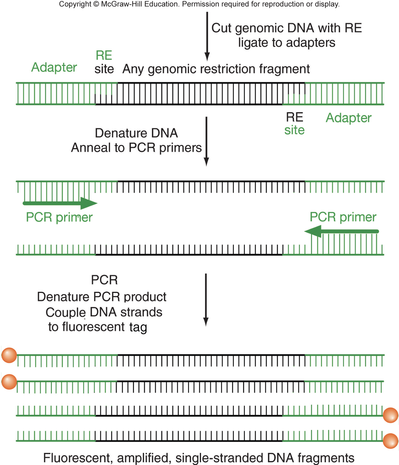
Genomic DNA used to probe the ASO chip (microarray)
- preparation of genomic DNA: fragmented, adapter attached, amplified by PCR and denatured to make single stranded, and fluorescent dye coupled to end of single-stranded DNA
- \
Fluorescent output is proportional to the number of copies of each allele
- up to 4 million loci can be genotyped simultaneously
- \
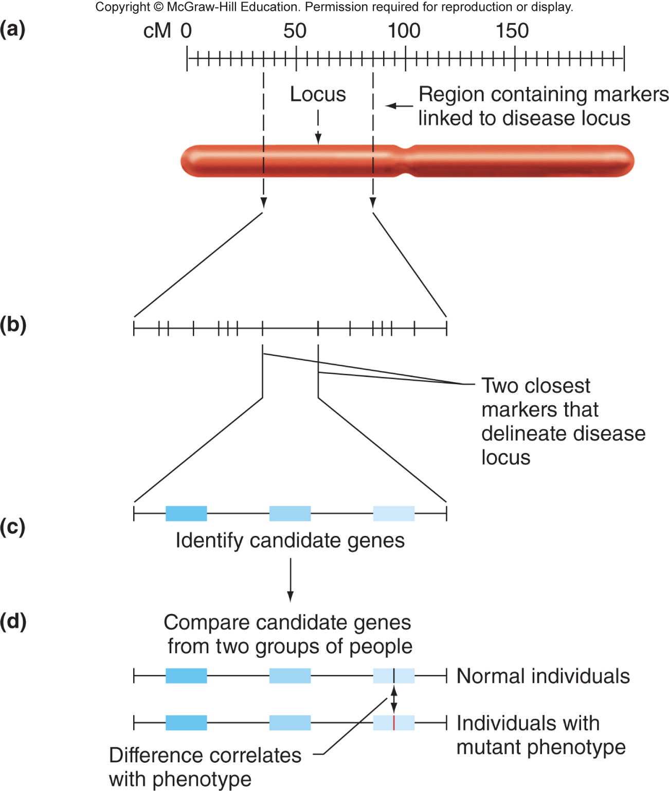
Positional cloning: from DNA markers to disease causing genes
- positional cloning: object is to identify disease-causing genes by genetic linkage to polymorphic loci
- strategy for positional cloning: Same as linkage analysis using two phenotypes, except one gene tracked by phenotype, the other by DNA genotype. Use microarrays to simultaneously analyze millions of two-point crosses, each one a test for linkage between a disease locus and a DNA marker.
- Steps for positional cloning: Region of interest narrowed by finding closely linked DNA markers. Candidate genes are located in the region of interest. Sequence and expression of candidate genes are determined in normal and diseased individuals.
- \
Genetic diseases can display allelic or locus heterogeneity
- allelic heterogeneity: disease caused by different mutations in the same gene
- compound heterozygote: individual with different mutant alleles of the same gene
- locus heterogeneity: disease caused by mutation in one of two or more different genes
Genome sequencing is becoming routine
sequencing an entire genome now costs about $400-$500
sequencing the whole exome is less expensive
high-throughput or massively parallel sequencing is like Sanger sequencing with a few modifications: individual DNA molecules anchored in place, each base is identified before the next one is added, increased sensitivity eliminates need for cloning or PCR
\
High-throughput breakthroughs
individual DNA molecules anchored in place
base additions controlled and known
some sensitivities are high enough to detect one DNA strand
Genome sequencing reveals a sea of variation
each individual differs at > 3 million locations from the RefSeq human genome
how can we tell which polymorphism causes a disease? transmission pattern, predicted effect on protein function, clues from other genome sequences
Chapter 11
Structure and function of Eukaryotic chromosome
Copy Cat (left) is clone of Rainbow (right)
\
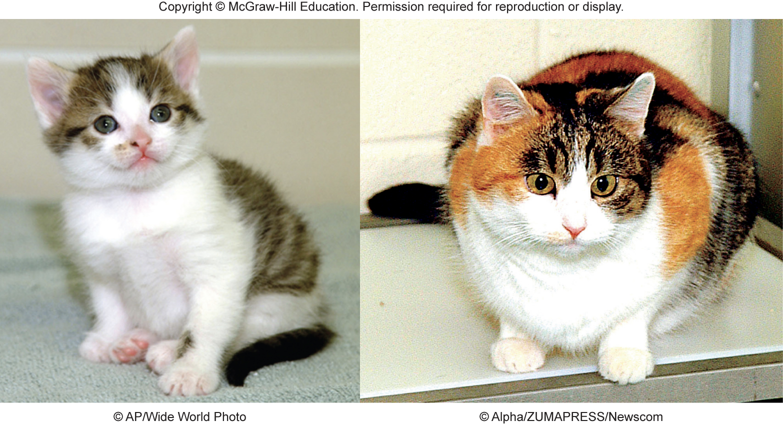
all cells have identical DNA
each cat has many types of cells
cats are dissimilar in many phenotypes
Chromosomal DNA and proteins
- chromosomes support packaging, replication, segregation, and expression of genetic information
- chromatin: generic term for any complex of DNA and protein found in a nucleus of a cell; 1/3 DNA, 1/3 histones, 1/3 non-histone proteins
- chromosomes: separate pieces of chromatin that behave as a unit during cell division
- DNA interaction with histones and non-histone proteins produces sufficient level of compaction to fit into a cell nucleus
Different levels of chromosome compaction
- if stretched out, total DNA in a single cell would be about 6 ft long
- compaction allows DNA to fit in cell nucleus
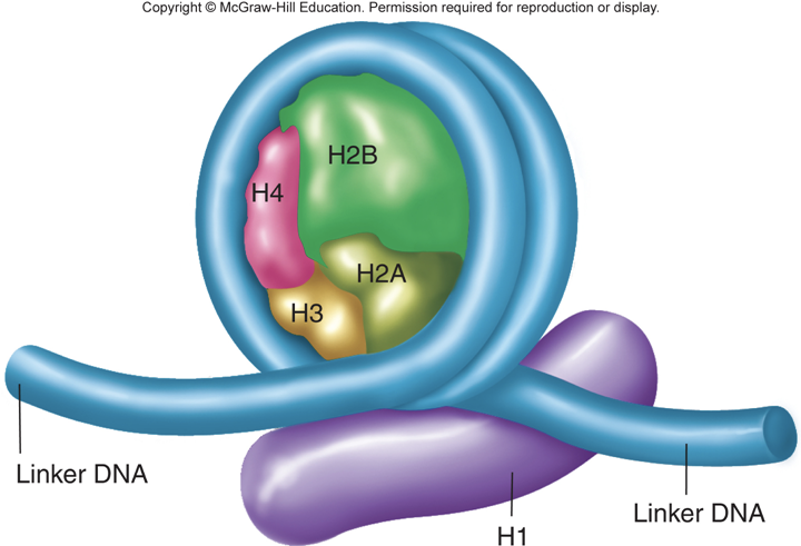
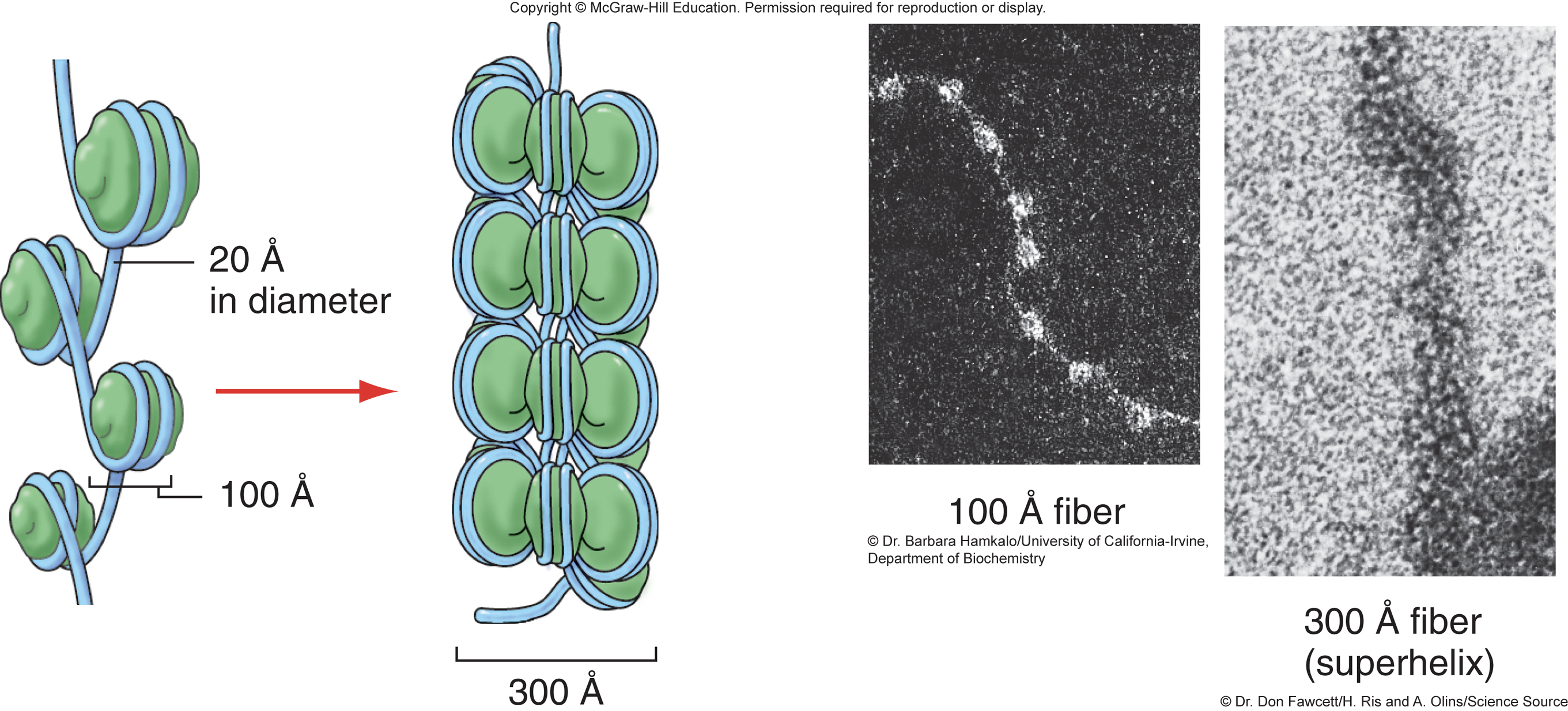
- Nucleosome: confirmed by crystal structure; condenses naked DNA 7-fold to a 100 A fiber
- Supercoiling: hypothetical model (300 A fiber predicted has been seen); causes additional 6-fold compaction, achieving a 40- to 50-fold condensation relative to naked DNA
- Redial loop-scaffold: hypothetical model (preliminary experimental support); through progressive compaction of 300 A fiber, condenses DNA to rodlike mitotic chromosomes that are 10,000 times more compact than naked DNA
Histone proteins
- histones: small, positively charged, highly conserved; bind to and neutralize negatively charged DNA
- ==5 types of histones: H1, H2A, H2B, H3, and H4==
- core histones 2x (H2A, H2B, H3, and H4) make up the nucleosome
- \
Non-histone proteins
- hundreds of other proteins that make up chromatin and are not histones
- 200-200,000 molecules of each kind of non-histone protein
- functions of non-histone proteins: structural role (chromosome scaffold), chromosome replication, chromosome segregation, active in transcription
Nucleosome is the fundamental unit of chromosomal packaging
- nucleosomes resemble beads on a string
- spacing and structure of nucleosomes affect genetic function
- accessibility for proteins that initiate transcription, replication, and further compaction
- arrangement along chromatid is highly defined and varies in different cell types and under different conditions
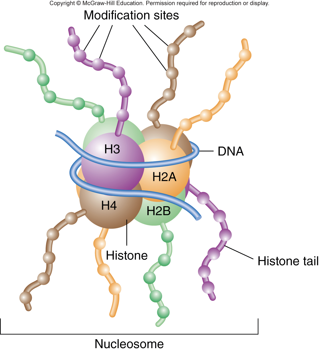
Nucleosome core is an octamer of 2 of each of histones H2A, H2B, H3, and H4
- 160 bp of DNA wraps twice around a nucleosome core
- 40 bp of linker DNA connects adjacent nucleosomes
- Histone H1 associates with linker DNA as it enters and leaves the nucleosome core
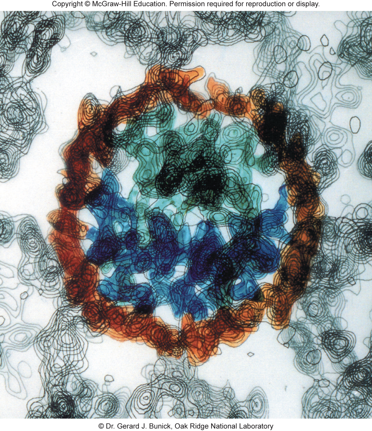
X-ray crystallography of a nucleosome
- DNA bends sharply at several places as it wraps around the core histone octamer
- base sequence dictates preferred nucleosome positions along the DNA
- \
Nucleosome supercoiling model of higher-order packaging
- 100 A nucleosomal chromatin is compacted into 300 A fiber by supercoiling
- \
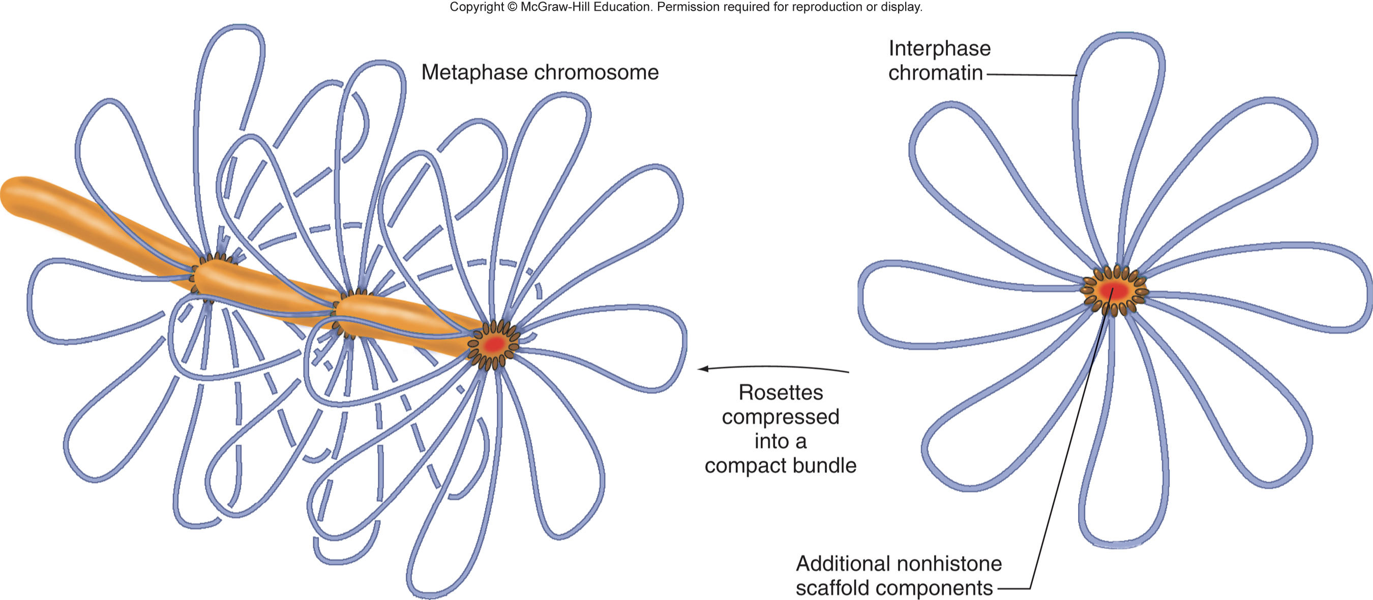
The ==radial loop-scaffold== model for higher level of compaction
several non-histone proteins (NHPs) bind to chromatin every 60-100 kb and tether the 300 A fiber into structural loops
other NHPs gather several loops together into daisy-like rosettes
\
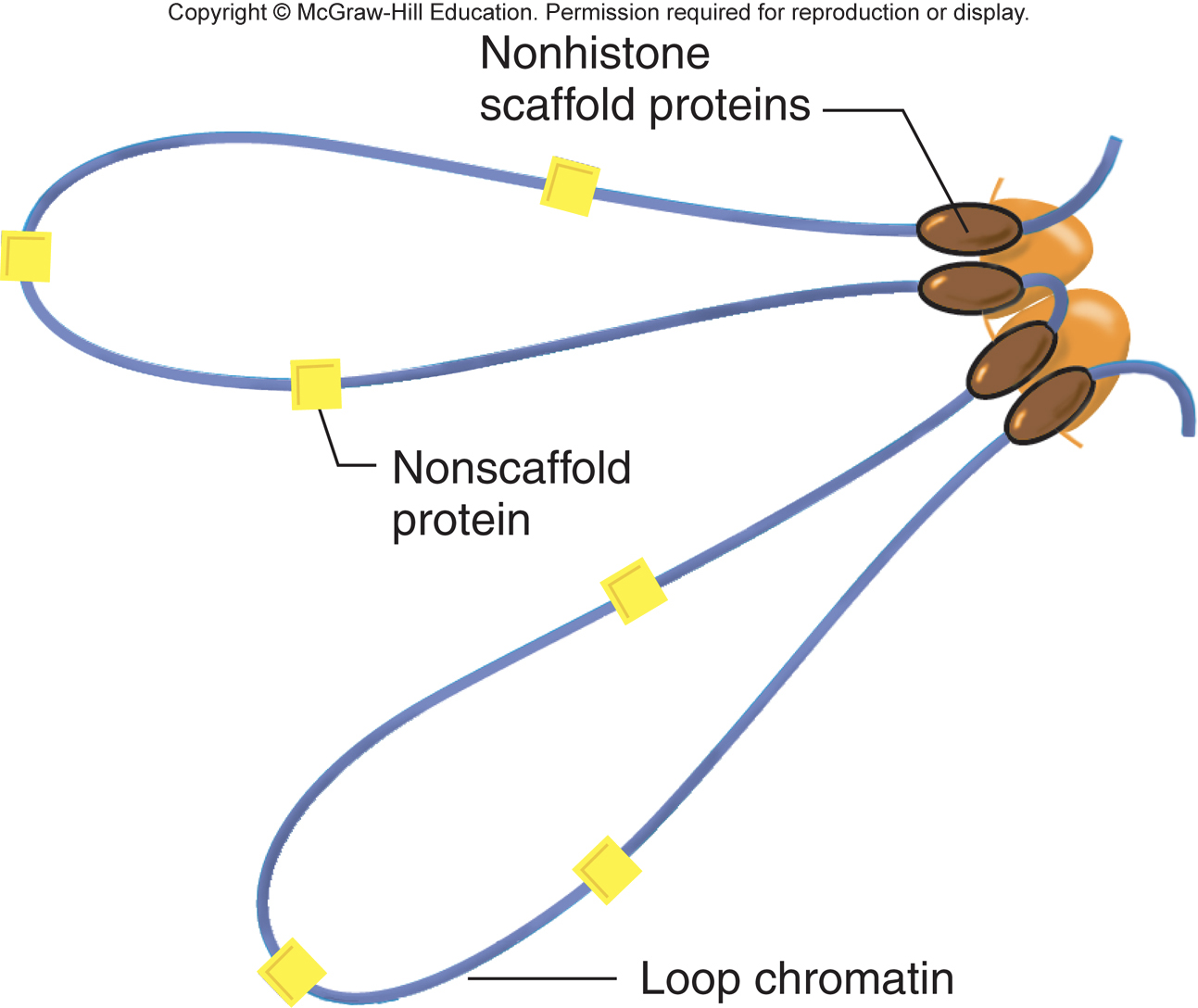
condensins may further condense chromosomes into a compact bundle for mitosis
\
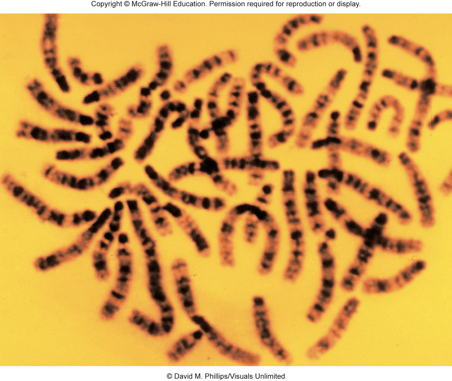
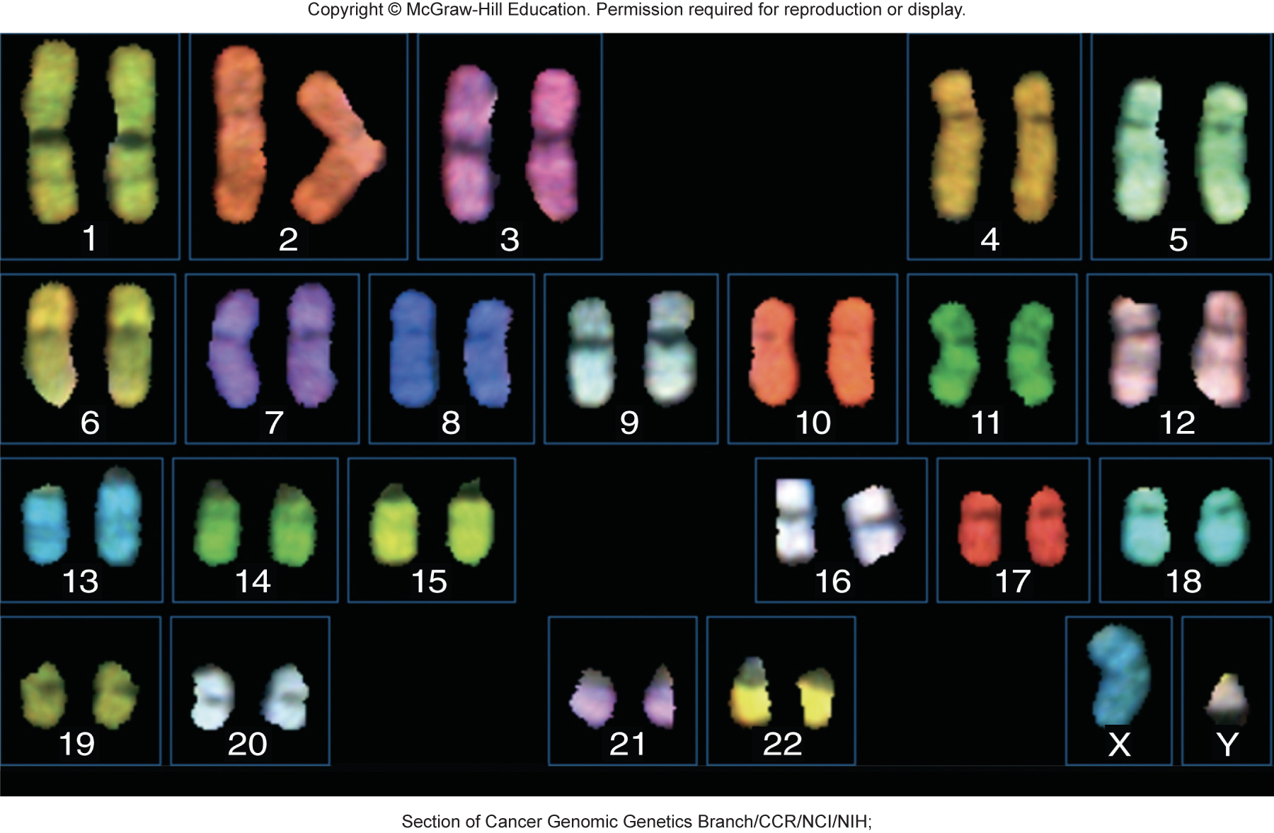
The karyotype of a human female examined by a high-resolution G-banding
- metaphase chromosomes stained with Giemsa stain have alternating bands of light and dark staining
- each band contains many DNA loops and ranges from 1 to 10 Mb in length
- banding patterns on each chromosome are highly reproducible
- \
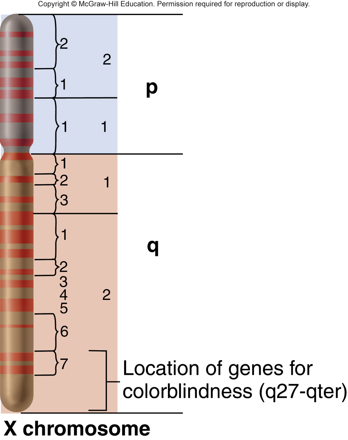
Locations of genes in relation to chromosomal bands
- short arm: p arm
- long arm: q arm
- within each arm, light and dark bands are numbered consecutively
- \
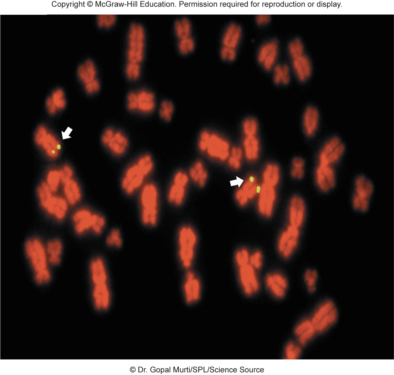
Fluorescent in situ hybridization (FISH) is used to characterize genomes
- FISH depends on hybridization between metaphase chromosomes and a labeled DNA sequence
- In FISH, chromosomes are spread on a glass slide and denatured to make them single stranded, a DNA sequence is labeled with a fluorescent tag to make a probe, and the probe hybridizes to chromosome at complementary region
- \
Spectral karyotyping (SKY) is a variation of FISH
- in SKY, probes specific for each chromosome are labeled with a different fluorescent dye
- \
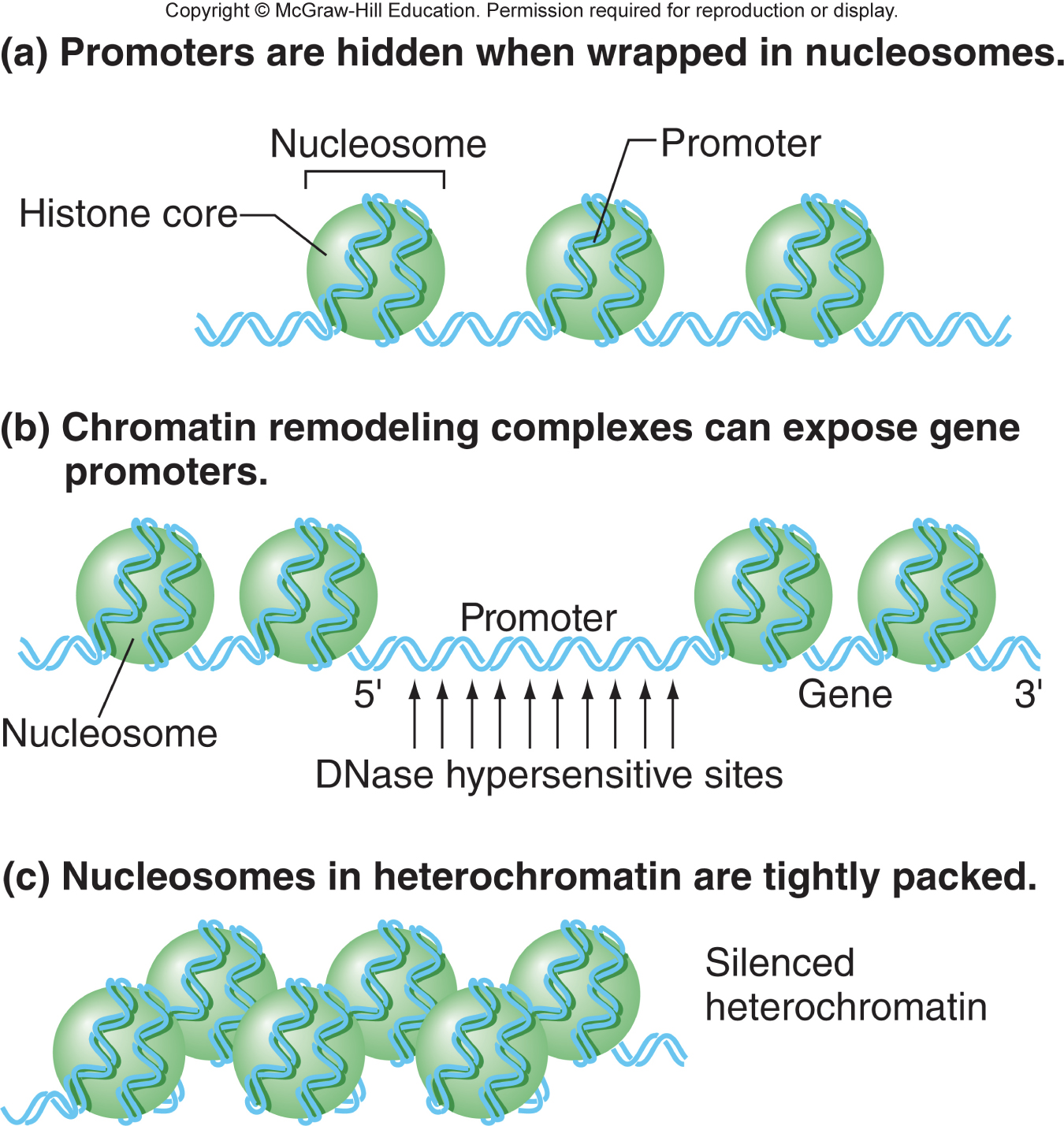
Chromosomal packaging and function
- heterochromatin: highly condensed, usually inactive transcriptionally
- darkly stained regions of chromosomes
- constitutive: condensed in all cells
- facultative: condensed in only some cells and relaxed in other cells
- Euchromatin: relaxed, usually active transcriptionally
- lightly stained regions of chromosomes
Transcription requires changes in chromatin structure
- promoters of inactive genes are hidden in nucleosomes
- to activate a gene, transcription factors bind to enhancers and recruit chromatin remodeling proteins
- promoters are exposed by removing or repositioning nucleosomes
- \
The four core histone tails can be modified with chemical groups
- tails extend outward from nucleosome
- enzymes can add chemical groups
- modified tails can alter nucleosomes and bind chromatin modifier proteins
- \
Histone tail modifications alter chromatin structure
methylation: histone methyltransferase adds methyl group to histone tails
methylation affect depends on specific amino acid modified
adding methyl group to H3 lysine 9 favors heterochromatin formation
methylation reversed by mehtyltransferases
\
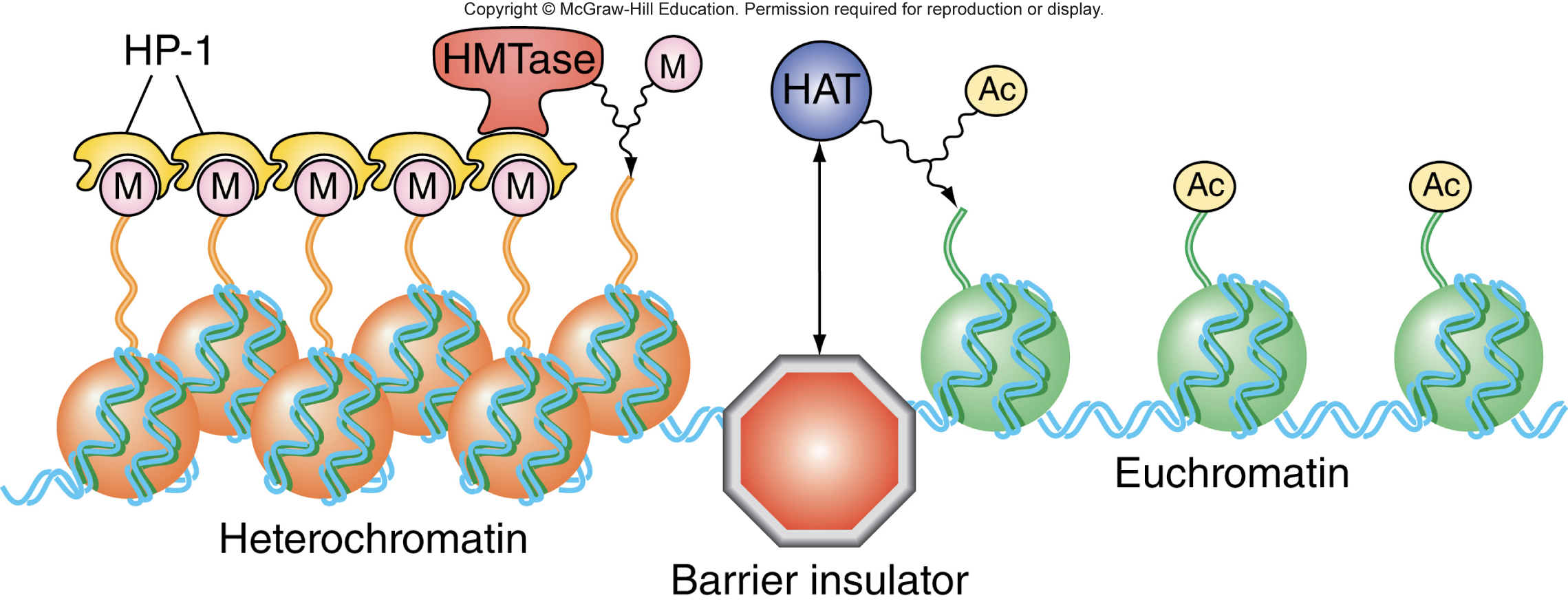
Acetylation: histone acetyltransferase adds acetyl group to histone tails
Acetylation prevents close packing of nucleosomes
Acetylation favors expression of genes in euchromatin
Acetylation reversed by deacetylases
\

X-chromosome inactivation in female mammals occurs through heterochromatin formation
- example of heterochromatin
- dosage compensation in mammals so that X-linked genes in XX and XY individuals are expressed at same level
- random inactivation of all except 1 X chromosome in each cell
Chapter 12
Chromosomal Rearrangements
Two main themes underlying the observations on chromosomal changes
- Karyotypes generally remain constant within a species
- most genetic imbalances result in a selective disadvantage
- Related species usually have different karyotypes
- closely-related species differ by only a few rearrangements on their karyotypes
- distantly-related species differ by many rearrangements on their karyotypes
- Correlation between karyotypic rearrangements and speciation
Four types of chromosomal rearrangement: deletion, duplication, inversion, and translocation
Fluorescent in situ hybridization can detect large chromosomal rearrangements using spectral karyotyping with probes specific for two different chromosomes showing chromosomal translocation
Phenotypic and genetic effects of deletions
- homozygosity for deletions is often lethal or harmful depending on size of deletions and affected genes
- deletion in heterozygotes can have a mutant phenotype due to gene dosage effects (haploinsufficiency)
- deletion in heterozygotes increases risk of altered phenotype due to mutation in the other copy of the gene, may uncover recessive mutant alleles
Transposable elements (TEs) are movable genetic elements
- TEs are any segment of DNA that has the ability to move from place to place within a genome
- Marcus Rhoades (1930s) and Barbara McClintock (1950s) inferred existence of TEs from genetic studies of corn
- TEs have now been found in all organisms with some functions beneficial to the host, can be present in hundreds of thousands of copies per genome
Instability of TE assiciated alleles
- a nonautonomous TE (Ds) inserted into gene C and disrupted its function. In kernals that have autonomous TE (AC), Ds can hop out, restoring gene function
- \
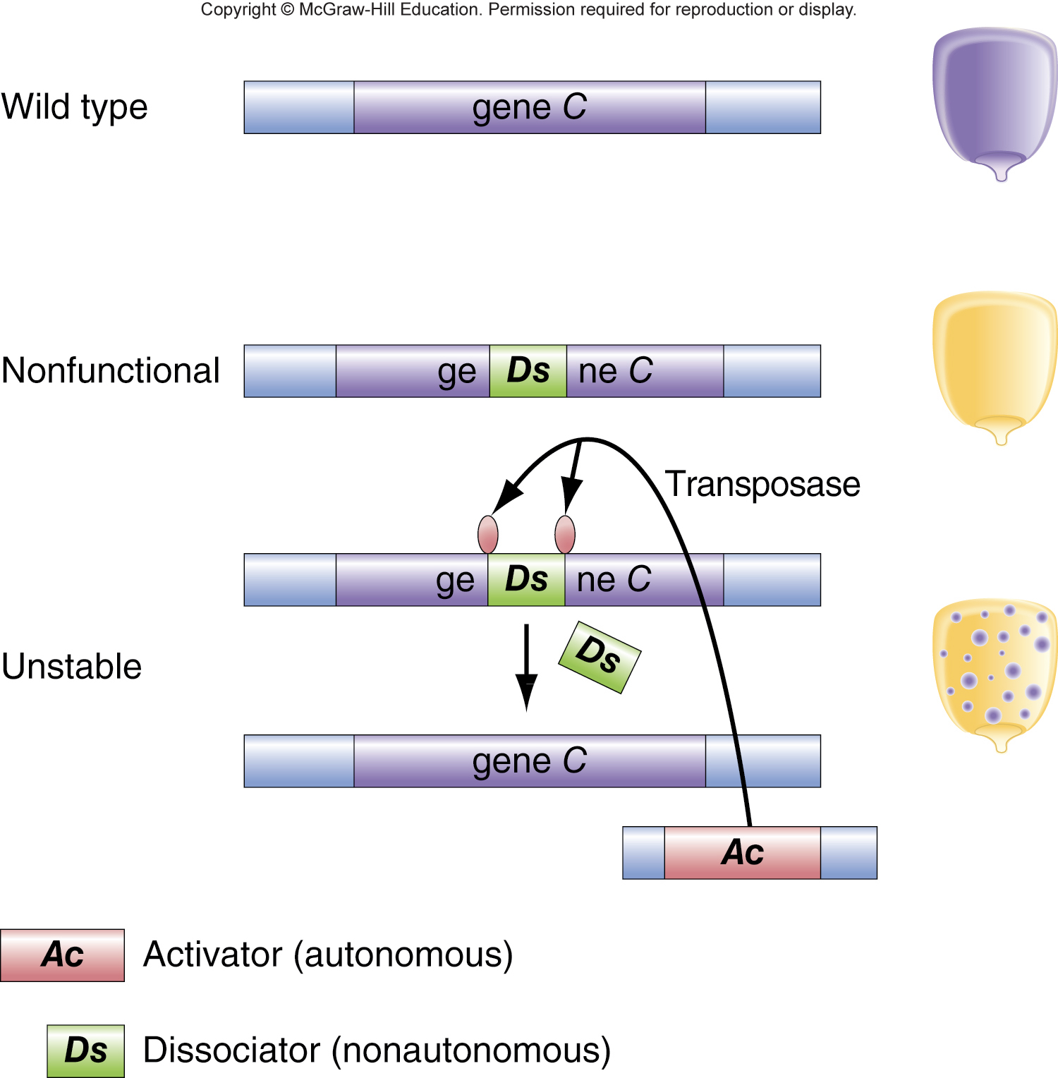
Structure of DNA transposons
- Most DNA transposons contain inverted repeats (IRs) of 10-200 bp long at each end, gene encoding trasnposase recocgnizes IRs and cuts at border between the IR and genomic DNA
TEs can disrupt genes and alter genomes
- TE insertion can result in altered phenotype. TE can insert within coding region of a gene or insert near a gene and affect its expression. TE-associated alleles can be unstable
- TEs can trigger spontaneous chromosomal rearragnements due to unequal crossing over between TEs
- Gene relocation due to transposition, formation of composite TE
Aneuploidy is loss or gain of one or more chromosomes
- Aneuploids: individuals whose chromosome number is not an exact multiple of the diploid number for that species
- Euploidy: 2n
- Nullisomy: 2n-2
- Monosomy: 2n-1
- Trisomy: 2n+1
Aneuploidy usually results in multiple abnormalities or lethality
- autosomal monosomy is usually lethal
- most trisomies are lethal; a few result in characteristic syndromes
- sex chromosome aneuploidy is tolerated due to X chromosome inactivation
- Trisomic 21: down syndrome
- Trisomic 13: Patau syndrome
- Trisomic 18: Edwards syndrome
- XO, monosomic: turner syndrome
- XYY trisomic, normal
- XXY: trisomic Klinefelter syndrome
- XXYY, XXXY: tetrasomic Klinefelter syndrome
- XXXXY: pentasomic Klinefelter syndrome
- XXXXXY: hexasomic Klinefelter syndrome
Aneuploidy is caused by nondisjunction
- nondisjunction: failure of chromosomes to segregate normally, can occur during meiosis I or meiosis II
Some euploid species are not diploid
- basic chromosome number (number of chromosomes in a single, complete set = x
- monoploidy and polyploidy are rarely observed in animals, except ants, bees, hermaphroditic worms, and some fish
Formation of a triploid (3x) organism
- diploid gametes may arise from a 4x parent or from a diploid with defects in meiosis (defect in spindle or at cytokinesis)
Meiosis in a triploid organism
- regardless of how chromosomes pair, there is no way to ensure that gametes contain a complete, balanced set of chromosomes. all polyploids with odd numbers of chromosome sets are sterile because they cannot produce balanced gametes.
Generation of tetraploid (4x) cells
- tetraploid cells occur during mitosis in a diploid when chromosomes fail to separate into two daughter cells
- if tetraploidy occurs in gamete precursors, diploid gametes are produced
- union of two diploid gates produces a tetraploid organism
- autopolyploid: all chromosome sets are derived from the same species
In tetraploid, pairing of chromosomes as bivalents generates balanced gametes
- four copies of each group of homologs pair two by two to form two bivalents
- successful tetraploids produce balanced 2X gametes and are fertile
Polyploids in agriculture
- 1/3 of all known flowering plant species are polyploid
- polyploidy often results in increased size and vigor in plants
- many polyploid plants have been selected for agricultural cultivation
- tetraploids: alfalfa, coffee, peanuts, Macintosh apples, Bartlett pear
- octaploids: strawberries
- Euploid: a complete set of chromosomes (usually diploid)
- Polyploid: a euploid species that carries 3+ complete sets of chromosomes
- Autopolyploid: a kind of polyploid that derives all its chromosome sets from the same species
- Allopolyploid: hybrids in which chromosome sets come from distict, but related, species. Usually infertile ebcause different chromosome sets cannot easily pair and segregate properly
- Amphidiploid: two diploid genomes, each from a different parental species
- Raphanobrassica: sterile F1 from crossing cabbages and radishes, has 18 chromosomes (9 from each parent)
Creation of the allopolyploid Triticale
- F1 hybrid of wheat and rye, sterile because there are no pairing partners for the rye chromosomes
- fertile Triticale can be created from infertile F1 hybrid Triticale
- some triticale hybrids combine high yield of wheat with ability of rye to grow in unfavorable environments
- some triticale hybrids combine high level of protein from wheat and high level of lysine from rye
Colchicine treatment prevents spindle formation and results in doubling of chromosome numbers
Duplicate genes may acquire new functions
- gene families: sets of related genes with slightly different functions
- genome in common ancestor of all cereal grasses was duplicated. The two copies are related, but have diverged in sequence and function
Rapid chromosomal evolution in house mice on the island of Madeira
- one population of mice introduced to island in 1400s; two populations evolved different sets of Robertsonian translocations, hybrid offspring are sterile
Chapter 13
Bacteria constitute one of the three major evolutionary lineages
- there are nine times as many bacterial cells as human cells
- most bacteria are in the intestines, but the skin, mouth, and respiratory tract are also homes to bacteria
- Bacteria aid human health and some cause disease
Characteristics of bacteria
- bacteria come in a variety of shapes and sizes
- bacteria lack a nucleus and membrane bound organelles
- bacteria have a singel genophore (prokaryotic chromosome) contained within a nucleoid
- most bacteria have a cell wall
A small fraction of bacteria are pathogens
- pathogen: bacterial strain that causes disease
- pathogenic bacteria": invade tissues, may produce toxins
- toxins: proteins that interfere with cell function or destroy cells
- tetanus toxin (Clostridium tetani) results in paralysis by interfering with communication between nerves and muscles
The typical bacterial genome is composed of once circular chromosome
- 4 to 5 Mb (million base pairs) of DNA is the most commonly studied bacterial species
- DNA molecule condenses by supercoiling and looping
- each bacterium replicates and then divides by binary fission into two daughter cells
Individual E. coli strains contain a subset of the E. coli pagenome
- Genomes of 100s of E. coli strains have been sequenced
- core genome: about 1000 genes that are found in all strains
- Pangenome: core genome plus all genes that are found in some strains and not in others (about 15,000 genes)
Plasmids carry additional DNA
- plasmids: small circles of double stranded DNA; may contain genes that benefit host bacterium or contribute to bacterial pathogenicity
- we use plasmids in molecular work as cloning vectors
Bacteria are monoploidL all mutations express their phenotype
- Altered colony morphology: large or small; shiny or dull; round or irregular
- Resistance to bactericides: antibiotics, bacteriophages
- Auxotrophs: unable to reproduce in minimal media because defective in enzymes required to synthesize complex compounds
Gene transfer in bacteria
- Transformation: lysis of donor cell releases DNA into medium, donor DNA is taken up by recipient cell
- Conjugation: Donor DNA is transferred directly to recipient through a connecting tube; contact and transfer are promoted by a specialized plasmid in the donor cell
- Transduction: bacteriophage infects cell, lysis of donor cell, donor DNA packaged in released bacteriophage, donor DNA transferred when phage particle infects recipient cell
Transformation: competent cells can take up DNA fragments from surrounding environment
- Natural transformation: bacteria take up DNA fragments spontaneously from their surroundings
- Artificial transformation: can be accomplished in the lab by making the cell membrane compromised; treat cells with calcium to make the cell walls and membranes permeable to DNA
Conjugation
Lederberg and Tatum, 1940s
Strain A: met- bio- thr+ leu+ thi+ no growth
Strain B: met+ bio+ thr- leu- thi- no growth
Mix of A and B: met+ bio+ thr+ leu+ thi+ grow in colonies
The F plsmid contains genes for synthesizing connections between donor and reicpient cells
F+ are the F plasmid donors
F- are the F plasmid recipients
\
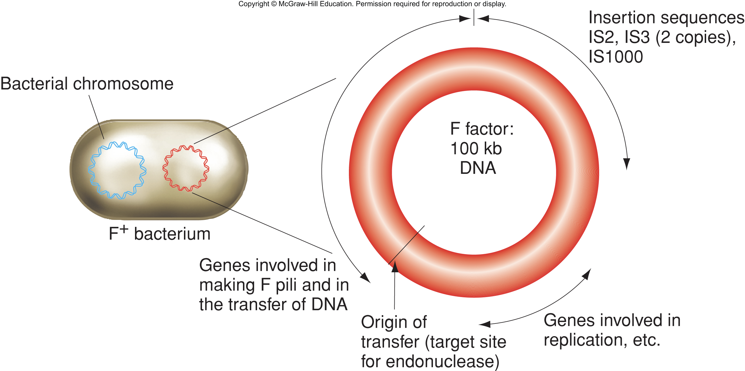
F pilus binds to F-cell wall; Pilus retracts and cells are drawn together; gene transfer; both cells are now F+
Formation of Hfr
F plasmid has 3 IS elements, which are identical to ES elements found at various positions on the bacterial chromosome
High frequency recombinant (Hfr) cells are formed when an F plasmid integrates into the bacterial chromosome through recombination between IS elements
Different Hfr chromosomes
episome: F plasmid that can integrate into the bacterial genome
Hfr strains differ in location and orientation of integrated episomes
Hfr strains retain all F plasmid functions and can be a donor for conjugation with an F- strain
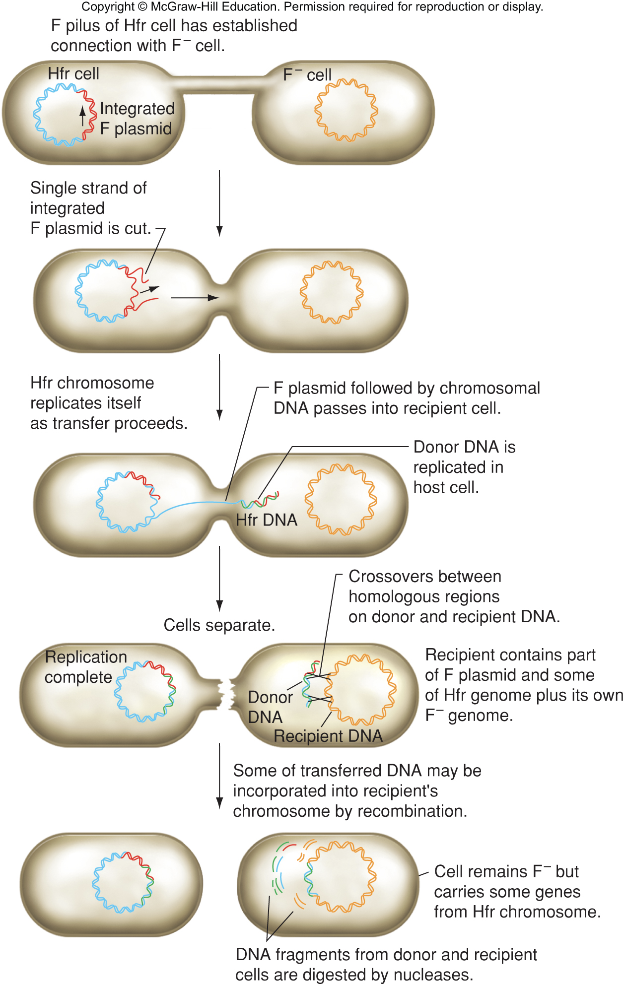
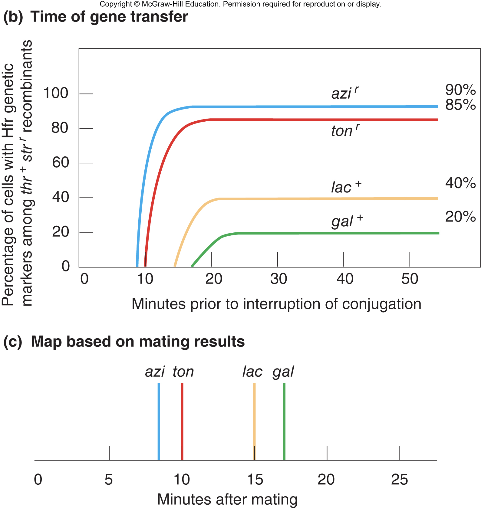
Gene transfer between Hfr donors and F- recipients
Transfer of DNA starts in the F plasmid at the origin of transfer
Chromosomal genes located next to F plasmid sequences are transferred to the recipient
transferred chromosomal DNA recombines into homologous DNA in recipient
\
\
\
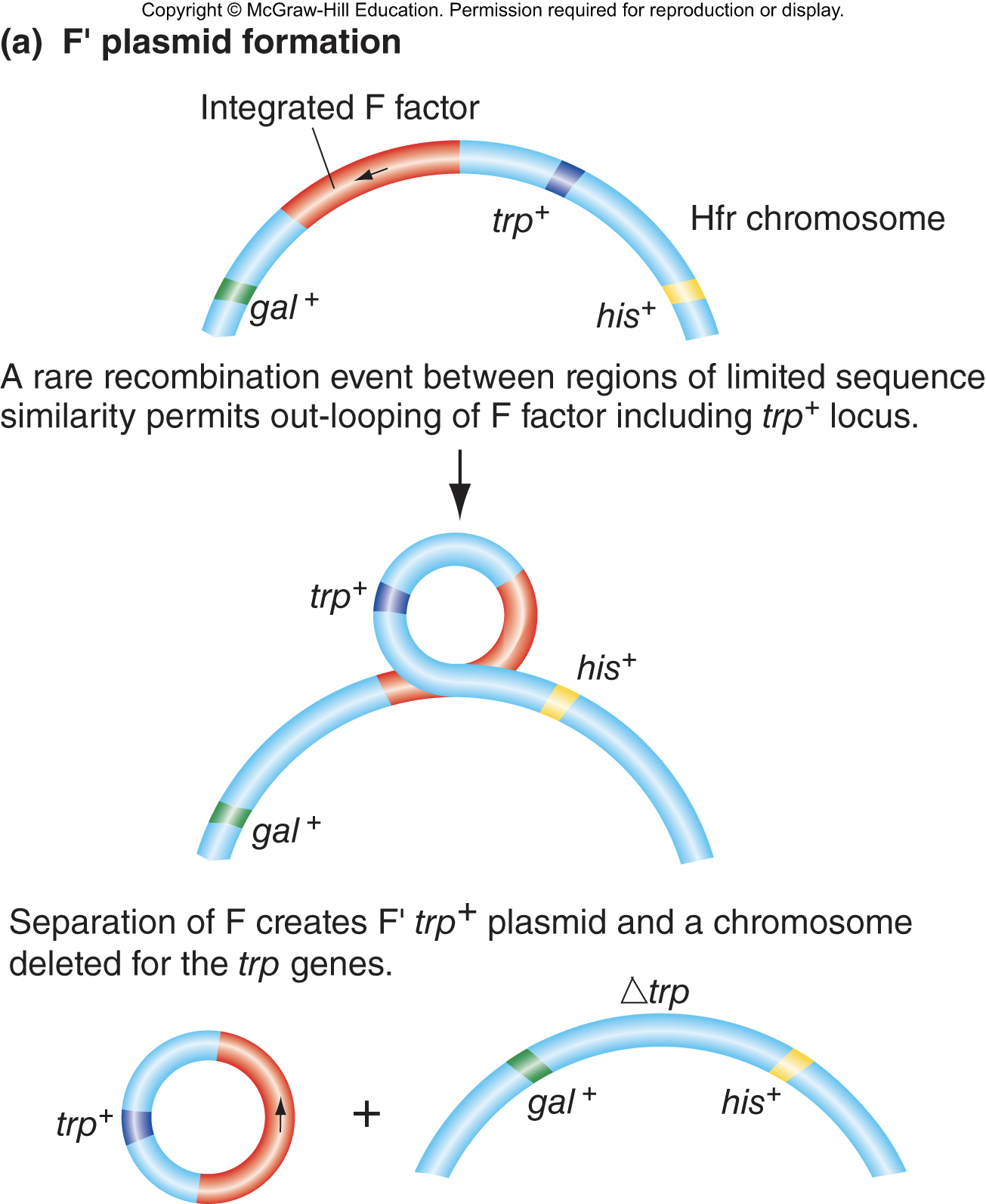
Formation of F’ plasmids by excision from an Hfr chromosome
F’ plasmid is formed by excision of F plasmid plus some adjacent bacterial chromosomal DNA; F’ plasmids replicate independently in bacterial cells
\
F’ plasmids can be transferred to F- cells by conjugation
\
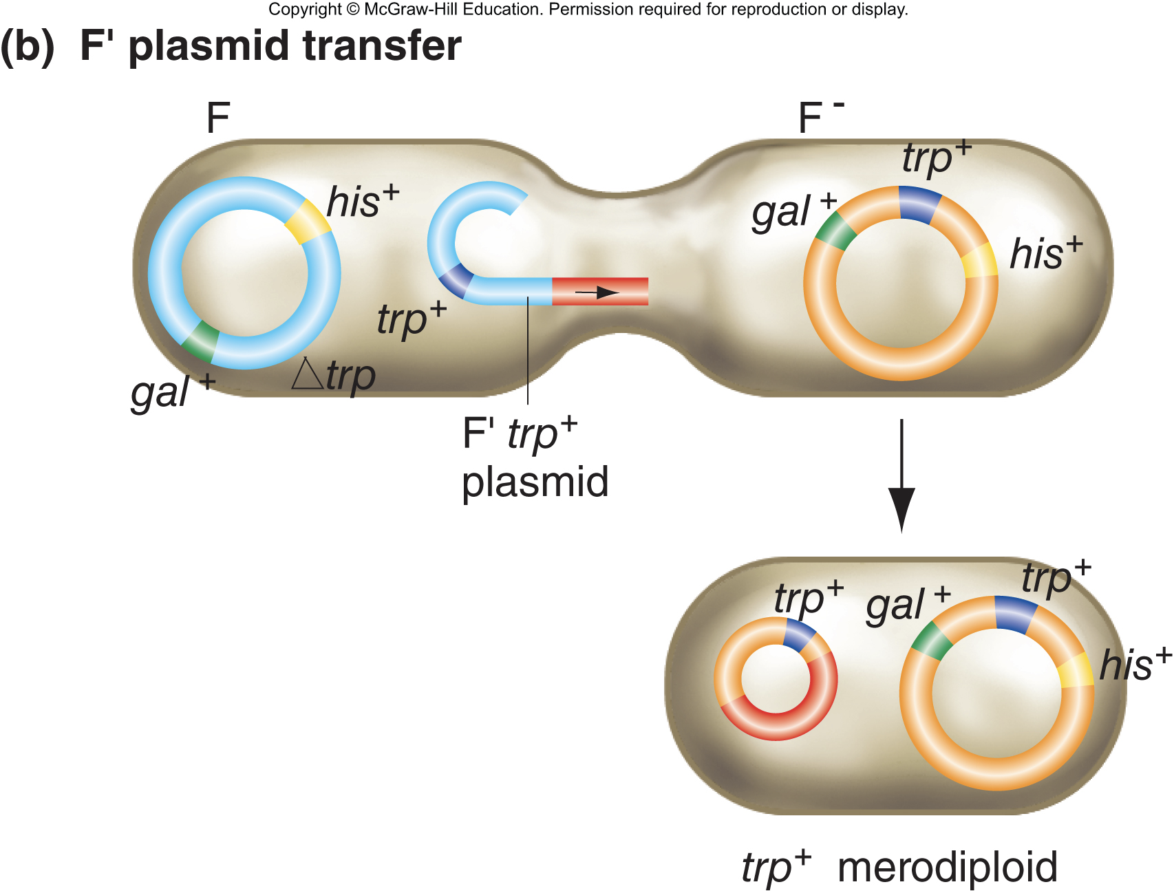
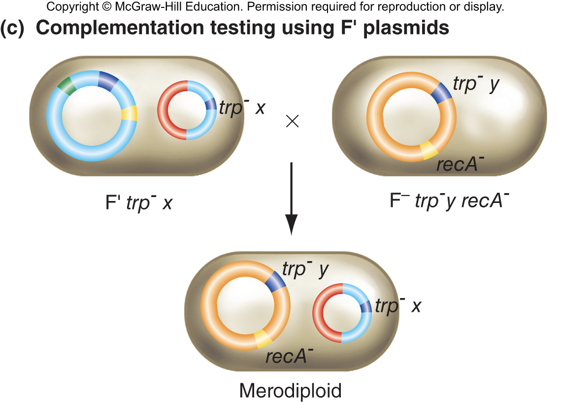
Use of F’ plasmids for complementation studies
conjugation with F’ plasmids can make partial diploids called merodiploids
if the merodiploid shown can grow without tryptophas, the two trp- mutations are in different genes
\
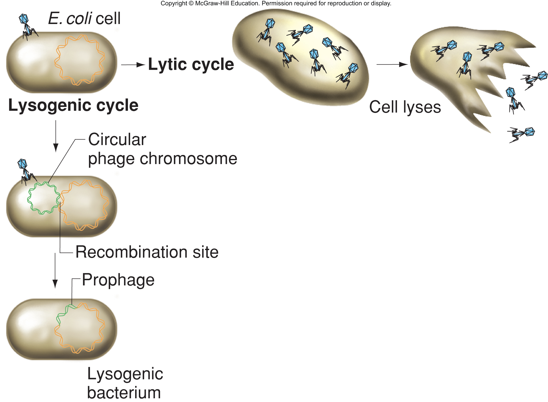
Transduction
In transduction, a phage transfers DNA from a donor bacterium to a recipient bacterium
bacteriophages: viruses that infect, multiply in, and kill various species of bacteria; widely distributed in nature; most bacteria are susceptible to 1 or more
Transduction: process by which a phage transfers DNA from one host cell to another host cell
Virulent phages: always enter lytic cycle after infecting cell, multiply cell, and kill cell
temperate phages: can enter either lytic cycle or enter an alternative lysogenic cycle
prophages: do not produce the viral proteins needed for more virus particles
lysogens: can be induced to enter lytic cycle
\
Generalized transduction
incorporation of random fragments of bacterial DNA from donor into bacteriophage particles
DNA from donor cell injected into infected recipient cell
transduced chromosomal DNA recombines into homologous DNA in recipient
Penicillin interferes with synthesis of bacterial cell wall
- penicillin binds to transpeptidase, inhibits its enzymatic activity, and prevents cross-linking
A second method to become penicillin resistant: mutation of chromosomal genes
- penA encodes traspeptidase, mutation decreases affinity for penicillin
- penB encodes a porin, a protein in the outer cell wall that regulates entry into periplasm; mutation decreases entry of penicillin to cell
- mtr encodes a repressor of an eflux pump; mutation results in increased pumping of penicillin out of cell
- \
Chapter 14
- Organelle genomes lead to non-Mendelian inheritance
- four-o’clocks have green or variegated leaves
- this trait is inherited from the mother (maternal inheritance), due to genes found on chloroplast genome.
- mitochondria and chloroplasts are nonnuclear organelles with their own small genomes
- Structure and function of mitochondria
- mitochondria: membrane bound cytoplasmic organelle
- many mitochondria in each eukaryotic cell
- outer membrane of mitochondria surrounds wrinkled inner membrane
- mitochondria produce energy packets (ATP) through the Krebs cycle and oxidative phosphorylation
- Human mitochondrial genome
- compact gene arrangement
- 16.5 kb genome
- 37 genes encoding tRNAs, rRNAs, and proteins for oxidative phosphorylation
- no introns
- Variation in mitochondrial genomes
- vary in size from 6-2400 kb
- some have introns and space between genes
- some have circular mtDNA (people, animals) and some have linear mtDNA (plants, fungi)
- some protozoan have a single mitochondrion (kinetoplast) with interlocking circular mtDNA
- Mitochondrial exceptions to the universal code
- mitochondrial genetic code varies in different organisms
- in humans, there are 5 differences between the universal and mitochondrial genetic codes
- UGA universal: stop; UGA mtDNA: Trp
- AGG universal: Arg; AGG mtDNA: Stop
- AGA universal: Arg; AGA mtDNA: Stop
- AUA universal: Ile; AUA mtDNA: Met
AUU universal: Ile; AUU mtDNA: Ile-elongation Met-initiation
Structure and function of chloroplasts
- chloroplasts: membrane bound cytoplasmic organelles in plants
- in corn, each cell has 40-50 chloroplasts
- outer membrane surrounds wrinkled inner membrane
- capture solar energy and store in chemical bonds of carbohydrates
- Chloroplast genomes
- most Chloroplast genomes between 120-160 kb long
- more than one copy of Chloroplast genome in each chloroplast
- compact gene arrangements (no introns) in Chloroplast genomes
- circular, linear, and branched forms in Chloroplast genomes
- Chloroplast genomes have more genes than mitochondria
Making transgenic chloroplast by biolistic transformation
- use gene gun: coat small metal particles (bullets) with DNA, shoot DNA bullets at cells, DNA rarely enters chloroplasts and recombines into genome
- transgenic cells identified using selectable marker
- transgenic cell cultured to produce a transplastomic plant
Mitochondria and chloroplasts have characteristics of prokaryotic cells
- Endosymbiont theory: mitochondria and chroloplasts descended from bacteria and fused with nucleated cells
- have their own DNA, like bacteria, mtDNA and cpDNA not arranged into nucleosomes
- inhibitors of bacterial translation block mitochondrial and chloroplast translation, but not eukaryotic translation
- comparisons of rRNA gene sequences suggest mitochondrial and chloroplast genomes derive from a common ancestor of non-sulfur and cyanobacteria, respectively
Cooperation between nuclear and organellular genomes
- mitochondria and chloroplasts require nuclear gene products to assemble and function
- cytochrome oxidase c: functions in mitochondrial electron transport; 7 subunits (3 encoded by mitochondrial genome genes, 4 by nuclear genes)
- nuclear genes encode majority of protein required for gene expression in mitochondria and chloroplasts
Implications of gene transfer between organelles and the nucleus
- organelles must function inside the cell
- organelle genes incorporated into nuclear genome
- organelle copy of the genes becomes redundant
- changes may make it non-functional
- different organelle genes transferred to nucleus in different lineages leading to organelle diversity
Mutations in organelle genes often produce whole-organism phenotypes
- mtDNA mutations may result in slow cell growth leading to small cell colonies or weak tissues
- cpDNA mutations may decrease chlorophyll production leading to a change in leaf color
- DNA polymorphisms can be followed by DNA sequencing
Mechanisms leading to maternal inheritance
- Gamete size:
- female gamete may be much larger
- zygote receives many female organelles and few paternal organelles
- paternal organelles may be actively excluded or destroyed
- paternal organelles may segregate into non-embryonic cells
- fertilization may prevent organelles from entering the egg
Inheritance of organellular genomes explains variegation in four-o’clocks
- variegated plants have green, white, and variegated branches
the plant is variegated because it has 2 types of chloroplasts
- wild-type cpDNA genes make chlorophyll
- mutant cpDNAs have a mutation that prevents chlorophyll production
- heteroplasmic cells: mix of organelle genomes
- homoplasmic cells: one type of organelle genome
Segregation of organelles during mitosis
- mitotic progeny of homoplasmic cells are also homoplasmic
- mitotic progeny of heteroplasmic cells can either be heteroplasmic, homoplasmic wild-type, or homoplasmic mutant
- uneven distribution of organellular genomes has distinct phenotypic consequences
Relationship between cytoplasmic segregation and variegation
- threshold effect: a certain fraction of wild-type organelles is sufficient for the normal phenotype
- in four-o’clocks, heteroplasmic cells make enough chlorophyll to be green
Characteristic pedigree for mitochondrial disease
- several diseases of the human nervous system are caused by mutations in the mitochondrial genome
- mutations are passed from mothers to children
- symptoms vary due to heteroplasmy
Mitochondrial mutations may have an impact on aging
- oxidative phosphorylation system in the mitochondria generates free radicals, which can damage DNA
- accumulation of mtDNA mutations over time may result in age-related decline in oxidative phosphorylation
- evidence in support of role of mtDNA and aging:
- percentage of heart tissue with a mitochondrial deletion increases with age
- brain cells of people with Alzheimer’s diseases have abnormally low energy metabolism
- 20%-35% of mitochondria in brain cells of most AD patients have mutations in cytochrome c oxidase genes, which may explain the low energy metabolism
Oocyte nuclear transplantation can sidestep transmission of mitochondrial disease
\
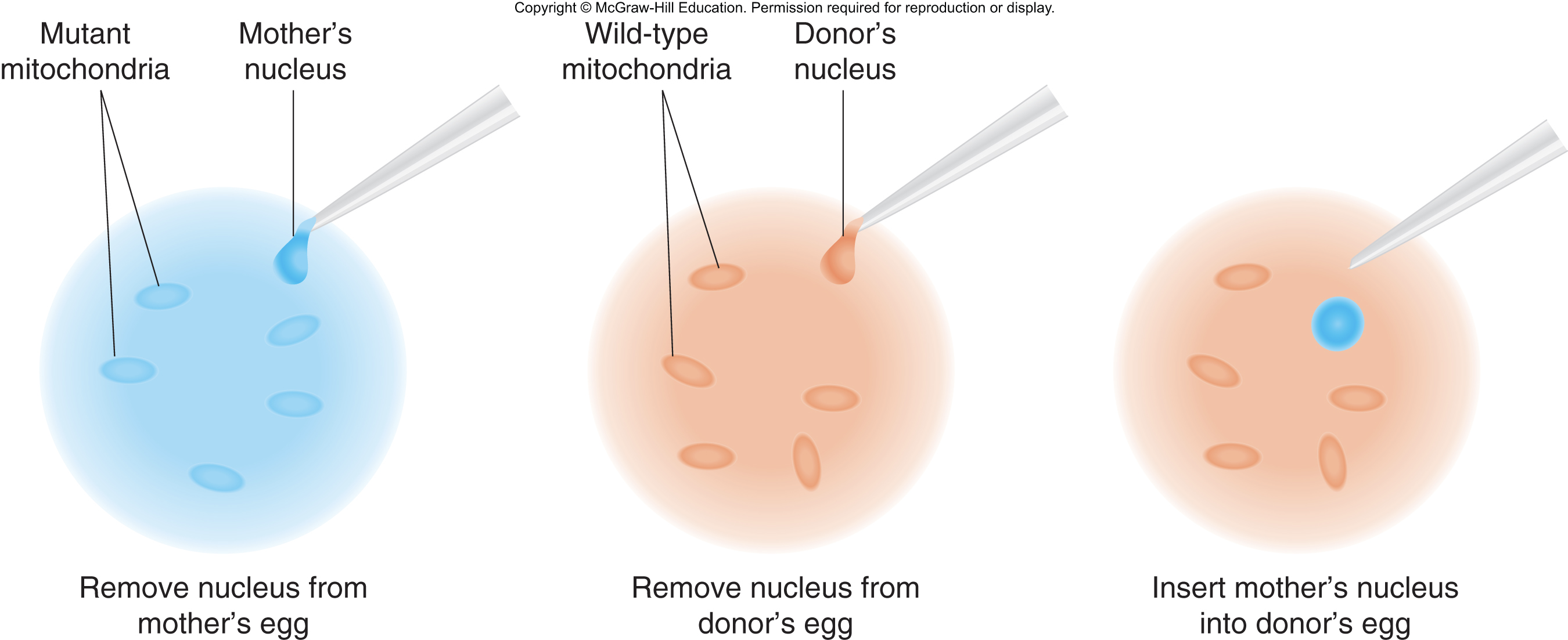
Oocyte nuclear transfer is possible in primates
From class:
- H1 is outside of the core and holds linker DNA around the core
- \