ephe midterm 1
Cells: Basic building blocks of all living things
Membrane
Lipid bilayer surrounding cell/organelle
Genetic Material
e.g., DNA
Cytosolic fluid
Tissues: Similar cells functioning together
4 types of tissues in the body (CMEN): Connective Tissue, Muscle Tissue, Epithelial Tissue, Nervous Tissue
Organ: 2 or more tissues that perform a specialized function
System: Group of organs functioning cooperatively
General Function | Name of Organelle |
|---|---|
BARRIER | PLASMA MEMBRANE |
POWER PLANT | MITOCHONDRIA |
TOWN HALL | NUCLEUS |
INDUSTRIAL SITES | ENDOPLASMIC RETICULUM* |
INDUSTRIAL WORKERS | RIBOSOMES (non-membranous) |
POSTAL FACILITY | GOLGI APPARATUS (close to nucleus) |
etc |
*Rough ER = Attached Ribosomes, Smooth ER = Detached Ribosomes
EPITHELIAL TISSUE
Epithelial cells = “covering” cell
Epitelial Tissue
Sheets of EC that cover all body surfaces,, lines all body cavities and organs, and forms most glands.
ET is almost always found close to CT (Connective Tissue)
ET is classified based on the type of layering
Simple (one layer)
Stratified (multiple layers)
…and based on the shape of the cells
EPITHELIAL TISSUE | SHAPE | SPECIFIC LOCATION | GENERAL FUNCTION |
|---|---|---|---|
Simple Squamous | 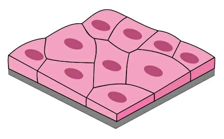 | Mesothelial cells; lining of ventral ventral body cavities, heart and blood vessels, alveoli (lungs), *lots of permeability | Reduce friction, control blood vessel permeability. Perform absorption and secretion. |
Simple Cuboidal | 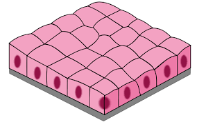 | Glands, ducts, portions of kidney tubules, thyroid gland | Limited protection, Absorption and Secretion |
Simple Columnar | 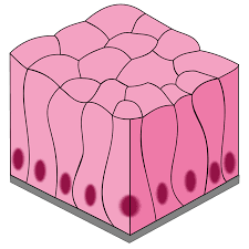 | Lining of the stomach intestine, gall bladder, uterine tubes, and collecting ducts of kidneys | Protection, Absorption and Secretion |
Stratified Squamous | 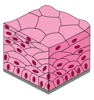 | Surface of the skin, lining of the mouth, throat, esophagus, rectum, anus, and vagina | Provides physical protection against abrasion, pathogens and chemical attacks |
Transitional | 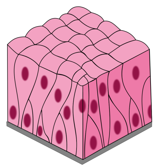 | Urinary bladder, renal pelvis, ureters | Permits expansion and recoil after stretching |
Pseudostratified Ciliated Columnar | 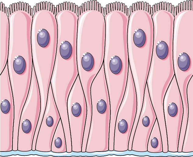 | Lining of the nasal cavity, trachea and bronchi, portions of the male reproductive tract | Protection and secretion |
GENERAL FUNCTIONS OF EPITHELIAL TISSUE
PROTECTION
e.g., Stratified squamous cells
SECRETION
ABSORPTION
EXCRETION/FILTRATION
DIFFUSION
e.g., Simple squamous cells
SENSATION
TRANSPORT
Consider the Following: Why are some cells..?
Larger vs Smaller | Flat vs Full | Cubed vs Round |
|---|---|---|
CHARACTERISTICS OF EPITHELIAL TISSUE
Cellularity: highly packed cells
Polarity: apical (top) and basal (bottom) surfaces
Attachment: Hemidesmosomes attach epithelial cells to protein fibers in basement membrane.
Avascular: without blood cells (EC must obtain nutrients through diffusion and absorption.
Regenerative: cells proliferate (multiply) quickly
ET is always set up with little space and very organized.
EPITHELIAL CANCERS
85% of cancers are cancers of the ET origin and are called carcinomas
EC are highly regenerative and therefore have greater chance of error
GLANDULAR TISSUE
organized collection of secretory EC
Types of Glandular Epithelium
ENDOCRINE GLAND | EXOCRINE GLAND |
|---|---|
|
|
|
|
*Epithelial ducts may release secretions unaltered or they may alter it through reabsorption, secretion, or counter transport.
CLASSIFICATION OF GLANDULAR EPITHELIUM
STRUCTURE OF GLAND | CHARACTERISTICS | EXAMPLE | LOCATION |
|---|---|---|---|
Unicellular |
|
| Respiratory/Digestive Tract |
Multicellular |
|
| Mouth/Skin |
TYPES OF GLAND SECRETIONS:
Serous gland: produces watery solutions mixed with enzymes.
Mucous gland: Secrete glycoproteins called mucins.
Mixed endocrine gland: Mixture of cells that secrete both serous and mucous secretions (e.g., pancreas secreting both insulin and glucagon.
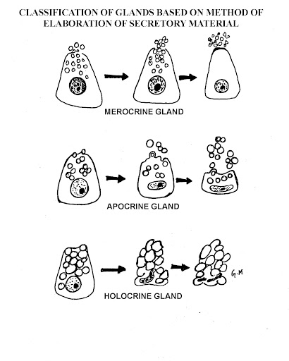
METHOD OF SECRETION
|  |
INTRO TO CONNECTIVE TISSUE
Learning Outcomes:
1. Describe the general functions of connective tissue (CT) and the structural elements found in all CT.
2. Recall the subtypes of CT and classification system.
3. Recognize structural elements common to all CT.
CT Proper
1. Recognize unique characteristics of each subtype of CT Proper including the relative proportion of
cells/proteins/ground substance.
2. Differentiate between fixed and wandering cells found in CT proper and describe functions of those discussed in lecture.
3. Recall and associate each subtype of CT proper with its respective functions and locations.
4. Using the integumentary system as an example, specify the type of epithelial tissue and connective tissue found in the epidermis, layers of the dermis, sebaceous glands, and sweat glands.
Membranes/Fascia
1. Compare and contrast the characteristics and locations of mucous, serous, cutaneous and synovial membranes.
2. Differentiate between superficial fascia, deep fascia, and subserous fascia in terms of location and structure.
Mind Map: Connective Tissue Proper
Central Idea: Connective Tissue Proper
Connective tissue proper is one of the four main types of connective tissue in the body.
It is responsible for providing structural support, connecting and protecting organs and tissues.
CT General Funegctions:
Provides structural framework for organs
Protects internal structure
Connects tissue types
Transports fluid and solutes
Stores energy
Assists the immune system.
EXAMPLES OF CELLS FOUND IN CT PROPER
Fibrocytes: Supports and maintains CT Proper
Fibroblasts: Makes and remodels CT Proper
Produces all connective tissue fibers.
Each fibroblast manufactures
Adipocytes: stores energy
Contains a single lipid droplet that occupies almost the entire cell.
Immune cells
Leukocytes: Various white blood cells
Mast Cells: Stimulate inf. response
Macrophages (clean up cells): phagocytize (process in which a cell uses it’s plasma membrane to engulf a large particle) pathogens and damaged cells
There are fixed and wandering macrophages
Fixed Macrophages: large, amoeboid cells scattered among the connective tissue fibers. They engulf damaged cells, dead cells, and pathogens that enter the tissue.
They release chemicals that attract wandering cells in the body’s defense mechanisms.
Wandering (Free) Macrophages: relatively large phagocytic cells that wander rapidly through the connective tissues of the body.
Termed “Monocytes” when circulating within the blood.
Act as a front-line defense that is reinforced by the arrival of free macrophages and other specialized cells.
Loose Connective Tissue | CHARACTERISTICS | IMAGES |
|---|---|---|
Areolar Connective Tissue
|
| 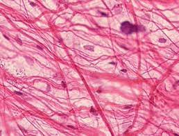 |
Adipose Tissue
|
| 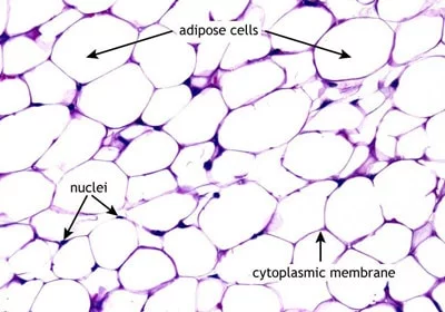 |
Reticular Connective Tissue
|
| 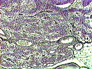 |
Dense Connective Tissue | CHARACTERISTICS | IMAGES |
|---|---|---|
Dense Regular Connective Tissue
|
| 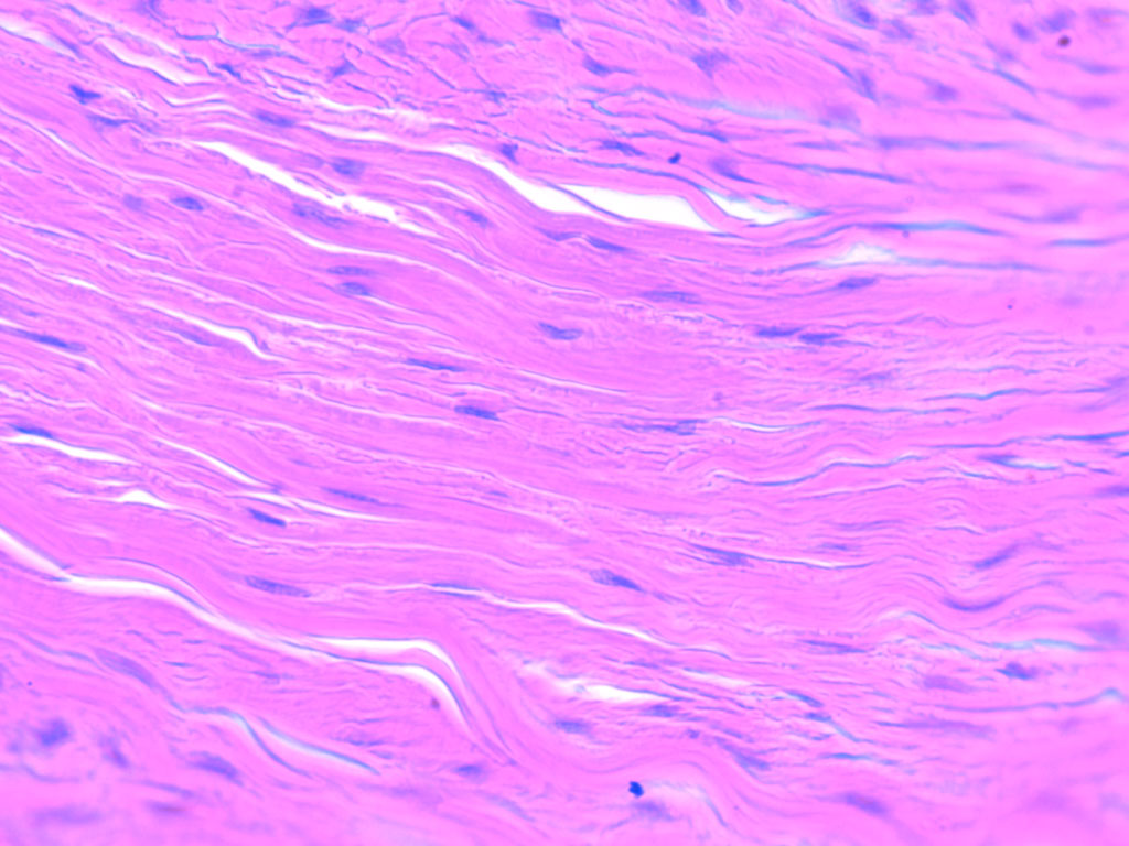 |
Dense Irregular Connective Tissue
|
| 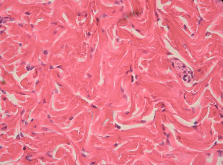 |
Elastic Connective Tissue
|
| 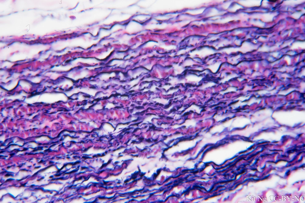 |
Get to Know the Following
Connective tissues are found throughout the body. They are never exposed to the environment outside the body.
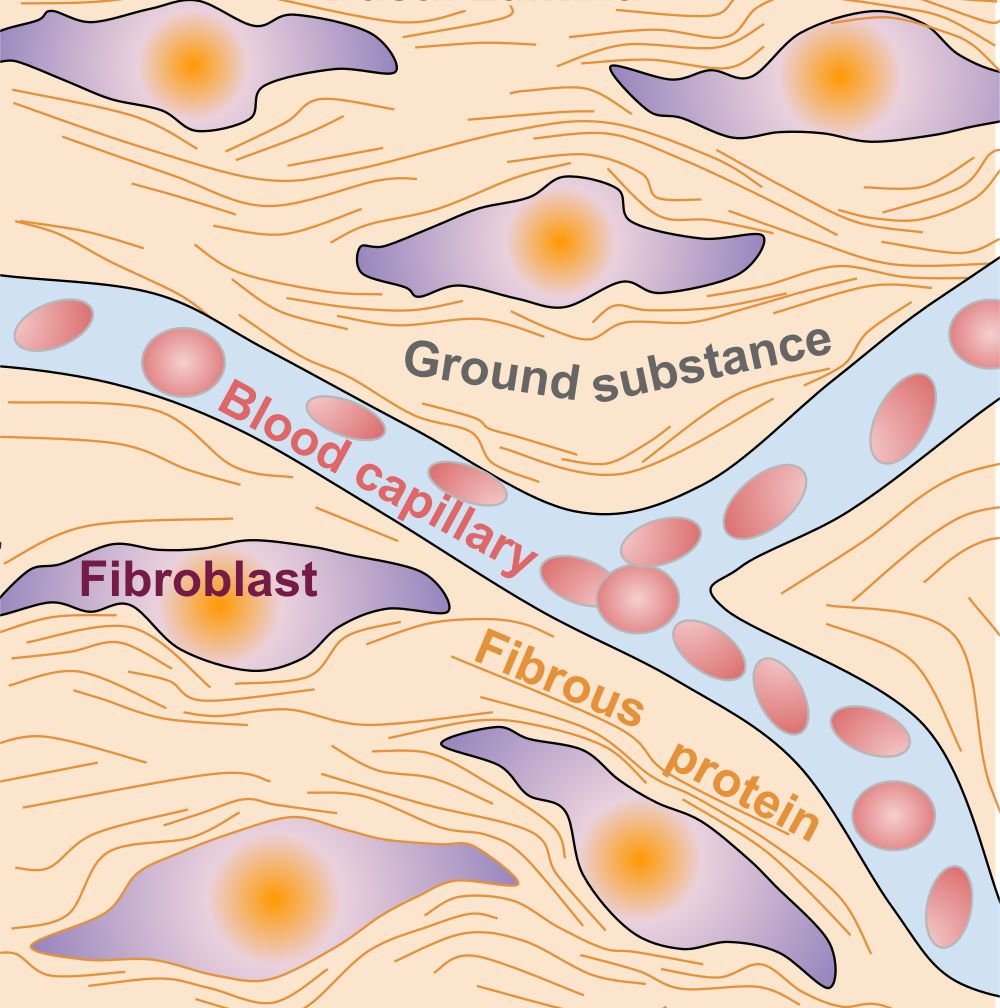
All CT have 3 basic components: (1) Specialized Cells, (2) Extracellular Protein Fibers, (3) A fluid known as Ground Substance.
The extracellular protein fibers and ground substance form the matrix that surrounds the cells.
CT consists almost entirely of extracellular matrix (ECM).
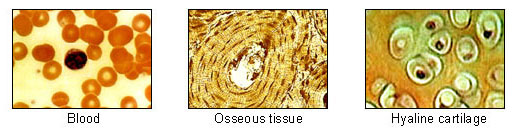
Specialized CT Cells: includes a variety of distinct tissues with specialized cells and unique ground substances that result in wide-ranging properties
adipose
cartilage
bone
blood
lymphatic
Extracellular Protein Fibers: The extracellular matrix (ECM) is the non-cellular component present within all tissues and organs, and provides not only essential physical scaffolding for the cellular constituents but also initiates crucial biochemical and biomechanical cues that are required for tissue morphogenesis, differentiation and homeostasis
collagen
STRENGTH
long, straight, unbranched.
reticulin
STRENGTH
fine collagen fibrils that are shorter, thinner, and lighter (but still strong).
elastic fibers
STRETCH
branching and wavy = stretch and recoil.
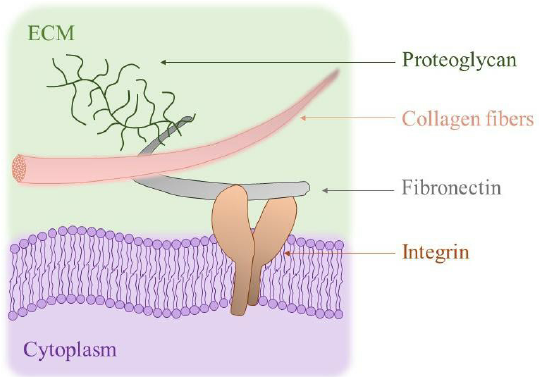
Ground Substance: Gelatinous substrate that lies between cells and fibers of the CT.
cartilage
umbilical cord
secreted by fibroblasts
contains a high concentration of glycosaminoglycans (i.e., hyaluronic acid)
Membranes; Fascia
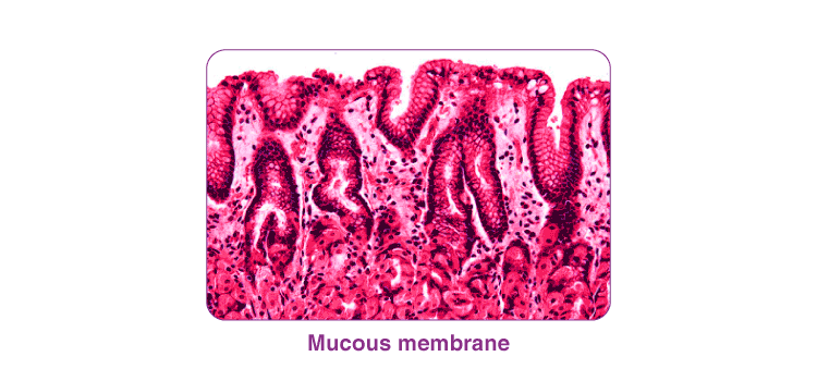
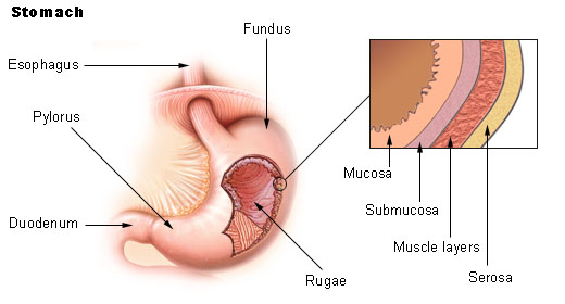
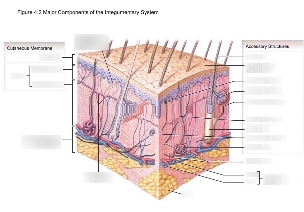
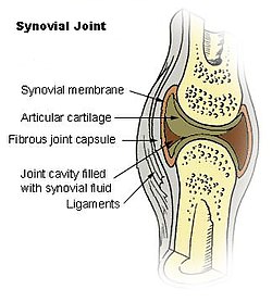
Epithelial Membranes: Epithelial sheet with an underlying CT layer. Forms an internal framework in body.
4 TYPES OF EPITHELIAL MEMBRANES | CHARACTERISTICS | IMAGES |
|---|---|---|
Mucous Membranes:
|
|  |
Serous Membranes:
|
|  |
Cutaneous Membrane (skin):
|
|  |
Synovial Membranes:
|
|  |
Fascia
TYPES OF FASCIA | CHARACTERISTICS |
|---|---|
Superficial Fascia |
|
Deep Fascia |
|
Subserous Fascia |
|
Cartilage
Type of Supporting Connective Tissue (cartilage, bone)
Tough, but flexible, CT that provides support
Key cells: chondrocytes
secrete collagen fibers and chondroitin sulfate (forms matrix)
found in lacunae
Predominant protein fibers: collagen
Matrix: firm gel; does not allow for easy diffusion of nutrients, gases
Avascular
TYPE OF CARTILAGE | FUNCTIONS | LOCATIONS/EXAMPLES |
|---|---|---|
Hyaline Cartilage |
|
|
Elastic Cartilage |
|
|
Fibrous Cartilage |
|
|
Perichondrium:
Separates cartilage from other tissues
Composed of 2 layers:
Perichondrium is vascular!
Fibrous layer: dense irreg. CT
Cellular layer: chondroblasts
Bone
Type of Supporting CT: Osseous Tissue, Strong and dynamic CT that provides support
Key cells: osteoprogenitor cells, osteoblasts, osteocytes, osteoclasts,
Predominant protein fiber: collagen
Matrix: calcium salts (e.g., calcium phosphate)
Vascular!
Bone Matrix:
60-70% bone minerals including insoluble crystals of
calcium phosphate (e.g., hydroxyapatite)~30-40% collagen
Functions: calcium phosphate interacts with calcium hydroxide to make the crystals. As the crystals form they incorporate other calcium salts and ions. The inorganic components enable bone to resist compression (hard and strong)
Collagen is tough but flexible. In bone, collagenous fibers and non-collagenous proteins provide framework for the formation of mineral crystals.
Info I stole from this youtube video:
Bones are composed primarily of an extracellular calcified material called the bone matrix or collagen matrix.
Three main types of cells in bones: osteocytes, osteoblasts, and osteoclasts.
Osteocytes are found in cavities or lacunae between the layers of the bone matrix and assist with bone nutrition.
Osteoblasts are found along the surface of bones and within the bone matrix, responsible for synthesizing and mediating mineralization of the bone matrix.
Osteoclasts are large multinucleated cells responsible for removing calcified bone matrix, allowing for bone turnover and remodeling.
Bones have two main types: compact or cortical bone and cancellous or trabecular bone.
Lamellar bone is arranged in sheets, while woven bone has randomly arranged collagen fibers.
TYPES OF BONE | CHARACTERISTICS |
|---|---|
Compact bone (aka cortical bone) |
|
Spongy bone (AKA trabecular/cancellous bone) |
|
Bone Histology
Functions of the parts of the bone
BONE PART NAME | FUNCTION |
|---|---|
Osteon |
|
Central Canal |
|
Perforating Canal |
|
Canaliculi |
|
Lamellae |
|
Periosteum: (NOTE: no periosteum within synovial joints)
covers bone surfaces & has a fibrous layer
that is superficial & cellular layer that is deep
Endosteum:
membrane lining the inner surface of the compact bone
Osteogenesis:
Fetal development (<8 weeks) begins as hyaline cartilage. After
~8 weeks the bony skeleton starts to to form.
Ossification – process of bone formation that completes early in
adulthood:
a. Intramembranous ossification: bone formed from undifferentiated
sheets of CT
b. Endochondral ossification: bone replaces a hyaline cartilage model
Intramembranous Ossification - bone formed from undifferentiated sheets of CT
Forms directly from sheets of mesenchymal connective tissue (i.e., undifferentiated tissue)
Process starts during fetal development at ~8 weeks in utero.
Specific to the clavicle, mandible, skull, and bones of the face
Ossification is mostly complete after birth with facial bones
ossifying at end of adolescent growth spurt.
Endochondral Ossification (bone replaces hyaline cartilage model)
Begins ~8-12 weeks in utero.
Chondroblasts form hyaline cartilage model - this serves as a template that will be replaced by new bone.
Specific to the bones of the limbs, vertebrae, and hips
Bones grow in length via endochondral ossification and grow in diameter by appositional growth
Endochondral Ossification - 2 main steps
1. Initiation
2. Increasing length
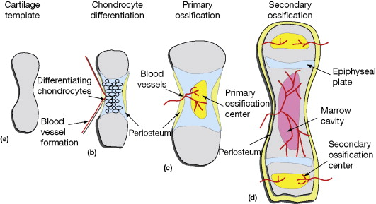
Growth Plates
made of hyaline cartilage
Hyaline cartilage is a flexible and smooth type of cartilage.
Hyaline cartilage provides a cushioning effect, reducing friction between bones.
Injuries to growth plates can lead to growth disturbances or deformities.
Hyaline cartilage lacks blood vessels, nerves, and lymphatics.
Appositional Growth: Increasing Diameter of Bone
Bone enlarges in diameter just deep to periosteum
Occurs concomitant with longitudinal growth AND continues after growth cease
As new bone added to outer surface by osteoblasts, osteoclasts resorb bone on inside enlarging medullary cavity
Factors Regulating Bone Growth
Diet: Calcium, phosphate salts, Vitamin A, C, D
Hormones: calcitriol, parathyroid hormone, calcitonin, growth hormone, thyroxine, sex hormones.
Weight bearing physical activity: particularly high-impact loading and resistance training!