Central Dogma, DNA Replication, and Cell Cycle Flashcards
Flow of Genetic Information
The Central Dogma of Molecular Biology: Describes the detailed pathway and directional flow of genetic information within a biological system.

Pathways of Genetic Information Flow:
DNA Replication: Synthesis of identical DNA copies catalyzed by .
Transcription: Transfer of genetic information from DNA to sense RNA catalyzed by .
Translation: Decoding of sense RNA into a specific functional protein sequence catalyzed by .
Reverse Transcription: Synthesis of complementary DNA from a sense RNA template catalyzed by $ rối\text{Reverse Transcriptase}.\n * **RNA Replication**: Replication of sense RNA to form antisense RNA and vice-versa catalyzed by \text{RNA Dependent RNA Polymerase (RDRP)}.\n \n\n* **Cellular Compartmentalization**:\n * Replication and Transcription occur within the cell nucleus of eukaryotic cells.\n * Processed mRNA is transported across the nuclear membrane into the cytoplasm.\n * Translation of mRNA into functional protein takes place on ribosomes located within the cytoplasm.\n\n# DNA Structure and Chromatin Organization\n\n* **Double Helix Geometry and Grooves**:\n \n * **Major Groove**:\n * Exposes nitrogenous base sequences directly to the external environment.\n * Facilitates sequence-specific binding of regulatory proteins, including transcription factors, polymerases, and nucleases.\n * Helical dimensions: A full helical turn spans \text{3.4}\,\text{nm}\text{10}\text{0.34}\,\text{nm}\text{2}\,\text{nm}.\n * **Minor Groove**:\n * Narrower groove that primarily binds non-sequence-specific molecular agents, including specific antibiotics and fluorescent dyes or stains.\n\n* **Law of Complementary Base Pairing**:\n \n * Operates on a strict lock-and-key chemical principle in DNA and RNA where a purine base selectively binds a specific pyrimidine base through hydrogen bonding.\n * **Purines**: Double-ring nitrogenous bases consisting of Adenine (\text{A}\text{G}).\n * **Pyrimidines**: Single-ring nitrogenous bases consisting of Thymine (\text{T}\text{U}\text{C}).\n * **Hydrogen Bonding Specificity**:\n * Adenine (\text{A}\text{T}\text{2} hydrogen bonds.\n * Guanine (\text{G}\text{C}\text{3} hydrogen bonds.\n * **Chargaff's Rule**: Total purines equal total pyrimidines (\text{Purines} = \text{Pyrimidines}\text{A} + \text{G} = \text{T} + \text{C}.\n\n* **Antiparallel Strand Orientation**:\n 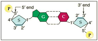\n * The two complementary DNA strands run parallel to one another but in opposite chemical directions (\text{5'} \rightarrow \text{3'}\text{3'} \rightarrow \text{5'}).\n * Chemical end designations are derived from carbon numbering on the deoxyribose sugar backbone:\n * \text{5'}\text{5'} carbon of deoxyribose.\n * \text{3'}\text{-OH}\text{3'} carbon of deoxyribose.\n * Enzymes responsible for nucleic acid synthesis (replication and transcription polymerases) exhibit mandatory directionality, appending nucleotides exclusively to the free \text{3'-OH} terminus.\n\n* **Chromatin Hierarchy and Nucleosome Structure**:\n \n * **Chromatin**: Dynamic nucleoprotein complex composed of DNA, histone proteins, and RNA that performs four core functions:\n 1. Packages genomic DNA into a small nuclear volume.\n 2. Structurally reinforces DNA to facilitate organized segregation during mitosis.\n 3. Prevents chemical and physical DNA damage.\n 4. Controls transcription and translation accessibility.\n * **Nucleosome Structure**:\n \n * The fundamental structural unit of chromatin.\n * **Histone Octamer**: Consists of a protein core containing two copies each of core histones \text{H2A}\text{H2B}\text{H3}\text{H4}.\n * Exactly \text{146}\,\text{bp}\text{1.65} turns around the histone octamer core particle.\n * **Linker DNA**: Spans \text{54}\,\text{bp}\text{H1}).\n * Histones are highly conserved across eukaryotic evolution.\n * Flexible N-terminal **histone tails** extend outward from the core and undergo post-translational modifications that regulate transcription and translation access.\n * **Levels of Structural Compaction**:\n 1. Short region of double-stranded DNA helix (\text{2}\,\text{nm} diameter).\n 2. "Beads-on-a-string" nucleosome chain (\text{11}\,\text{nm} diameter).\n 3. Packed nucleosome chromatin fiber (\text{30}\,\text{nm} diameter).\n 4. Extended chromosome loops (\text{300}\,\text{nm} diameter).\n 5. Condensed section of chromosome (\text{700}\,\text{nm} diameter).\n 6. Entire condensed mitotic chromosome (\text{1,400}\,\text{nm} total width across sister chromatids).\n\n# Protein-Coding Gene Structure and Transcription Mechanics\n\n* **Structure of a Protein-Coding Gene**:\n \n * **Enhancers (Distal Control Elements)**: Regulatory DNA sequences located upstream or downstream of the promoter that bind activator proteins to stimulate transcription initiation.\n * **Proximal Control Elements**: Control elements positioned adjacent to the promoter that modulate transcription initiation efficiency.\n * **Promoter Region**: Binds \text{RNA Polymerase}\text{TATAAA}) where local unwinding of DNA occurs.\n * **Exons**: DNA coding regions that are preserved in mature mRNA and translated into protein sequences.\n * **Introns**: Non-coding DNA sequences intervening between exons that are excised during post-transcriptional RNA processing.\n * **Poly-A Signal Sequence**: Sequence (\text{AATAAA}) providing the signal for transcript cleavage and transcription termination.\n\n* **Transcription Initiation and Chromatin Remodeling**:\n \n * Transcription initiates when the cell receives specific signaling indicating a required protein.\n * Chromatin must decondense to permit transcription factor binding and enzyme access:\n * **Decondensed Chromatin (Transcriptional Activation)**:\n * DNA demethylation catalyzed by demethylase enzymes.\n * Histone acetylation catalyzed by Histone Acetyltransferases (\text{HATs}), neutralizing positive histone charges to loosen DNA wrapping.\n * Activating histone methylation marks (e.g., \text{H3K4me3}\text{H3K36me3}\text{HMTs}).\n * **Condensed Chromatin (Transcriptional Repression)**:\n * DNA methylation at cytosines catalyzed by methyltransferase enzymes.\n * Histone deacetylation catalyzed by Histone Deacetylases (\text{HDACs}).\n * Repressive histone methylation marks (e.g., \text{H3K9me3}\text{H3K27me3}\text{HMTs}\text{HDMs}).\n * **Assembly of Pre-Initiation Complex**:\n \n 1. Activator proteins bind specific enhancer sequences, triggering DNA bending.\n 2. Activators interact with coactivators (\text{SWI/SNF}\text{HATs}) to decondense local chromatin.\n 3. Activators bind the Mediator complex, triggering assembly of \text{RNA Polymerase}\text{TFIID}\text{TFIIB}\text{TFIIF}\text{TFIIE}\text{TFIIH}) at the core promoter.\n\n* **Transcription Elongation**:\n \n * Double-stranded DNA enters \text{RNA Polymerase} through the DNA entry channel and unwinds inside the enzyme.\n * \text{RNA Polymerase}\text{3'} \rightarrow \text{5'} direction.\n * The complementary \text{5'} \rightarrow \text{3'} strand is termed the coding strand because it carries the identical sequence as the synthesized pre-mRNA (except Uracil replaces Thymine).\n * Ribonucleotide triphosphates (rNTPs) enter via the rNTP entry channel and supply energy for synthesis.\n * rNTPs are incorporated into the transcript through synthesis reactions releasing pyrophosphate (\text{PP}_i).\n * Uracil (\text{U}\text{A}\text{T}).\n * The newly synthesized pre-mRNA transcript exits through the RNA exit channel in the \text{5'} \rightarrow \text{3'} direction.\n\n* **Transcription Termination**:\n * The Poly-A Signal Sequence directs \text{RNA Polymerase} to halt transcription and release from DNA.\n * The primary transcript undergoes cleavage downstream of the polyadenylation signal.\n\n* **Post-Transcriptional RNA Processing**:\n * **5' Capping**: Addition of a \text{7-methylguanosine}\text{m}^7\text{G}\text{5'} end.\n * **3' Polyadenylation**: Enzymatic addition of a Poly-A tail comprising \text{50}\text{250}\text{3'} end.\n * **Cap and Tail Functions**: Regulate mRNA export from the nucleus, prevent enzymatic degradation, and promote ribosome binding during translation initiation.\n * **Splicing and Alternative Splicing**:\n \n * Non-coding intron sequences are excised, and coding exons are joined together.\n * Alternative Splicing allows a single gene to code for multiple unique protein isoforms by varying which exons are included in the mature mRNA.\n\n# Translation and Protein Synthesis\n\n* **Ribosomes and Transfer RNA (tRNA)**:\n 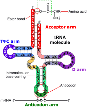\n * **Ribosomes**:\n * Ribonucleoprotein structures composed of ribosomal RNA (rRNA) and structural proteins.\n * Designated as ribozymes because peptidyl transferase activity resides in catalytic rRNA.\n * Composed of a large subunit and a small subunit.\n * Contains three functional tRNA binding sites:\n * \text{A} site (aminoacyl site): Accepts incoming aminoacyl-tRNA.\n * \text{P} site (peptidyl site): Holds peptidyl-tRNA attached to the growing polypeptide chain.\n * \text{E} site (exit site): Releases uncharged tRNA.\n * **tRNA Structure**:\n * Folded RNA molecule conjugated to a specific amino acid via an ester bond at its \text{3'}\text{CCA-3'} sequence).\n * Contains distinct functional loops: Acceptor arm, \text{D}\text{T}\Psi\text{C} arm, and Anticodon arm.\n * Contains a 3-nucleotide anticodon sequence that forms complementary hydrogen bonds with mRNA codons.\n\n* **The Genetic Code**:\n \n * Arranged in triplet codons: sequence of \text{3}\text{1} amino acid.\n * **Degeneracy**: Multiple distinct triplet codons can encode the exact same amino acid (e.g., UCU, UCC, UCA, and UCG all encode Serine).\n * **Start Codon**: \text{AUG} (codes for Methionine).\n * **Stop Codons**: \text{UAA}\text{UAG}\text{UGA}.\n\n* **Translation Initiation Modes**:\n \n * **Cap-Dependent Initiation**:\n 1. Small ribosomal subunit (\text{40S}\text{eIF1}\text{eIF1A}\text{eIF3}\text{eIF5}\text{eIF2-GTP}\text{Met-tRNA}_i^{\text{Met}}\text{43S} pre-initiation complex.\n 2. Cap-binding complex (\text{eIF4E}\text{eIF4G}\text{eIF4A}\text{eIF4B}\text{5'}\text{m}^7\text{G} cap of mRNA.\n 3. The \text{43S}\text{5'} \rightarrow \text{3'}\text{AUG}).\n 4. Initiation factors dissociate, and the large ribosomal subunit (\text{60S}\text{80S}\text{Met-tRNA}\text{P} site.\n * **Cap-Independent Initiation**:\n * Protein complexes bind directly to an Internal Ribosome Entry Site (IRES) near the start codon without \text{5'} cap scanning.\n * Operates under cellular stress conditions (e.g., starvation, viral infection, hypoxia) that normally suppress cap-dependent translation.\n * Mechanisms involve direct RNA binding, ITAF-mediated recruitment, or modified initiation factor combinations (\text{eIF2/5B}\text{Ligatin}\text{MCT-1/DENR}).\n\n* **Translation Elongation Mechanics**:\n \n * Translation proceeds along mRNA strictly from the \text{5'}\text{3'} end.\n * **Step 1**: Initiator tRNA carrying Methionine (\text{Met}\text{P}\text{eEF1}\alpha\text{-GTP}\text{A}\text{eEF1}\alpha\text{-GDP} + \text{P}_i$.
Step 2 (Peptide Bond Formation):

Peptide bonds are synthesized via dehydration synthesis (condensation reaction releasing ).
A peptide bond () forms between the carboxyl group () of the amino acid in the site and the amino group () of the amino acid in the site.
Step 3 (Translocation):
Elongation factor (driven by GTP hydrolysis) advances the ribosome one codon forward.
Uncharged tRNA moves from site to site and exits; peptidyl-tRNA moves from site to site.
Translation Termination:
Occurs when a stop codon (, , or ) enters the site.
Eukaryotic release factor 1 () binds directly to the stop codon.
Release factor promotes ester bond hydrolysis, releasing the completed polypeptide chain.
Ribosomal subunits, mRNA, and uncharged tRNAs dissociate completely.
Post-Translational Modifications (PTM):
Covalent structural modifications appended to cellular proteins following translation:
Acetylation (): Addition of acetyl groups.
Methylation (): Addition of methyl groups.
Phosphorylation (): Addition of phosphate groups to amino acid side chains.
Sulfation (): Addition of sulfate groups.
Glycosylation (): Attachment of carbohydrate oligosaccharide chains.
DNA Replication Mechanics
Overview and Origin Recognition:

DNA replication initiates at specific AT-rich sequences called replication origins (which unwind easily due to hydrogen bonds per pair versus per pair).
Initiator proteins separate double strands, creating replication bubbles flanked by active replication forks.
Replication Initiation and Assembly:
The Pre-Replication Complex (pre-RC) assembles at origins during phase:
Origin Recognition Complex (ORC): Hexameric protein complex that recognizes origin sequences.
Cdc6 and Cdt1: Recruited by ORC to load helicases.
Minichromosome Maintenance Complex (MCM2-7): Hexameric helicase loaded onto DNA with unwinding activity.
Replisome Architecture and Elongation:

Active replisomes assemble at each fork containing MCM helicases, , , and specialized DNA polymerases (, , ).
Single-stranded binding protein (RPA) coats unwound single strands to prevent re-annealing.
Chemical Step:

catalyzes nucleophilic attack of the group on the incoming deoxyribonucleoside triphosphate (dNTP), attaching the nucleotide and releasing pyrophosphate (), which is hydrolyzed to \text{2P}_i$.\n * **Asymmetric Fork Polymerization**:\n \n * \text{DNA Polymerase}\text{3'} \rightarrow \text{5'}\text{5'} \rightarrow \text{3'}.\n * **Leading Strand**: Template runs \text{3'} \rightarrow \text{5'}\text{DNA Polymerase }\epsilon$.
Lagging Strand: Template runs toward the fork; synthesized discontinuously away from the fork by in short Okazaki fragments.
Lagging Strand Mechanism: Template DNA loops around so polymerases read . Primase inserts short RNA primers; extends DNA from primers. RNA primers are removed, gaps filled, and nicks sealed by DNA ligase.
Replication Termination and Telomere Maintenance:

End-Replication Problem: Removal of the terminal RNA primer on the lagging strand leaves an un-replicated gap at the end of newly synthesized DNA because polymerases require a primer. Without intervention, linear chromosomes shorten progressively with each cell division cycle.
Telomerase Action:

Telomerase (a specialized reverse transcriptase carrying its own integral RNA template) binds repetitive telomeric sequences on the overhang of the template strand.
Telomerase synthesizes additional repetitive DNA repeats on the template strand end, expanding it outward.
This extension allows Primase and to complete lagging strand synthesis without deleting vital genomic coding sequences.
Genetic Mutation Categories and Effects
Mutational Inheritance:

Germline Mutations:
Occur in germ cells/gametes (sperm or egg).
Transmitted to offspring; present in every cell of the resulting organism.
Passes mutation to of the organism's gametes.
Somatic Cell Mutations:
Occur in non-germline somatic body cells.
Affects only a localized patch of tissue in the individual organism.
Cannot be inherited by offspring (none of the gametes carry the mutation).
Mutational Origins:
Spontaneous Mutations: Result from intrinsic errors, base mispairings, or tautomeric shifts during DNA replication.
Induced Mutations: Caused by external environmental mutagens (e.g., radiation or chemical agents).

Intercalating chemicals (e.g., Acridine) slip between adjacent base pairs, distorting the double helix and causing insertion or deletion errors during replication.
Base Substitution Point Mutations:

Silent Mutation: Base substitution alters codon sequence, but due to genetic code degeneracy, it codes for the identical amino acid (e.g., TTC TTT; both code for Lysine). Protein structure remains unchanged.
Nonsense Mutation: Base substitution converts an amino acid codon into a premature stop codon (e.g., ATC mRNA UAG = STOP). Leads to truncated, typically non-functional proteins.
Missense Mutation: Base substitution changes codon to specify a different amino acid.
Conservative Missense: Replaces amino acid with one sharing similar chemical properties (e.g., TTC TCC; Lysine [basic] Arginine [basic]). Functional impact is minimal.
Non-conservative Missense: Replaces amino acid with one having distinct chemical properties (e.g., TTC TGC; Lysine [basic] Threonine [polar]). Frequently disrupts folding and protein function.
Frameshift Mutations:
Insertion Mutation:

Insertion of single or multiple nucleotides (not multiples of ) shifts downstream codon reading frames, altering all subsequent amino acids (e.g., His-His-His His-His-His-Thr-Ser-Ser-Ser).
Deletion Mutation:

Deletion of nucleotides shifts reading frames downstream, changing amino acid translation (e.g., His-His-His His-His-His-Leu-Ile-Ile-Ile) and generating nonfunctional proteins.
Eukaryotic Cell Cycle and Regulation
Subphases of the Cell Cycle:
Interphase: Comprises three subphases covering cell growth and genome duplication:
Phase (Gap 1): Duration . Cell active in metabolism and growth, accumulating materials, ATP, and enzymes required for DNA replication. DNA content is .
Phase (Resting Phase): Quiescent state entered from by terminally differentiated or non-dividing cells. Can re-enter upon mitogenic signals.
Phase (Synthesis Phase): Duration . DNA replication duplicates entire genome, doubling DNA content from . Centrioles are duplicated.
Phase (Gap 2): Duration . Complete centriole maturation, synthesis of structural components, and production of mitotic control proteins. DNA content remains .
M Phase (Mitosis and Cytokinesis): Duration . Nuclear division (mitosis) and cytoplasmic division (cytokinesis) separate duplicated chromosomes into two daughter cells, returning DNA content from .
Cell Cycle Control and Kinase Regulation:

Driven by Cyclin proteins and Cyclin-Dependent Kinases (CDKs):
Phase: regulates initiation of pre-replication complex assembly and centrosome duplication.
Phase: activates origin firing and blocks re-replication.
Transition: (Mitosis Promoting Factor / MPF) triggers entry into mitosis.
Cell Cycle Checkpoints:

Checkpoint: Blocks entrance into phase if DNA damage is detected.
Checkpoint: Halts DNA replication if genomic damage or stalled forks occur.
Checkpoint: Blocks entrance into phase if DNA replication is incomplete or damaged.
Checkpoint (Spindle Assembly Checkpoint): Blocks anaphase onset if sister chromatids are improperly assembled on the mitotic spindle.
Mitosis Stages and Cytokinesis Mechanics
Overview of Mitosis Stages:

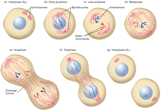
Nuclear division divided into five continuous phases: Prophase, Prometaphase, Metaphase, Anaphase, and Telophase.
Prophase and Prometaphase:

Prophase:
Chromatin coils and condenses tightly into distinct, visible chromosomes consisting of two identical sister chromatids joined at centromeres.
Nucleolus breaks down and disappears.
Centrosomes migrate toward opposite cell poles, radiating aster microtubules and forming the early mitotic spindle.
Prometaphase:
Nuclear envelope breaks down into membrane fragments.
Spindle microtubules enter the nuclear region.
Kinetochore protein complexes assemble at chromosome centromeres and attach to kinetochore microtubules.
Chromosome Compaction Structure:

DNA double helix wraps around histones forms chromatin chain coils into supercoiled loops condenses into duplicated mitotic chromosome () flanked by telomeres at end tips and centromere at center junction.
Mitotic Spindle Architecture:

Organized by two centrosome poles containing centriole pairs.
Three distinct microtubule sets:
Astral Microtubules: Anchor spindle poles to the cell cortex.
Kinetochore Microtubules: Attach directly to kinetochore protein complexes on chromatids.
Polar (Non-kinetochore) Microtubules: Overlap at the cell equator to push poles apart.
Metaphase and Spindle Assembly Checkpoint (SAC):

Metaphase Alignment: Chromosomes align in single file along the metaphase plate (equatorial plane).
Mitotic Checkpoint Regulation:

Unattached kinetochores accumulate inhibitory proteins (Mad1, Mad2, Bub1, Bub3, BubR1, CENP-E) that bind and sequester Cdc20, keeping Anaphase Promoting Complex (APC) inactive.
When all kinetochores are attached under bipolar tension, Mad2/Bub complexes dissociate, releasing Cdc20 to activate APC ().
Active APC ubiquitinates securin for degradation, activating separase enzyme to cleave cohesin rings holding sister chromatids together.
Anaphase:

Anaphase A: Cohesin cleavage allows sister chromatids (now individual daughter chromosomes) to separate. Kinetochore microtubules depolymerize at kinetochore ends (losing tubulin subunits), pulling chromosomes toward opposite poles powered by motor proteins.
Anaphase B: Polar microtubules slide past each other and elongate, increasing separation between spindle poles.
Telophase and Cytokinesis:

Telophase: Daughter chromosomes reach opposite poles and decondense into diffuse chromatin. Nuclear envelope re-forms around each genome set; nucleoli reappear, and spindle fibers disassemble.
Cytokinesis: Cytoplasm divides along the metaphase plate driven by a contractile ring composed of actin and myosin filaments, pinching the parent cell into two genetically identical daughter cells.