Kinesiology Lesson 7 and 8
Definitions
Myocardium - Specialized muscle tissue that forms the heart → is a double pump that has the left and right sides.
Pulmonary Circulation - main function of the RIGHT SIDE of the heart, pumps the deoxygenated blood, (has returned) to the LUNGS.
Systemic Circulation - Role of the LEFT SIDE of the heart to pump the OXYGENATED blood, (returned from the lungs) to the BODY.
Arteries - blood vessels that carry blood Away from the heart, they are used in both pulmonary (deoxygenated blood is carried by arteries to the lungs from the right side) and systemic circulation (oxygenated to the body from the left side of the lungs)
Arterioles - they are smaller branches of the arteries that lead to capillaries (gas exchange) THEY ARE SURROUNDED BY SMOOTH MUSCLE WHICH MEANS THAT THEY ARE THE PRIMARY SITE OF VASCULAR RESISTANCE
Veins - blood vessels that carry blood towards the heart, they are used in both pulmonary (oxygenated from lungs to the left side) and systemic (deoxygenated from the body to the right side)
Capillaries - SMALLEST OF THE blood vessels, they help to enable exchange of gases such as CO2, O2, H2O, waste and nutrients substances between the blood and tissues of the body. There are millions of capillaries in the lungs and all over the body.
Components of Blood
Plasma - fluid component of blood (mostly water), it keeps the blood from sticking up
There are 3 types of blood cells
Red blood cells (Called erythrocytes) - they are made in the bone marrow and transports the O2 and the CO2 and transports waste and nutrients. They also contain hemoglobin.
Hemoglobin - very important for oxygen transportation
White Blood Cells (Called leukocytes) - they destroy foreign element (protect the body and are very important in the immune system)
Platelets - they regulate blood clotting, they are the mesh material hat stops blood flow from out of the body
The Cardiac Cycle
This is the series of events that occurs through one heart beat.
2 Phases:
Diastole - relaxation and the ventricle is filling with blood
Systole - contraction is when the heart contracts and ejects the blood
Blood Pressure
force exerted by the blood against the walls of the arteries and the vascular vessels. MEASURED IN THE 2 PHASES AND IN MILLIMETRES OF MERCURY (mmHg)
This is systole (maximum pressure when contracting) over diastolic (minimum pressure when relaxing)
Skeletal Muscle Pump - low pressure in veins (going up and does not have the heart to pump blood around) → problem for the cardiovascular system, so they’re are skeletal muscle pumps that helps to return the blood to the heart, with each contraction the blood is pushed back to the heart
Flow of Blood Through the Heart
https://www.youtube.com/watch?v=jBt5jZSWhMI&t=46s
Superior and inferior vena cava → right atrium → tricuspid valve → right ventricle → pulmonary semi-lunar valve → out of pulmonary arteries → lungs → returns through pulmonary veins → left atrium → bicuspid valve → left ventricle → pumped out through aortic semilunar valve → into aorta → body through systemic circulation → gas exchange in the body → back to the heart through pulmonary circulation
Electrical Conduction System of the Heart
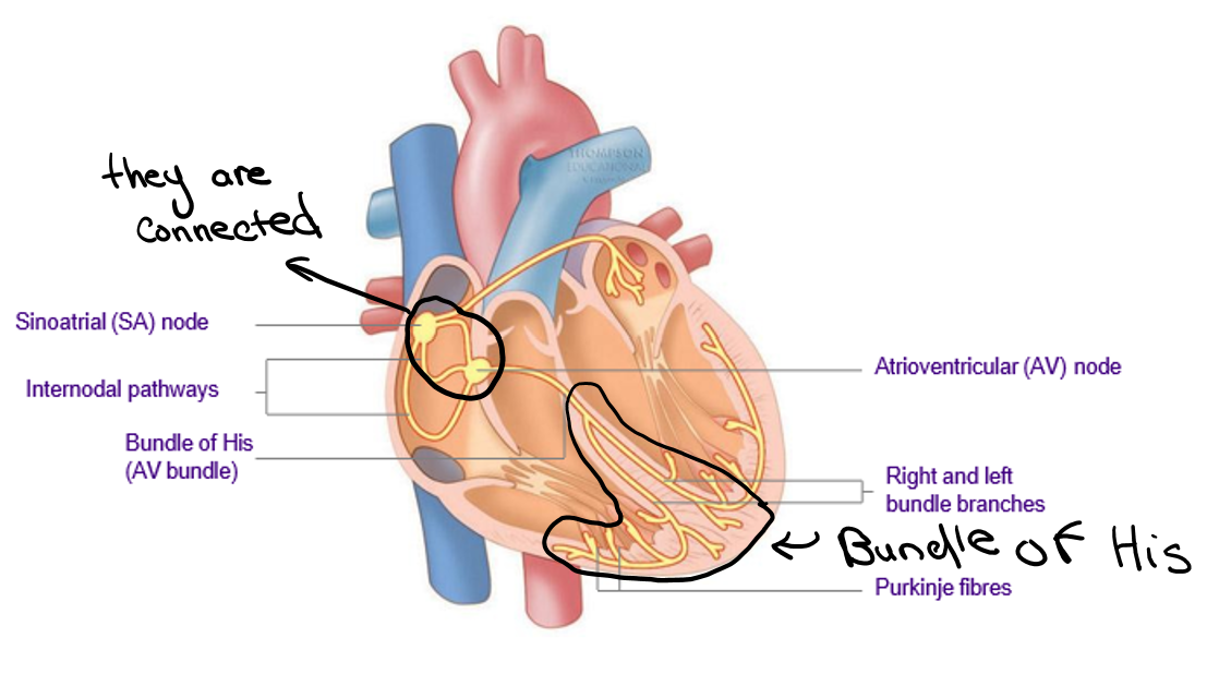
All cardiac muscle cells are excitable, with electrical stimulation they will all contract → SPECIALIZED TISSUES IN THE HEART THAT ARE IMPORTANT FOR REGULATION AND COORDINATION OF THE ELECTRICAL ACTIVITY, this contracts the heart which leads to the pumping of blood
Definitions
Sinoatrial node (SA node) - specialized region of tissue that is found in the RIGHT atrium where electrical signals that lead to contraction are initiated, this makes the top of the heart to contract and force blood to the lower heart which then gets the AV node to contract from there → this is what makes the heart start to contract (from 60 to 100 bpm for an adult) → THE PACE MAKER OF THE HEART
Atrioventricular node (AV node) → Specialized tissue that transmits the electrical signal from the top (atria) to the bottom (ventricles) and and to the Ventricular septum (the thing circled at the bottom), which has the bundle of his in it. The bundle of his allows for synchronized contraction of ventricles. (There are branches in both the right and left ventricles)
Electrocardiogram (ECG) - graphical representation of the electrical sequence of events occurring with each contraction → show how your blood is flowing, there are different waves (under Electrical Activity of the Heart)
Depolarization: The discharge of energy that accompanies the transfer of ions across the cell membrane, this is the heart getting ready to contract. This is the wave of the SA node that starts this all (the p wave) . They are getting less negative and contracting. This is when the electrical impulse is generated, (because sodium ions are rushing into the cells, which makes the cell more positive because it was negative before hand).
Repolarization: The cells of the heart return to the negatively charged state which means that it is relaxing (this is because the positive k+ is leaving the cell making it more negative) (https://www.google.com/search?sca_esv=68708e1df62cd149&sxsrf=ADLYWIL72n1akTJfWNbW6p0ROsoohVpXsg:1737597507705&q=depolarization+vs+repolarization&udm=7&fbs=AEQNm0Aa4sjWe7Rqy32pFwRj0UkWd8nbOJfsBGGB5IQQO6L3J7pRxUp2pI1mXV9fBsfh39LpAWJ-Nb3mi2m4EiVUszBibUgYBXGmUct3yVHr_9JSnE288fo6RrD78oXAmOKgB50q3R9TnL6GiT-TSc4e50gY12NCMPCqq1khedYIKxEeV2qq9hPSLEOebufY8kQJKNoKafpkMpjAihYoouDfUWxmeoLDTg&sa=X&ved=2ahUKEwidjNaR34qLAxV1F1kFHdKfDN4QtKgLegQIEBAB&biw=718&bih=823&dpr=2#fpstate=ive&vld=cid:2fbbe5ce,vid:6ulvh_ItKcw,st:0)
Bradycardia: easily observed characterizes after training which is a heart beat that is 60bpm or less
Tachycardia: heart rate that is more the 100bpm at rest
Electrical Activity of the Heart

P wave: this is the depolarization through the atria (creates the action potential for the heart to contract) → to get the blood to the ventricles
P-R interval: this is the small delay that is happening when the electronic charge is moving from the SA node to the AV node, then the bundle of his which makes the ventricles to contract which leads to the QRS complex → leads to ventricular depolarization
QRS complex: this is the depolarization of the ventricles (bottom), contracts up to allow for the blood to get to the lungs and body
ST Segment: this is when the repolarization happens and allow for the heart to become negatively charged again
T-Wave: the period of recovery where the SA node recharges and allows for another cycle to begin
T wave: this represents the repolarization of the ventricles (relaxation, now contracted it allows the atria to fill up again and allows for the SA node to recharge to send a new signal)
Effects of Exercise
Regular aerobic (in oxygen) exercises → improvement of the efficiency of the system at rest and in exercise
During exercise there are cardiovascular dynamics (which is the dramatic changes that occur) → the heart and vessels are constantly trying to adapt to the ever-changing requirements of the body during exercise
some factors of cardiovascular dynamics are:
cardiac output (Q) - amount of blood pumped by the heart per minute
blood pressure (BP)
distribution of blood flow (where it goes)
oxygen consumption (VO2)
If you have a larger heart, there are an increase number of capillaries, increased diameter of the coronary arteries, and increase in blood volume
Coronary Circulation
this is the system of vessels that supply white and red and essential material via the blood to the heart and the muscles itself
Serios medical health issues occur if there isa narrowing or blockage of blood vessel which would restrict the blood flow
Heart attack - myocardial infarction is a result of part of the section of the heart muscle become blocked due to PLAQUE BUILD UP or another reason
Atherosclerosis - is a coronary artery disease that involves a narrowing if the coronary arteries the is an accumulation of plaque (hard deposit of cholesterol), on/in-between the lining of the blood vessels
Causes of Coronary Artery Disease (italic is controllable)
poor diet
smoking
elevated blood lipids
hypertension
family history
physical inactivity
stress
excessive alcohol
Each one increases the risk independently but when combined the chance is magnified
The Respiratory System
Main Function and 3 main parts
nose/mouth, trachea, and lungs
supplies O2 to blood (gas exchange in the capillaries, using diffusion)
removes the CO2 from the blood (gas exchange in the capillaries)
regulate blood pH by removing the CO2 so there will not become the carbonic acids
Definitions
External Respiration - what occurs with the lungs of the exchange of O2 and CO2
Internal Respiration - the exchange of gases at the tissue level → through diffusion of the O2 being delivered and the CO2 being removed
Cellular Respiration - the process that the cells do in the mitochondria using O2 to create energy
2 Main Zones of the Respiratory System (all controlled by smooth muscle)
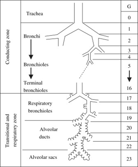
Conductive zone - filters air into the lungs
consists of - mouth, nose, pharynx, larynx, trachea, primary and secondary bronchi, and tertiary bronchiole, and terminal bronchioles
Respiratory zone - where the gas exchange occurs
consists of - bronchioles, alveolar ducts, and alveolar sacs
Mechanics of Breathing
Inspiration - ACTIVE PROCESS, there are contractions of many respiratory muscles thus there is an expenditure of a significant amount of energy, this increases the lung volume and follows the contraction of the diaphragm and intercostal muscles (muscles in-between the lungs).
Expiration - PASSIVE PROCESS, as in quietly breathing out and does not use as much energy or ACTIVE as in forcing the air out of your lungs, this just the relaxation of your diaphragm and intercostal muscles (muscles in-between the lungs).
Gas Exchange - there are about 300 million alveolar sacs that are surrounded by a web of capillaries → they are 1 cell thick so there is a very short distance for gases to diffuse
Control of Ventilation → this is just breathing (controlled by contraction and relaxation of muscles) → they are stimulated from the central nervous system (CNS) → Everything is associated with the overall need of O2, metabolic processes, muscle activity, and production of CO2 → very complex and need many forms of feedback from specialized sensory systems to the neural control centers within the brain
Diffusion → high to low gradient → concentration gradient
Oxygen Transport (ALL A PROCESS)
O2 → absorbed into lungs and carried to the peripheral tissues
CO2 → moves from the blood into the alveoli then is exhaled from the body
VO2 → difference between the amount of CO2 in the artery and vein reflects the amount of the O2 that is delivered to the muscle
The Cardiorespiratory Effects of Exercise
Rest-To-Exercise Transition
The delivery of the O2 to the working skeletal muscle is through a combination of physiological mechanisms → NOT INSTANTEOUS, there is a lag which is when the O2 deficit occur→ YOU THEN HAVE TO RELY ON THE ANAREROBIC (NOT O2) when you are getting transitioning to the steady-state of oxygen consumption then is achieved.
There are steps to getting to the steady state from where you were and in that lag there is a O2 deficit
O2 Deficit - as you start to work out your muscle uses more O2 which brings you into a deficit (This is the difference between the O2 required to preform a task and the actually oxygen consumed before reaching the new stead state → trained get to this steady state faster)
Transitions
The Oxygen is required to perform a task → this is the amount of oxygen that is needed for your body to sustain a specific level of activity, ie running → your muscles need a specific amount of oxygen
Oxygen is actually consumed prior to reaching the new steady state: when you start an activity it take time for your body to catch up to the oxygen demand so your breathing, heart, and oxygen have a lag and an adjustment period called the transition phase.
Ventilatory Threshold - a state in which ventilation increases much more rapidly than workload (this is with the heavy breathing)
Lactate Threshold - this is the point where blood lactate concentration begins to increase, the amount of lactate that is produced is still the same amount as your body can remove
Onset of Blood Lactate Accumulation
this is when the levels of lactate begin to accumulate rapidly in the blood, this is when the lactate is now being produced at a higher rate then it being removed at, when you are trained then this would shift to the right on a graph which means that your body is more efficient.
VO2 Max
the maximal rate of oxygen consumption
Is the maximum volume (V) of O2 that the human body can use in 1 minute per kilogram of weight, when breathing air at sea level
Asthma
spasm of smooth muscle that is line of the respiratory system
over secretion of mucous
swelling of the cell lining in the respiratory tract
Factors that can lead to an attack:
exercise
allergic reaction
contaminates
stress
Asthma can be controlled by different medications → you can still be an Olympic-level athlete and able to compete with it
COPD
general term that describes a family of diseases that leads to a dramatic reduction in the airflow through the respiratory system → there would be a plaque build up in the lungs that can be caused by smoking
people with COPD have shortness of breath (dyspnea) when doing normal activities
Treatment:
medications
For more extreme cases: supplemental oxygen therapy and respiratory muscle training