Topic 3: Neurons and Neurotransmission
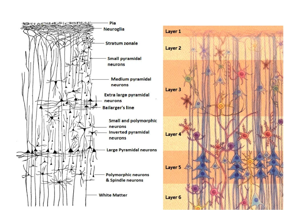
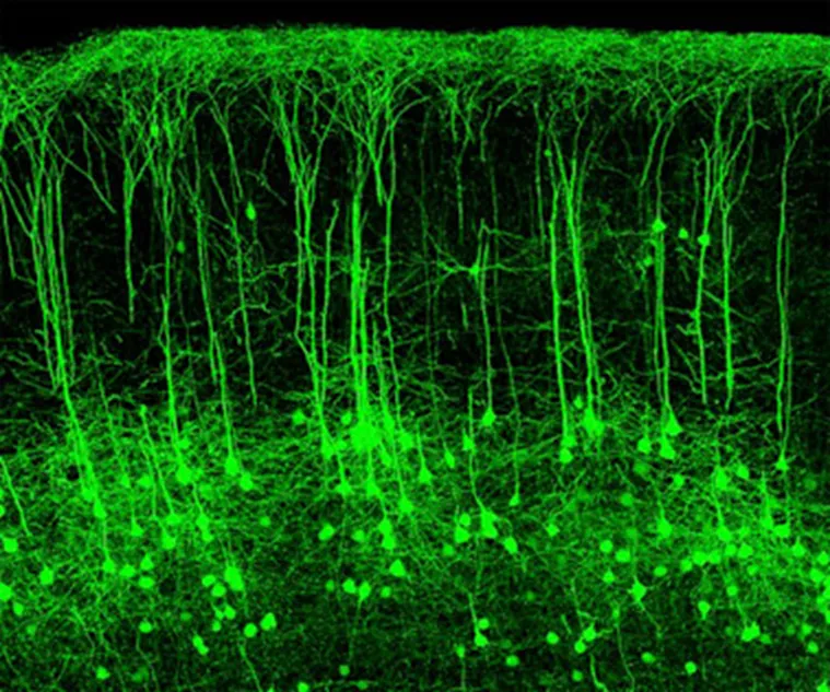
Has 5 layers, neurons concentrated 5mm from surface of brain.
Layer one is most dorsal, layer 5 is most ventral.
Layer 4 receives input mainly from the thalamus, which collects sensory information.
Layers 5 and 6 are the output neurons, send information to other parts of the brain.
Neurons
Neurons are located:
Packed a 5mm from cortical surface.
Also in brain regions beneath the cortical surface, such as the caudate putamen or striatum.
Neurons consist of
The cell body, or soma.
Dendrites (Latin for tree branches), which receive incoming signals from other neurons.
Axon hillock, where the soma connects to the axon.
Axon, which initiates and propagates action potentials or nerve impulses toward the axon terminals.
Ribosomes can be found here, suggesting protein synthesis can take place in an axon
Axon terminals, which releases neurotransmitters when activated.
Some neurotransmitters can even release two neurotransmitters, but by convention neurons are referred to by their dominant neurotransmitters, for example glutamatergic.
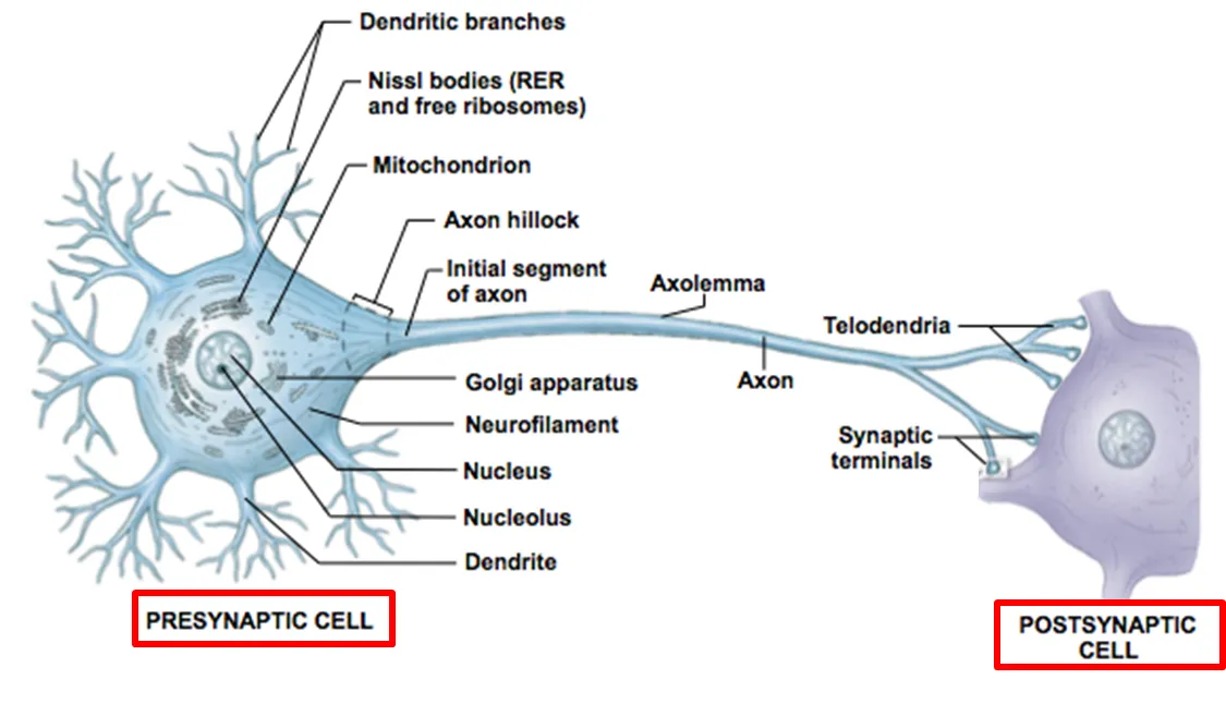
Presynaptic neuron is the term for the first neuron in the sequence, which releases neurotransmitters to the postsynaptic neuron.
Terms are relative.
Each spine on a neuron is a connection to another neuron.
Action potential generation
At rest, the inside of neurons are more negatively charged than the outside, ranging from around -80 to -50.
This is known as hyperpolarisation.
This hyperpolarisation is maintained by, using transport proteins:
Active ion transport - Axon’s actively transport two ions, K+ and Na+, against their concentrations gradients.
This is done using sodium-potassium exchange pumps, which are transmembrane proteins that bring K+ ions in and pump out Na+ ions.
They have ATPase activity.
For every 3 Na+ ions pumped out, only 2 K+ ions are brought in, contributing to the uneven distribution.
Facilitated diffusion - The active transport causes a high K+ concentration inside the axon, and a high NA+ concentration in the tissue fluid outside the axon. This leads to facilitated diffusion occurring.
However, the membrane is selectively permeable, and is about 100x more permeable to K+ ions.
This is due to the channels that diffuse the ions being able to open and close in response to voltages across the membrane (known as voltage-gated), and during resting potential most of the Na+ channels are closed, while only some K+ channels are.
Hodgkin and Huxley worked discovered the action potential mechanism using squids.
Won Nobel prize in Medicine and Physiology in 1963.
Discovered this using ion movements.
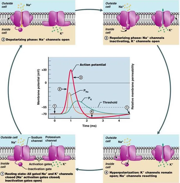
1 - A stimulus generates energy, and once over the -55 mV threshold value the voltage-gated sodium channels in the axon membrane open.
2 - Depolarisation: An increasing amount of Na+ ions enter the axon via chemiosmosis, causing the positivity of the axon to increase. This opens more voltage-gated sodium channels, and therefore even more Na+ ions can enter.
Depolarisation is the temporary reversal of potential difference, where the inside is less negative than the outside as an action potential is transferred.
3 - The potential difference reaches +40 mV from -70 mV, which is the action potential.
The Na+ is aiming to reach it’s equilibrium point, but it never does this.
4 - Repolarisation: Once this +40 mV is reached, the voltage-gated sodium channels close and K+ ions diffuse out via their concentration gradient. The positivity inside the membrane decreases.
These work based on a ball-and-chain mechanism, and are inactivated/ blocked from functioning once membrane is fully depolarised.
5 - Hyperpolarisation - More K+ ions diffuse out than Na+ diffuse in, causing the potential difference to be more negative than resting potential.
The sodium-potassium exchange pump pumps K+ ions back in and Na+ ions back out, restoring the resting potential.
At the original action potential site, sodium channels are inactivated and unable to reopen until resting potential is re-established.
This prevents a new action potential from being triggered, therefore preventing the action potential from moving backwards in the axon, keeping in in one direction.
This is the absolute refractory period.
For the next 5-10 ms, strong enough impulses can cause an action potential to start.
This occurs while sodium-potassium transfer pumps are still restoring resting potential.
This is known as the relative refractory period.
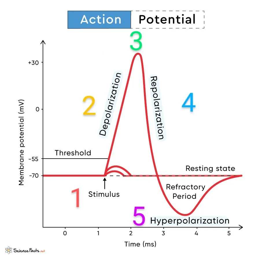
There are also graded potentials which disobey the ‘all or nothing‘ rule by increasing on size based on stimulus.
These can be found in the retina.
Signal transmission
The all or nothing law states that:
If stimulus intensity does not reach the threshold value, no action potential is generated.
If initiated, however, it will always be the same size of and always remains this size as no energy is lost in transmission.
Increases in stimulus intensity don’t increase action potential, but increase the frequency of action potential generation.
This is done to allow neurons to act as a filter for minor stimuli, therefore not overloading the brain with nervous impulses.
There are types of summation, when action potentials are built up to:
Temporal summation - Where multiple action potentials are passed on, building up to reach the threshold value.
Spatial summation - Several pre-synaptic neurons contribute to the same post-synaptic neuron, building up to an action potential.
These can occur simultaneously.
Limiting factors
Speed of conduction:
Myelination - Insulates the axon, therefore increasing transmission.
Myelinated nerve fibres only depolarise at areas of low resistance, which is the nodes of Ranvier, which is where the voltage-gated ion channels occur. This is therefore where Na+ ions enter.
This leads the action potential to seemingly jump from node to node, which is known as saltatory conduction.
This also prevents ions from leaking out of the membrane, as myelin traps them in.
It is rapid due to nodes of Ranvier being 1 mm apart.
Multiple sclerosis is a demyelinating disorder.
Axon diameter - Bigger axons have less resistance on the inside, allowing for faster ion movement.
Dendrites are limited by:
If a charge is injected, charge is lost easily due to leakiness.
The length constant is the distance over which voltage decreases by 63%
Time constant - Time for the voltage to increase or decrease by 63%.
Synapses
There are two types of synapse:
Electrical - these neurons directly touch and have what is referred to as openable windows.
They are attached by gap junctions, and can share transmission via passing on of sodium ions.
These can be found in the retina.
Extremely fast, but allow for no modulation or complex transmission.
Chemical - the gap is 20 nm, which is too large for the impulse to jump.
It is the majority of synapses.
Axon branches lie close to dendrites of other neurons without touching, and the impulse is instead transmitted via neurotransmitters.
These chemicals diffuse across the synaptic cleft, from the pre-synaptic membrane of one neuron to the post-synaptic neuron of an adjacent neuron, which transmits the impulse.
Slower, but allow for modulation and more complex communication.
Chemical synapses
This has 4 steps:
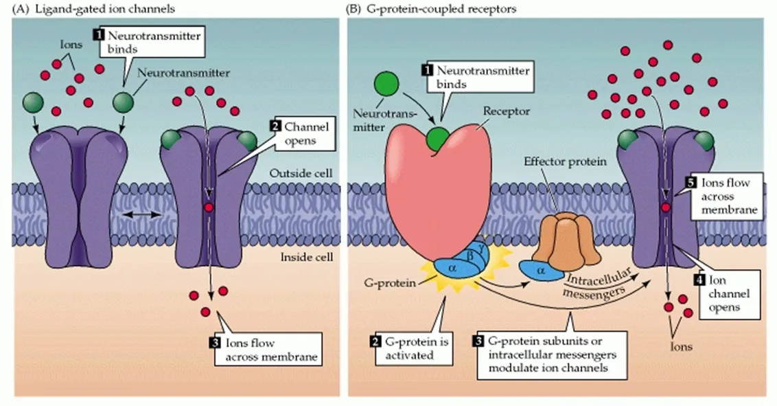
When an action potential reaches the pre-synaptic end bulb, the membrane permeability is altered, which opens voltage-dependent calcium channels.
These diffuse into the end of the bulb, down a concentration gradient from the synaptic cleft.
This influx of Ca+ causes snare proteins which attach synaptic vesicles in the pre-synaptic bulb to the membrane to activate, bringing vesicles closer until fusion occurs.
This releases the neurotransmitter via exocytosis, into the synaptic cleft.
This then diffuses across the cleft to bind to a receptor - an intrinsic protein - in the post-synaptic membrane.
This causes the receptor protein to change shape, which opens a channel to allow Na+ ions to diffuse in down a concentration gradient, from the synaptic cleft.
This depolarises the post-synaptic neuron, and once depolarised above the threshold value an action potential is generated.
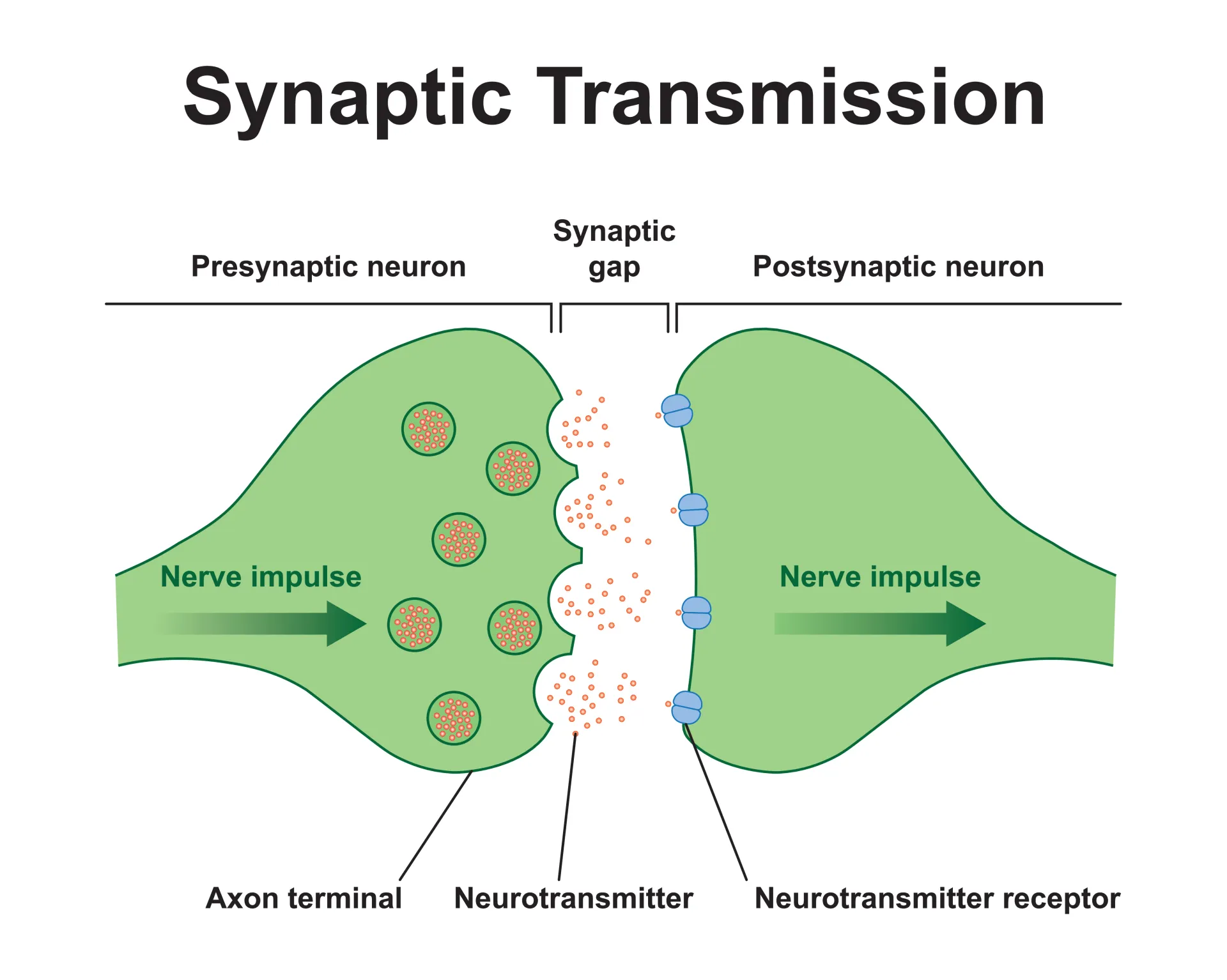
There are two types of receptors:
Ligand (neurotransmitter)-gated ion channels/ ionotropic - There are 5 of these on a membrane. They become ion channels that allow, normally sodium, ions to flood in.
This depolarises/hyperpolarises the axon.
Faster, but less range of actions.
G-protein-coupled receptor/ metabotropic: The G protein is activated and can either interact with a separate effector protein to open a separate ion channel or activate secondary messengers.
5% of human genome codes for GPCRs.
Incredibly useful as they amplify signals, therefore not much neurotransmitter binding is needed.
For example, 1 neurotransmitter can activate secondary and tertiary messengers.
Adenine monophosphate (AMP) is an example of a secondary messenger that causes sleepiness, it’s attachment to receptors is blocked by caffeine.
Targeted by 45% of industry drugs, such as SSRIs.
Slower, but more range of actions.
There are multiple methods to stop neurotransmitter action:
Active reuptake - A molecule in presynaptic neuron takes in any surplus neurotransmitters.
Prozac and other SSRIs blocks these reuptake proteins.
Enzymatic degradation - The neurotransmitter is broken down in the cleft.
Cholinergic synapses breaks down acetylcholine via acetylcholinesterase, and choline and acetate is recycled back into presynaptic cleft to make more acetylcholine.
Types of neurotransmitters
Glutamatergic neurons package and transmit glutamate.
Glutamate binds to metabotropic receptors.
NMDA receptors are widely studied, as they have many special features:
They are ligand-gated.
They have multiple binding sites for other ions and neurotransmitters than glutamate.
At rest, they are blocked by magnesium and ions.
They generate EPSPs.
They are involved in synaptic plasticity.
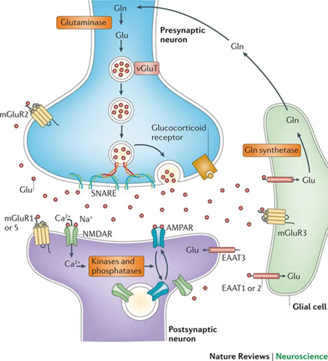

GABA is similar to glutamate as it relies on glial cells to absorb the excess, and this is used to make glutamate.
Glutamate is also a precursor for GABA creation.
GABA-A has 5 subunits, and GABA binding causes Cl- to move into the neuron.
This hyperpolarises the neuron, causing the threshold value to be harder to reach.
Uses an ionotropic receptor.
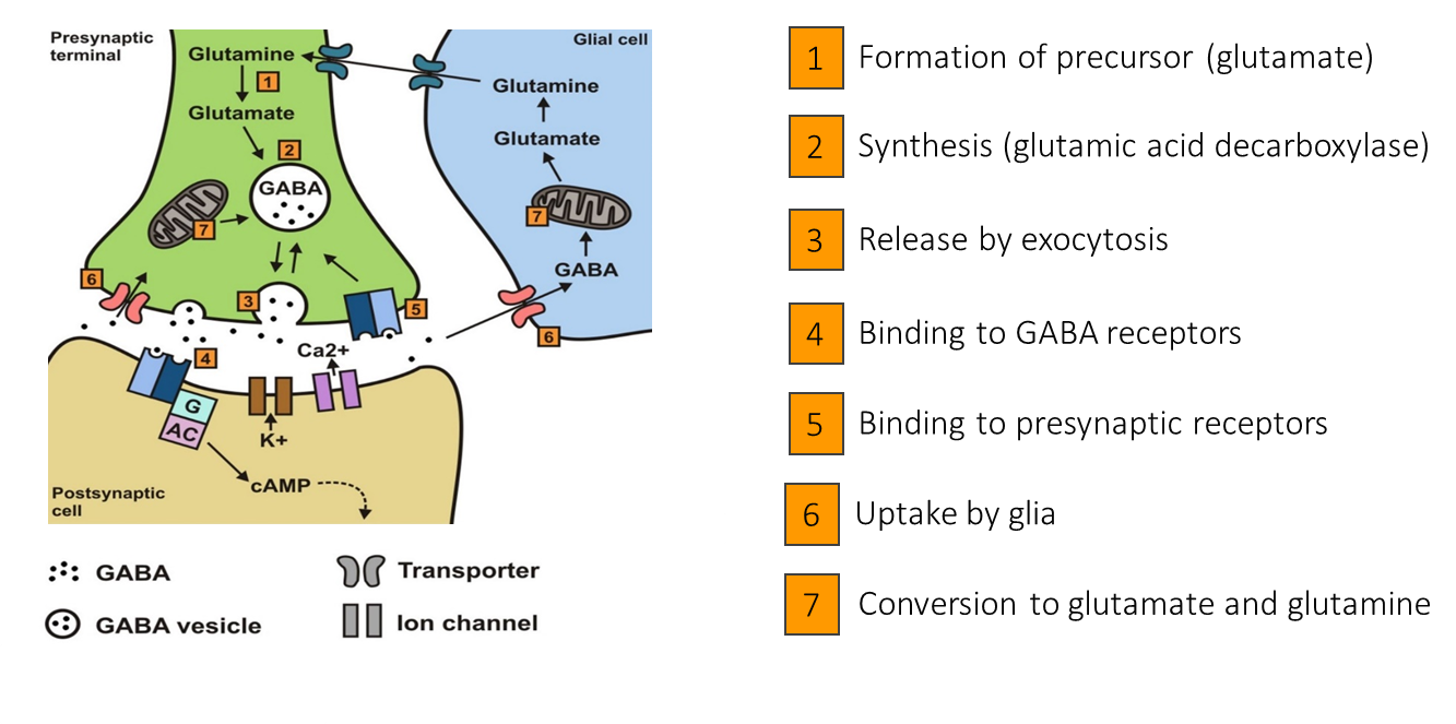
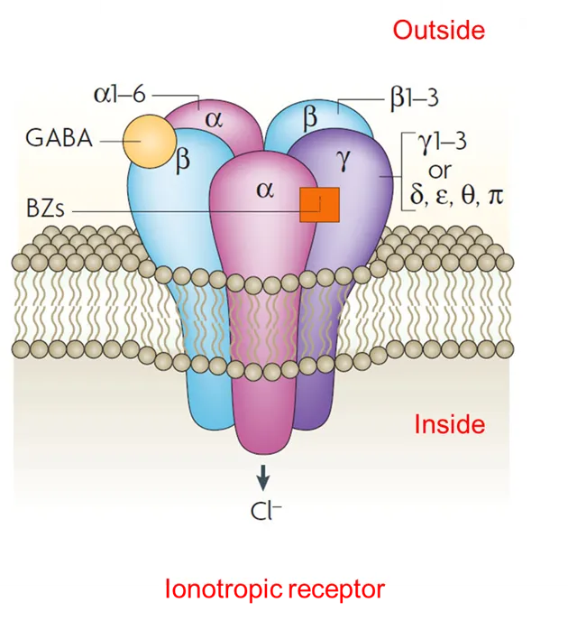

Dopamine can modulate GABA and glutamate activity.
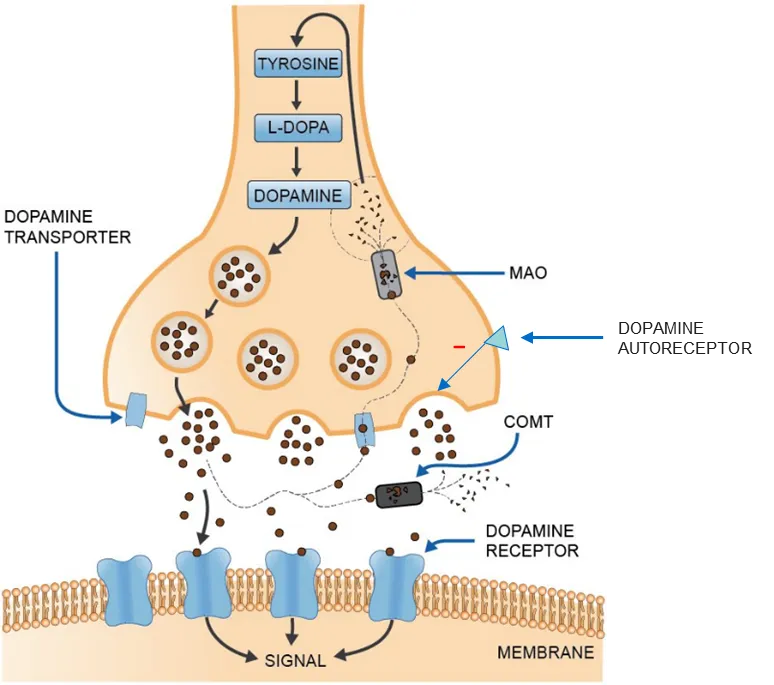
Dopamine is reabsorbed to lower presence.
Degeneration of dopamine neurons in substrata nigra occurs in Parkinson’s.
Parkinson's vs normal brain.
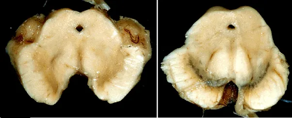
Dopamine is involved in reward learning (Schultz):
Reward causes high motivational response, and high amounts of dopamine are released.
Once learned, the conditioned stimulus generates dopamine.
This is used for predicting rewards.
If reward doesn’t come, dopamine activity is reduced.
This is what causes prediction errors, which stops dopamine from activating when this stimulus is presented again.
Dopamine is involved working memory, such as phone numbers.
It has been found to be important in short term spatial memory as well as the maintenance and manipulation of information in working memory.
Excitation: inhibition
Across cerebral cortex, there is a balance between inhibition and excitation.
Blue is excitation, red is inhibition.
A constant ratio.
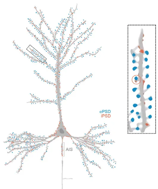
Reduction of GABAergic neurons causes overexcitation, and in post mortem examinations this has been found in SCZ patients.
This has also been found in those with ASD.
When born, GABA is excitatory and remains this way for first 2 weeks of life.
This shift is known as the polarity shift.
Caused by delayed expression of a transporter protein.
Chloride is high inside neurons at birth, and leaves the neuron when GABA attaches, which causes EPSPs.
Babies therefore have overactive brains, making seizures more common and less serious.
Prenatal infections can prolong this shift, causing issues.
Types of neurons
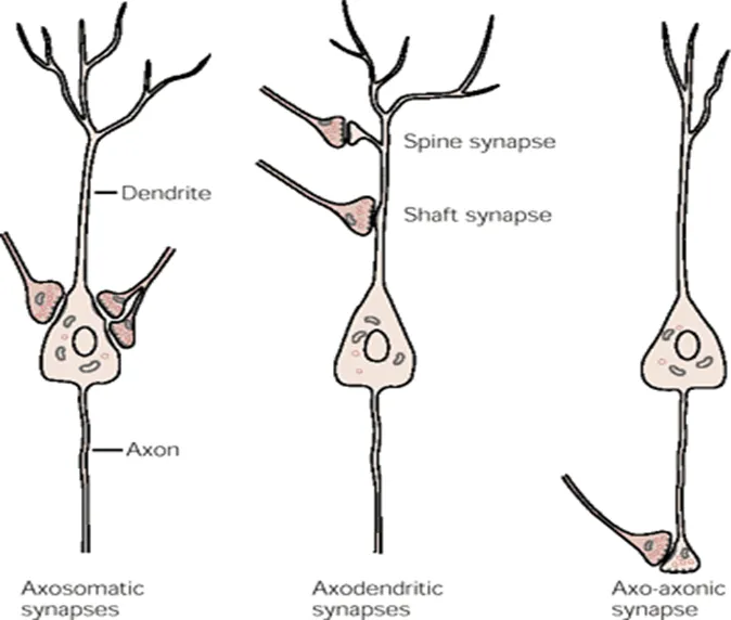
Three types of neurons based on locations of synapses:
Axosomatic - Occurs if the axon synapses directly with the soma of the postsynaptic neuron.
Axodendritic - Occurs when the axon synapses with a dendrite. Most common.
Can either inhibit by decreasing postsynaptic membrane potential, or excite by increasing postsynaptic membrane potential.
This is known respectively as inhibitory post-synaptic potential (IPSP), or excitatory post-synaptic potential (EPSP).
Axoaxonic - Occurs when the axon synapses another axon, often inhibitory.
Suppress neurotransmitter release and therefore neurotransmission.
This is known as inhibitory postsynaptic potential, or IPSP.
Neuron growth
It is still not known whether neurotransmitters can be replaced.
If this occurs, it must be via stem cells as neurons cannot replicate.
Some areas of the brain have been found to generate new neurons, such as the hippocampus.
Neurons are actually pruned in teenage years, and this pruning can be faulted leading to an imbalance in excitation and inhibition.
This has been linked to conditions such as ASD and SCZ.
Visual system
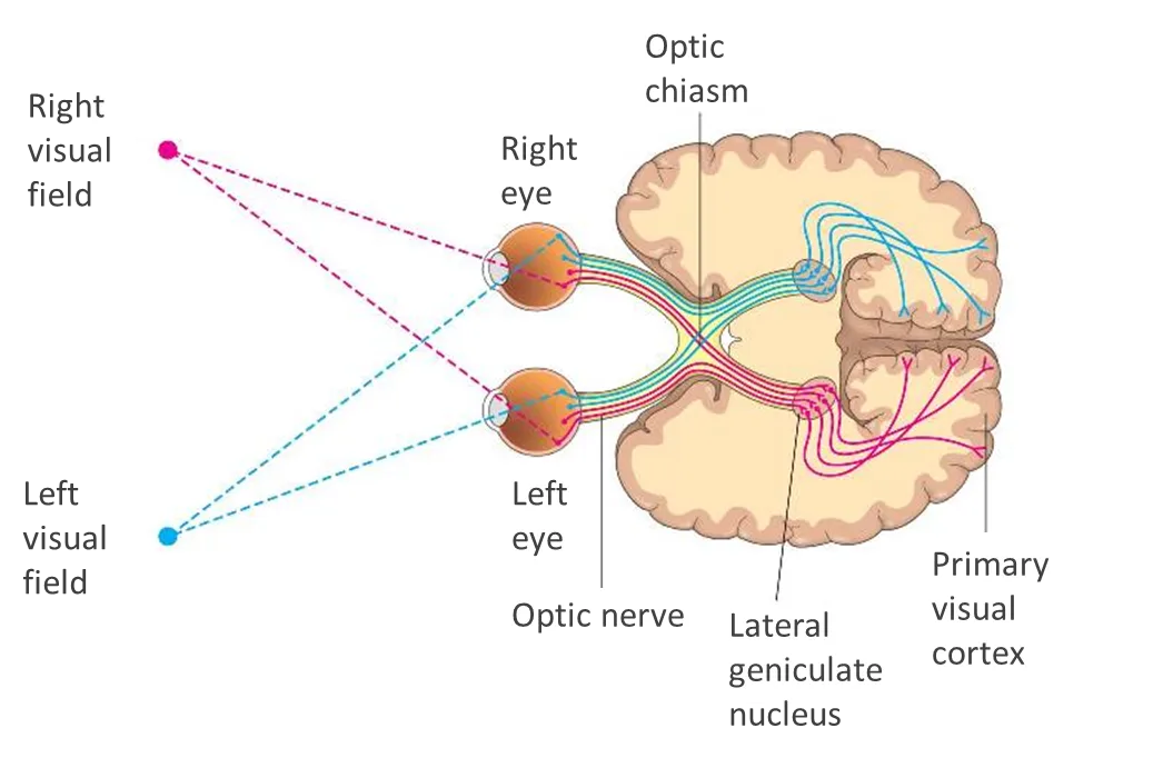
Information conveyed through optic nerve, makes an obligatory synapse in the thalamus, in a region known as the lateral geniculate nucleus.
Map is created in the primary visual cortex.
Special neurons in the primary visual cortex:
Simple neurons responds with action potentials when it stimulus moving in one direction.
Known as orientation columns, in cerebral cortex that are tuned to direction or orientation of stimuli.
Complex neurons are activated when stimuli move in multiple directions.
Noradrenaline is released from the locus coeruleus when we need to pay attention.
Schultz discovered that dopamine is released to the frontal lobe and in the basal ganglia when we receive a reward for the first time.
With classical conditioning, the conditioned stimulus also releases dopamine, believing there will be a reward.
Hebb discovered that neurons that fire together have strengthened connections, known as a Hebbian process.
One theory is plasticity related proteins (PRP), which are released between neurons to strengthen connections.
This is the cause of brain plasticity.