Exam 1
1. Fundamentals of Anatomy and Physiology
Levels of Organization
Atoms, Molecules, Cells, Tissues, Organs, Organ Systems, Organisms
Characteristics of Life
Reproduction: Passing genetic information to offspring.
Growth and Differentiation: Increasing cell size or number; cell specialization in multicellular organisms.
Metabolism: Acquiring and utilizing energy to fuel cellular processes.
Response: Processing internal and external environmental stimuli.
Biomolecules
Carbohydrates: Composed of sugar molecules; used in cellular respiration to produce ATP energy.
Proteins: Composed of amino acids; function in transport, structure, enzymatic activity, and support.
Lipids: Fats used for connective tissue, insulation, and long-term energy storage.
Nucleic Acids: Composed of nucleotides; store and process genetic information.
Anatomical Terminology
Directional Terms:
Anterior: Toward the front.
Posterior: Toward the back/behind.
Medial: Toward the body's midline.
Lateral: Away from the body's midline.
Proximal: Toward an attachment site.
Distal: Away from an attachment site.
Anatomical Planes:
Midsagittal: Divides the body into equal left and right halves along the midline.
Parasagittal: A cut parallel to the midsagittal plane.
Frontal: Divides the body into anterior and posterior sections.
Transverse: Divides the body into superior and inferior sections.
Body Cavities:
Cranial Cavity: Holds the brain.
Vertebral Canal: Holds the spinal cord.
Thoracic Cavity: Chest region; includes the pleural cavities (lungs) and pericardial cavity (heart).
Abdominal Cavity: Holds the stomach, liver, spleen, and intestines.
Pelvic Cavity: Holds the urinary bladder and reproductive organs.
2. Cytology and Mitosis
Cell Theory
Cells are the building blocks of all life.
Cells are produced by the division of pre-existing cells.
Cells are the smallest structural units performing all vital functions.
Cell Structure and Organelles
Cell Types:
Prokaryotic Cells: Contain only non-membranous organelles.
Eukaryotic Cells: Contain both membranous and non-membranous organelles (found in animals, plants, and fungi).
Organelle Functions:
Nucleus: Houses DNA, produces mRNA and ribosomes.
Ribosomes: Read mRNA to synthesize proteins.
Rough Endoplasmic Reticulum (RER): Primary site of protein synthesis.
Smooth Endoplasmic Reticulum (SER): Synthesizes lipids and detoxifies toxins.
Golgi Apparatus: Modifies, packages, and directs proteins for secretion or intracellular use.
Mitochondria: Performs cellular respiration to generate ATP from glucose.
Cytoskeleton: Network of protein fibers providing structure, shape, movement, and transport.
Plasma Membrane and Transport
Structure: Phospholipid bilayer with hydrophilic phosphate heads and hydrophobic lipid tails. Projections called microvilli increase surface area.
Integral Proteins:
Channel: Allows specific ions to enter or exit.
Gated Channel: Requires specific activation to open for ion transport.
Receptor: Binds to extracellular chemical messengers (e.g., hormones).
Enzyme: Catalyzes breakdown of chemical messengers and other molecules.
Cell Identity Marker: Distinguishes self-cells from foreign cells.
Cell Adhesion Molecule (CAM): Binds cells together to form tissues.
Transport Mechanisms:
Passive Transport: Movement down a concentration gradient (high to low concentration) without energy input. Includes diffusion and osmosis (movement of water).
Active Transport: Movement against a concentration gradient (low to high concentration), requiring ATP energy.
Selective Permeability: Regulates entry and exit of molecules through membrane mechanisms.
Intercellular Attachments
Tight Junction: Interlocking cell adhesion proteins fuse membranes with minimal space.
Desmosome: Strong connection utilizing plaques, anchoring cytoskeleton, and CAMs, maintaining a small intercellular gap.
Gap Junction: Intercellular connection with direct pore channels allowing rapid passage of ions and molecules.
Mitosis and Cell Division (Ordered Process)
S Phase: DNA duplication occurs.
Prophase: Nuclear membrane breaks down to release chromatin/DNA.
Metaphase: Chromosomes align along the equatorial middle of the cell.
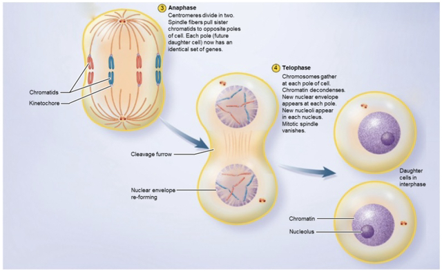
Anaphase: Cytoskeletal spindle fibers pull chromosomes to opposite poles.
Telophase: Two new nuclei form around separated chromosomes.
Cytokinesis: Cytoplasm divides, separating into two individual daughter cells.
3. Histology: Tissues, Glands, and Membranes
Primary Tissue Types
Epithelial: Functions in protection, absorption, and secretion.
Connective: Supports, binds, and protects body structures.
Muscle: Contracts to generate mechanical movement.
Nervous: Transmits rapid electrical signals for sensory processing and motor commands.
Epithelial Tissue Classifications
Cell Shapes:
Squamous: Irregular, flattened shape.
Cuboidal: Cube or square shape.
Columnar: Tall, pillar-like shape.
Layering:
Simple: Single layer of cells.
Stratified: Multiple layers of cells.
Surfaces & Basement Membrane:
Apical Surface: Faces the lumen or external environment.
Basal Surface: Underside of epithelial tissue attached to basement membrane.
Lateral Surface: Intercellular boundaries between adjacent epithelial cells.
Basement Membrane: Connects epithelium to underlying tissue; contains the basal lamina (secreted by epithelium) and reticular lamina (secreted by connective tissue).
Glands and Secretion Methods
Gland Types:
Exocrine Glands: Release secretions onto surfaces or into lumens.
Endocrine Glands: Release hormones into interstitial fluid or bloodstream via exocytosis.
Unicellular Glands: Single cell secretors (e.g., goblet cells).
Multicellular Glands: Multi-celled glands forming sheet or tube structures.
Serous Exocrine Glands: Secrete watery solutions filling body cavities.
Mucous Exocrine Glands: Secrete mucins to form slippery mucus.
Secretion Mechanisms:
Eccrine (Merocrine): Direct secretion via exocytosis without cell loss or damage.
Apocrine: Product is shed bound in vesicles from apical cytoplasm.
Holocrine: Secretion builds up until entire cell lyses and dies.
Connective Tissue Classifications
Connective Tissue Proper:
Loose Connective Tissue:
Areolar: Ground substance rich; composes upper papillary dermis.
Adipose: Fat storage tissue.
Reticular: Reticular fiber matrix supporting organs like the liver and spleen.
Dense Connective Tissue:
Regular: Collagen fibers aligned in parallel arrangements.
Irregular: Unpatterned, multidirectional collagen fibers (dermis).
Fluid Connective Tissue:
Blood: Watery plasma matrix carrying RBCs, WBCs, and platelets.
Lymph: Interstitial fluid matrix carrying lymphocytes and macrophages.
Supporting Connective Tissue:
Cartilage: Chondroblasts harden matrix with sulfate (elastic, hyaline, fibrous).
Bone: Osteoblasts mineralize matrix with calcium phosphate.
Tissue Membranes
Membrane Composition: Epithelium supported by underlying connective tissue.
Cutaneous Membrane: Skin; 5-layered epithelial boundary providing protection, absorption, and secretion.
Mucous Membrane: Lines respiratory and digestive tracts; secretes mucus to trap pathogens or protect against autodigestion.
Serous Membrane: Lines internal cavities; secretes serous fluid to diminish friction.
Synovial Membrane: Lines joint cavities; secretes lubricating synovial fluid.
4. Integumentary System
Layers of the Epidermis (Deep to Superficial)
Stratum Basale: Deepest mitotic layer.
Stratum Spinosum: Interlocking spiny layer.
Stratum Granulosum: Keratin production layer.
Stratum Lucidum: Clear layer found exclusively in thick skin (palms and soles).
Stratum Corneum: Superficial layer composed of flat, dead keratinized cells.
Epidermal and Dermal Cells
Keratinocytes: Main epidermal cell; produces tough, waterproof keratin protein.
Melanocytes: Produce melanin to shield nuclei from UV radiation.
Tactile (Merkel) Cells: Sensory cells involved in perceiving light touch.
Dendritic (Langerhans) Cells: Mobile macrophages carrying out phagocytosis.
Fibroblasts: Produce collagen fibers; essential for tissue repair and scar formation.
Mast Cells: Produce histamine to dilate blood vessels and trigger inflammation.
Dermal and Subcutaneous Structures
Dermis: Middle layer of connective tissue.
Papillary Layer: Superficial region with dermal papillae creating epidermal ridges (fingerprints).
Reticular Layer: Deep, dense connective tissue layer above hypodermis.
Hypodermis: Subcutaneous adipose tissue layer.
Integumentary Structures:
Sebaceous Gland: Produces sebum/oil for skin lubrication.
Arrector Pili Muscle: Smooth muscle contracting to erect hair for thermal insulation.
Hair Follicle & Hair Receptor: Hair structure embedded in tissue wrapped with sensory nerves to detect movement.
Sensory Nerve Fibers: Communicate tactile, thermal, vibrational, and pain stimuli.
Motor Nerve Fibers: Send brain signal commands to cutaneous glands and smooth muscles.