muscular system Ai generated
articulations(joints)- where 2 bones interconnect
bones are relatively inflexible so necessary to allow movement
reflect compromise between the need for strength verses the need for mobility
A. three functional categories
synarthrosis- no movement
ex: sutures and gomphosis (teeth to mandible)
amphiarthrosis -little movement
ex: symphysis & sendesmosis
synarthrotic and amphiarthrotic joints- relatively simple structure
diarthrosis- free movement
ex: synovial (long bones in upper and lower bones)
complex in structure
symphysis- cartilage between 2 pubic bones
syndesmosis- ligament between tibia and fibula
Lt1- synovial joints are freely moveable diatheses containing synovial fluid components of synovial joints (greatest ROM)
A. articular cartilages
resemble hyaline cartilages- matrix contains mire water cimparatively
have no perichondrium
slick and smooth, to reduce friction
are separated by thin film of synovial fluid
B. synovial fluid
similar in composition to group substances
produced at the synovial membrane
circulates from areolar tissue to joint cavity
percolates through articular cartilages
C. joint capsule
dense and fibrous
may be reinforces with accessory structures (tendons & ligaments)
continuous with periosteum of each bone
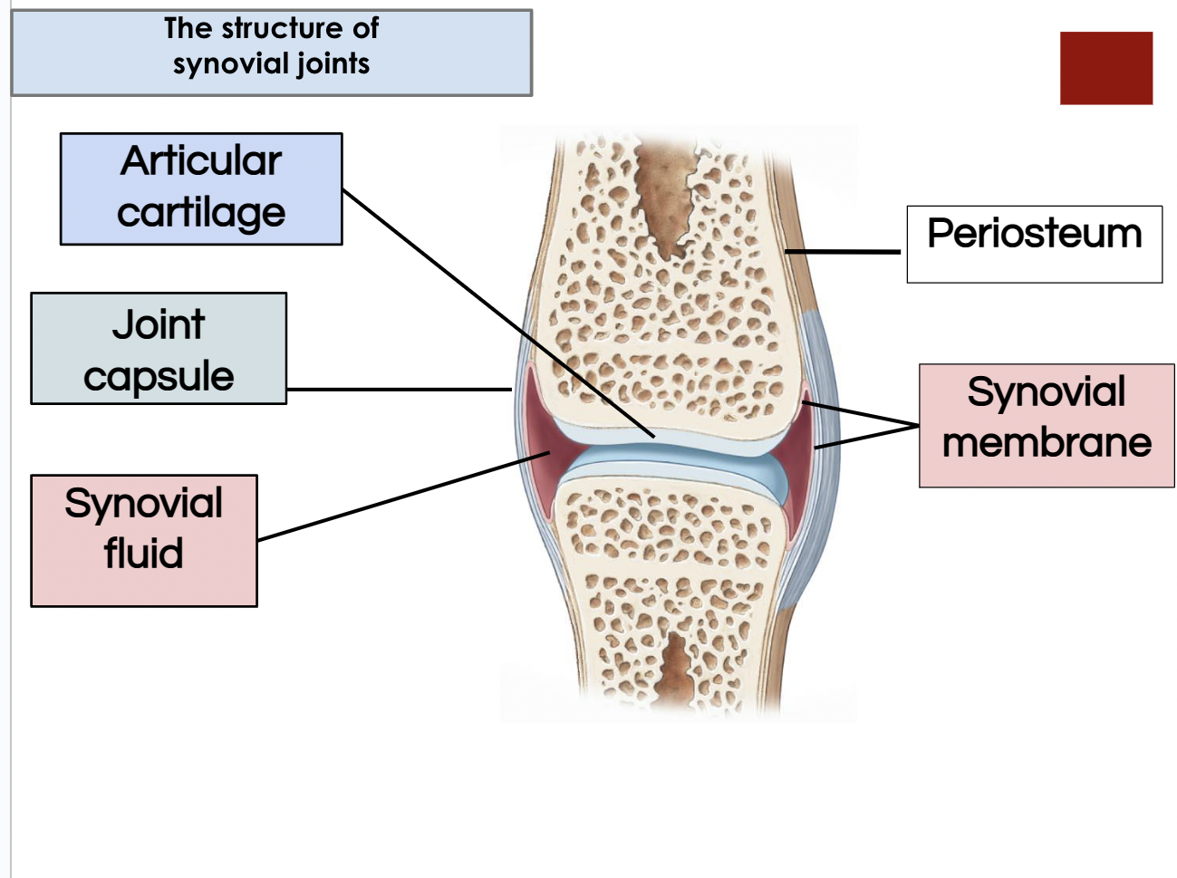
II Function of synovial fluid
A. lubrication- with articular cartilage compression, synovial fluid is squeezed out and reduces friction between moving surfaces
B. nutrient distribution- provide nutrients and O2 ad well as waste disposal for the chondrocytes of articular cartilages
compression and expansion of articular cartilages pump synovial fluid in and out of cartilage matrix
C. shock absorption- distributes compression forces across articular surfaces and outward to joint capsule
III Accessory structures- provide support and additional stability
not all are included in every joint- most are seen in the knee
accessory structures in the knee
tendons of quadriceps- pass across joint
limit movement & provide mechanical support
A. Bursa (a pouch)- small pocket filled with synovial fluid
often form in areas where tendon or ligament rub against other tissues
reduce friction and act as shock absorbers
B. fat pads- adipose tissue covered by synovial fluid
protect articular cartilages
act as packing material for joint
C. meniscus (a crescent)- pad of fibrous cartilage between bones
D. accessory ligaments- strengthen and support
intrinsic ligaments- localized thickening of joint capsule
ex: capsular ligament
extrinsic ligaments- separate from joint capsule
may pass inside (intracapsular) or outside (extra-capsular) the joint capsule
intracapsular example: cruciate ligaments
extra-capsular example: patellar ligaments
LT 5- arrangement of fascicles (bundles of muscle cells) in the various types of muscles
I Fascicle organization
A. parallel muscle
ex: biceps brachii
fascicles parallel to muscle long axis
most skeletal muscles in the body are parallel
some flat with broad attachments (aponeurosis), or pump and cylindrical with tendon attachments
muscle has central body (belly)
can contract until shortened about 30% (due to fiber shortening)
B. convergent muscle
ex: pectoralis major
fascicles extending over broad area coverage on common attachment site
versatile action
different portions can contract to produce different actions
pulls less on attachment compared to parallel
C. Pennate muscle (penna, feather)
fascicles form common angle with tendon
pull at an angle on the tendon
shorter movement of tendon verses parallel
contains more fibers than parallel of same size= more tension
three types
unipennate- all fibers on one side on tendon
ex: extensor digitorum
Bipennate- fibers insert on both sides of tendon
ex: rectus femorus
multipennate- tendon branches within pennate muscle
ex: deltoid
D. sphincter muscle= circular muscle
ex: orbicularis oris
LT 6- the name of a muscle can help identify its location, appearance, or function
I individual muscle parts
A. Origin- fixed attachment