Session 1 (extra) Tissue Resources
Epithelial Tissue
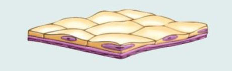
Simple Squamous Epithelium (Figure 4.3a)
Description: Single layer of flattened cells, disc-shaped central nuclei, sparse cytoplasm, simplest epithelium.
Function: Allows passage of materials via diffusion and filtration in areas where protection isn't crucial; secretes lubricating substances in serosae.
Location: Kidney glomeruli, air sacs of lungs, lining of heart, blood vessels, lymphatic vessels, ventral body cavity (serosae).
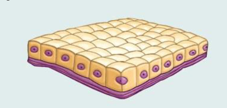
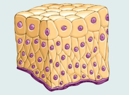
Simple Cuboidal Epithelium (Figure 4.3b)
Description: Single layer of cube-like cells with large, spherical, central nuclei.
Function: Secretion and absorption.
Location: Kidney tubules, ducts and secretory portions of small glands, ovary surface.
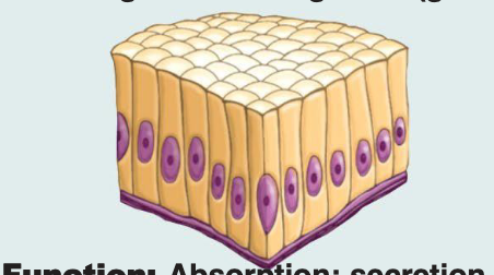
Simple Columnar Epithelium (Figure 4.3c)
Description: Single layer of tall cells with round to oval nuclei; some cells bear cilia; may contain mucus-secreting unicellular glands (goblet cells).
Function: Absorption; secretion of mucus, enzymes, and other substances; ciliated type propels mucus (or reproductive cells) via ciliary action.
Location: Nonciliated type lines most of the digestive tract (stomach to anal canal), gallbladder, and excretory ducts of some glands; ciliated variety lines small bronchi, uterine tubes, and some regions of the uterus.
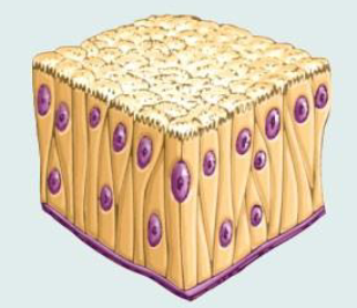
Pseudostratified Columnar Epithelium (Figure 4.3d)
Description: Single layer of cells of differing heights, some not reaching the free surface; nuclei seen at different levels; may contain mucus-secreting cells and bear cilia.
Function: Secretion, particularly of mucus; propulsion of mucus by ciliary action.
Location: Nonciliated type in male's sperm-carrying ducts and ducts of large glands; ciliated variety lines the trachea, most of the upper respiratory tract.
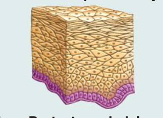
Stratified Squamous Epithelium (Figure 4.3e)
Description: Thick membrane of several cell layers; basal cells are cuboidal or columnar and metabolically active; surface cells are flattened (squamous); in the keratinized type, surface cells are full of keratin and dead; basal cells are active in mitosis, producing cells of superficial layers.
Function: Protects underlying tissues in areas subjected to abrasion.
Location: Nonkeratinized type forms moist linings of the esophagus, mouth, and vagina; keratinized variety forms the epidermis of the skin (a dry membrane).
Transitional Epithelium (Figure 4.3f)
Description: Resembles stratified squamous and stratified cuboidal; basal cells cuboidal or columnar; surface cells dome-shaped or squamous-like, depending on the degree of organ stretch.
Function: Stretches readily and permits distension of the urinary organ by contained urine.
Location: Lines the ureters, urinary bladder, and part of the urethra.
Connective Tissue
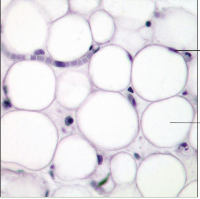
Loose Connective Tissue: Adipose (Figure 4.8b)
Description: Matrix as in areolar, but very sparse; closely packed adipocytes (fat cells) have nucleus pushed to the side by a large fat droplet.
Function: Provides reserve food fuel; insulates against heat loss; supports and protects organs.
Location: Under skin in the hypodermis; around kidneys and eyeballs; within the abdomen; in breasts.
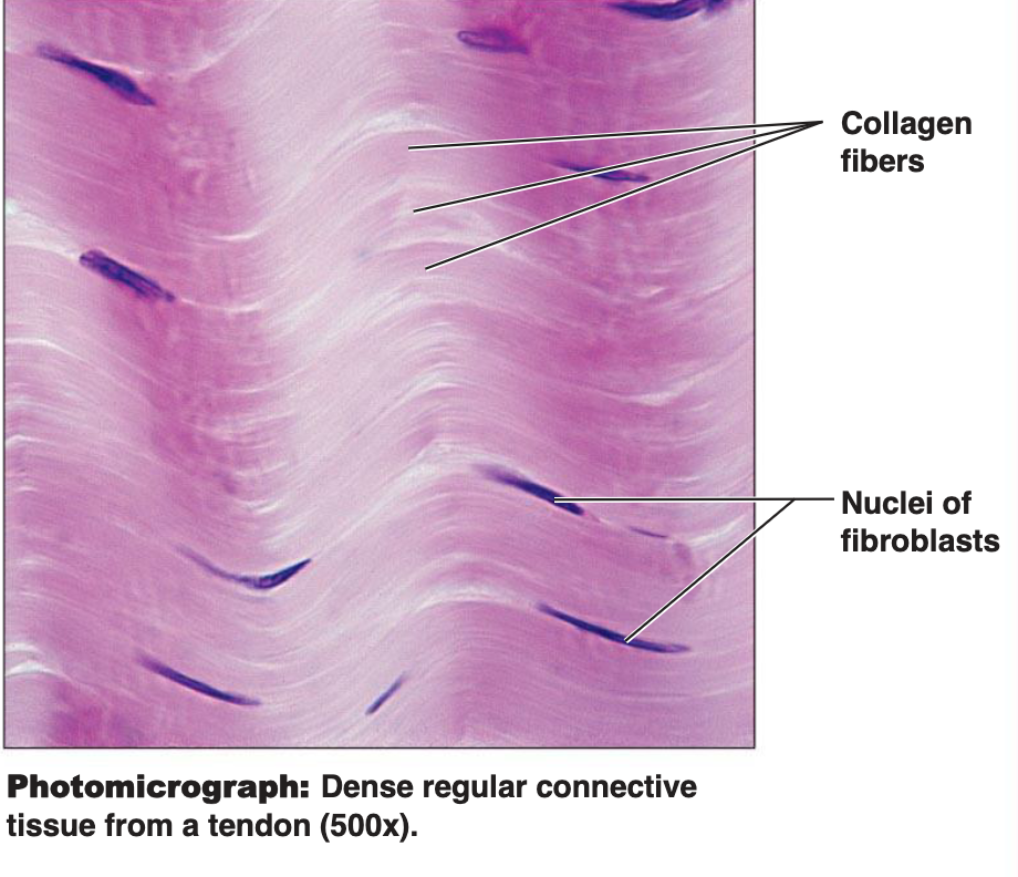
Dense Connective Tissue: Dense Regular (Figure 4.8d)
Description: Primarily parallel collagen fibers; a few elastic fibers; the major cell type is the fibroblast.
Function: Attaches muscles to bones or to muscles; attaches bones to bones; withstands great tensile stress when pulling force is applied in one direction.
Location: Tendons, most ligaments, aponeuroses.
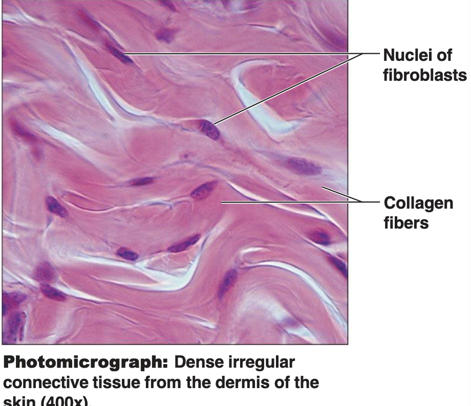
Dense Connective Tissue: Dense Irregular (Figure 4.8e)
Description: Primarily irregularly arranged collagen fibers; some elastic fibers; the major cell type is the fibroblast.
Function: Able to withstand tension exerted in many directions; provides structural strength.
Location: Fibrous capsules of organs and joints; dermis of the skin; submucosa of the digestive tract.
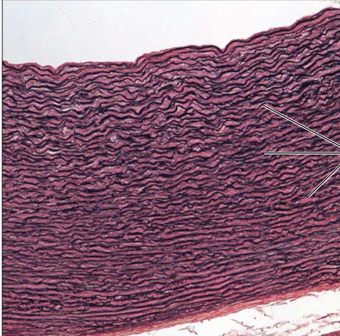
Dense Connective Tissue: Elastic (Figure 4.8f)
Description: Dense regular connective tissue containing a high proportion of elastic fibers.
Function: Allows recoil of tissue following stretching; maintains pulsatile flow of blood through arteries; aids passive recoil of lungs following inspiration.
Location: Walls of large arteries; within certain ligaments associated with the vertebral column; within the walls of the bronchial tubes.
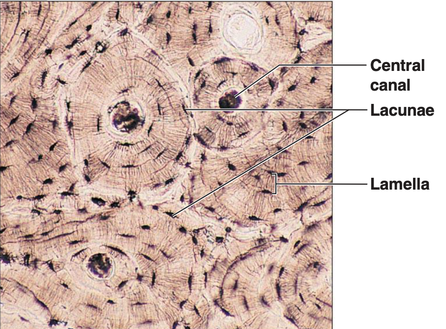
Bone (Osseous Tissue) (Figure 4.8j)
one is a specialised connective tissue (CT)
Distinguished from other CT by its mineralised matrix
Bones are organs – they contain several tissue types eg bone, cartilage, adipose tissue, blood vessels and nerves
Classified as compact or spongy/cancellous
Covered by periosteum (except where they articulate with other bones)
Description: Hard, calcified matrix containing many collagen fibers; osteocytes lie in lacunae; very well vascularized.
Function: Supports and protects (by enclosing); provides levers for muscles to act on; stores calcium, other minerals, and fat; marrow inside bones is the site of blood cell formation (hematopoiesis).
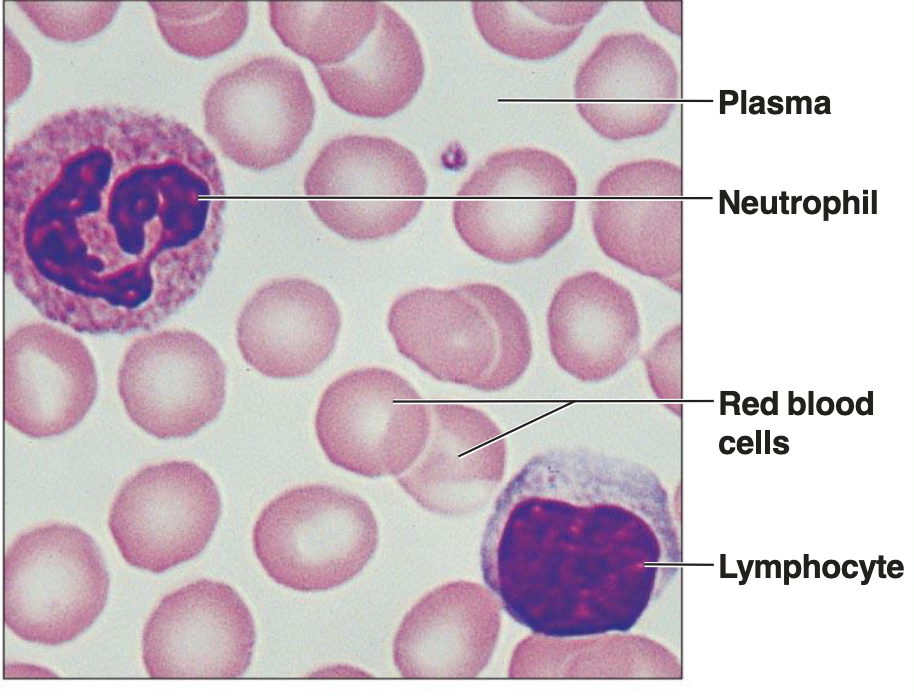
Blood (Figure 4.8k)
Description: Red and white blood cells in a fluid matrix (plasma).
Function: Transports respiratory gases, nutrients, wastes, and other substances.
Location: Contained within blood vessels.
Cartilage
Specialized connective tissue distinguished by a mineralized matrix.
Bones are organs containing bone, cartilage, adipose tissue, blood vessels, and nerves.
Classified as compact or spongy/cancellous.
Covered by periosteum (except where they articulate with other bones).
Lacks nerve fibers and is avascular (receives nutrients via diffusion).
Receives nutrients from a vascular connective tissue membrane – perichondrium.
Chondroblasts produce extracellular matrix (ECM); when mature, these cells become chondrocytes.
Chondrocytes typically grouped together, exist in small cavities called lacunae.
Rigid connective tissue providing a supportive framework.
Cartilage cells (chondrocytes) lie within lacunae in the gel-like fluid matrix.
Cartilaginous structures are enclosed within a connective tissue perichondrium.
Integumentary System (Skin)
Components: Epidermis, Dermis, Hypodermis
Other structures: Arrector pili muscle, Hair follicle, Sebaceous (oil) gland, Hair root, Hair follicle receptor, Adipose tissue, Sensory nerve fiber, Hair shaft, Pore of sweat gland duct, Eccrine sweat gland, Pacinian corpuscle, Cutaneous vascular plexus
Muscle & Nervous Tissue
Will be covered in separate workshops.
Types of Muscle Tissue: Skeletal muscle, Cardiac muscle, Smooth muscle
Nervous Tissue Structures: Neuron (cell body), Nucleus, Dendrite, Axon, Axon terminals, Astrocyte (glial cell), Oligodendrocyte (glial cell)