Genetic code, DNA and genome
The Genome
What is a genome?
A genome is the complete set of genetic material in an organism. It includes all of the organism’s DNA (or RNA in some viruses), which contains the instructions needed to build and maintain that organism.
The genome of human cells consists of the DNA found in the cell nucleus and in the mitochondria.
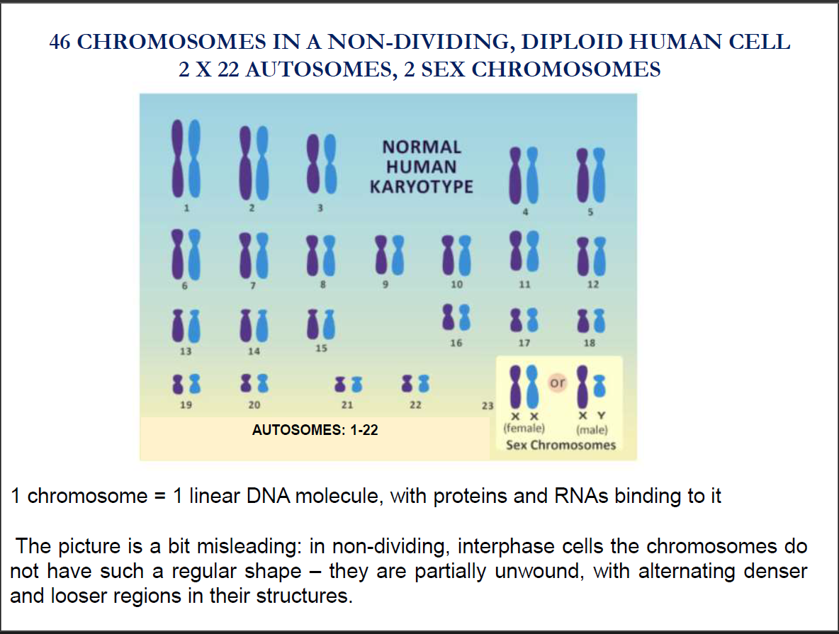
The nuclear genome is diploid and organised in linear molecules named chromosomes.
The mitochondrial genome is polyploid and circular.
Note: Polyploid means “more than 1 circular DNA molecule per mitochondrion”.
DNA is the physical carrier of inherited traits
Griffith & Avery Experiments
Main idea: DNA (not proteins) carries genetic information.
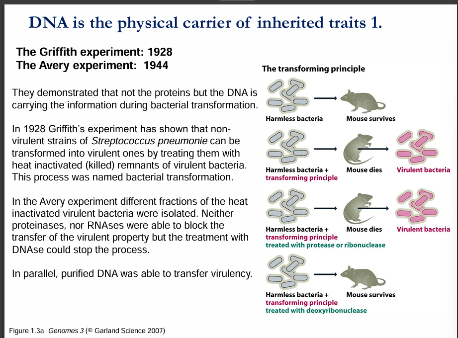
Griffith’s Experiment (1928):
He worked with Streptococcus pneumoniae bacteria.
There are two types:
Virulent (disease-causing) → kills mice.
Non-virulent (harmless) → mice survive.
When he mixed dead virulent bacteria (dead disease-causing bacteria) with live harmless bacteria, the harmless ones became virulent → the mice died.
This showed that some "transforming principle" was passed from the dead bacteria to the live ones, turning them dangerous.
👉 This process was called bacterial transformation.
Avery’s Experiment (1944):
He wanted to find out what the "transforming principle" actually was.
He separated the components of the dead virulent bacteria:
Destroyed proteins → transformation still happened.
Destroyed RNA → transformation still happened.
Destroyed DNA → transformation did not happen.
Conclusion: DNA was the molecule responsible for carrying genetic traits.
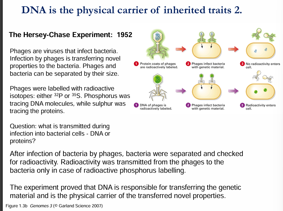
Hershey–Chase Experiment (1952)
Main idea: DNA, not protein, is the genetic material in viruses.
They used bacteriophages (viruses that infect bacteria). A phage is basically DNA inside a protein coat.
They labelled:
Protein coat with sulfur-35 (³⁵S) (since proteins contain sulfur).
DNA with phosphorus-32 (³²P) (since DNA contains phosphorus).
Then, they let phages infect bacteria and looked at what entered the bacterial cells:
When proteins were labelled → radioactivity stayed outside the bacteria (so proteins didn’t enter).
When DNA was labelled → radioactivity was found inside the bacteria (so DNA entered and passed on instructions).
Conclusion: DNA is the genetic material that viruses inject into bacteria to reproduce.
✅ Big Picture from both slides:
Griffith discovered transformation (something passes traits between bacteria).
Avery showed that this “something” is DNA, not proteins or RNA.
Hershey–Chase confirmed with viruses that DNA is the hereditary material that carries instructions for life.
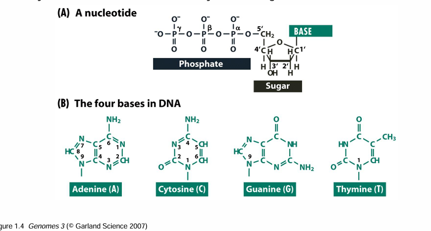
Components of the DNA
Chemical analysis has shown that DNA is built of four different nucleotides.
Nucleotides are made of phosphates, deoxyribose sugar, and 4 nitrogenous bases (adenine, thymine, cytosine and guanine).
The ratio of these nucleotides is different in different species, but these ratios follow some specific rules; namely, the molar ratio of adenine and thymine is always the same, and this is true for cytosine and guanine too.
DNA is a polymer
The DNA in the cell is in the form of a linear polymer.
The units of the polymer are repeated, but in a non-periodic manner.
The units are connected through their sugar and phosphate groups with phosphodiester bonds.
By numbering the carbons of the deoxyribose, we can find the phosphate groups at the fifth carbon, the so-called 5' end.
At the third carbon of the deoxyribose, on the so-called 3’ end, we can find the hydroxyl group.
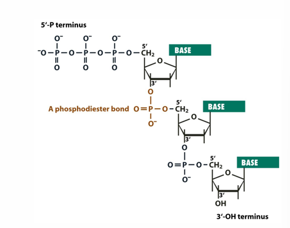
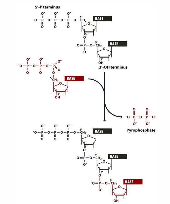
The DNA polymer has a specific direction
In biological processes, the direction of polymerisation of the DNA is from the 5’ to the 3’ end.
The new nucleotides are always linked to the 3’ end of the polymer.
The direction of the polymer is always: 5’—>3’
When a new nucleotide is added to the 3’ end, a pyrophosphate group leaves the reaction from the nucleotide triphosphate.
SEE THE DIAGRAM BELOW:
Important note:
✅ So the new DNA strand grows 5′ → 3′, but each nucleotide is added to the 3′ end.
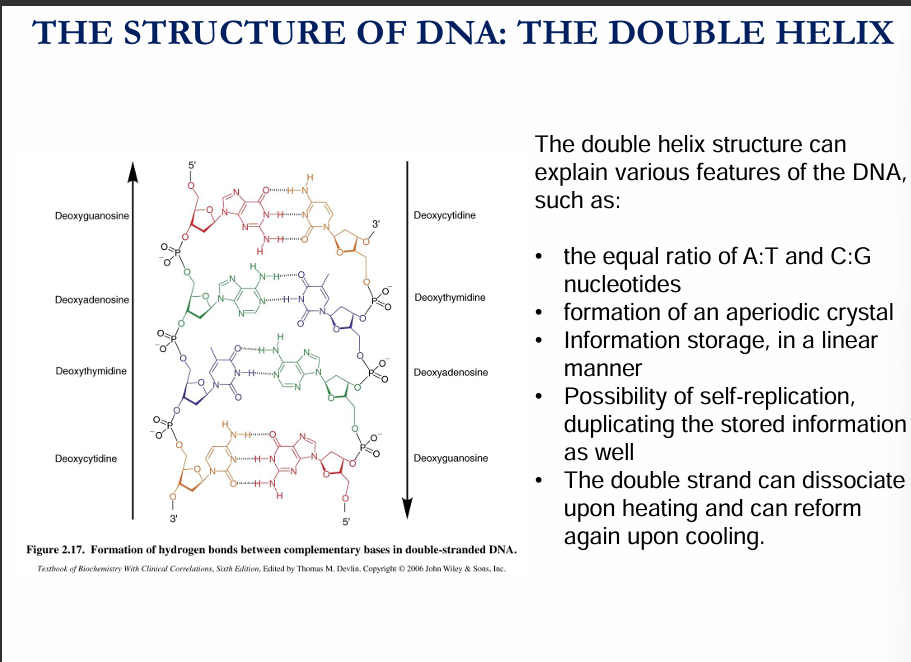
DNA bases can form hydrogen bonds with each other.
The nucleotides of the DNA can form pairs.
Adenine is paired with thymine through two hydrogen bonds, and cytosine can bind to guanine through three hydrogen bonds.
Hydrogen bonds confer stability to the structure.
Different treatments can disrupt these hydrogen bonds— heating to high temperature is one example. More hydrogen bonds will need a higher temperature to disrupt them.
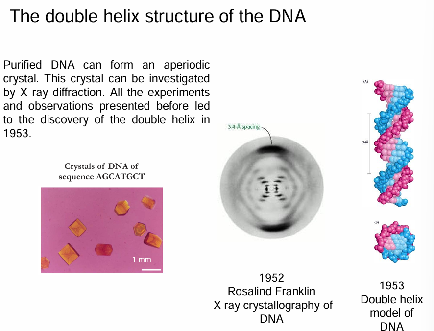
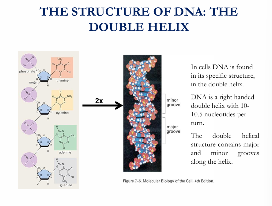
The double helix structure of the DNA.
Chromatin
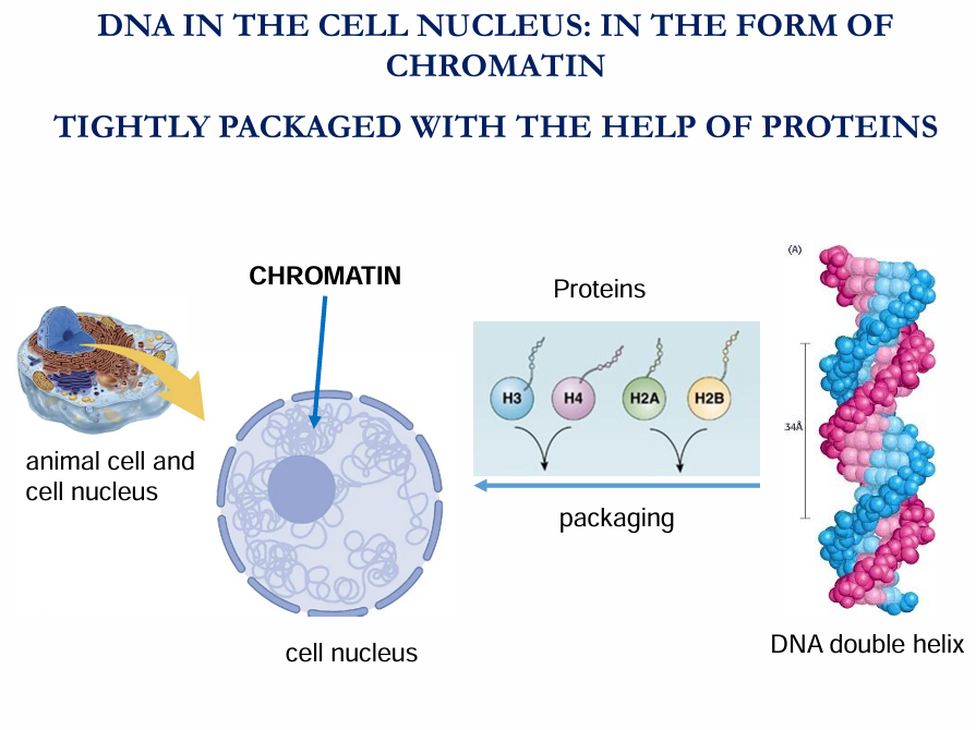
Chromatin is the form that DNA takes inside the nucleus of eukaryotic cells (plants, animals, fungi).
🔹 Definition:
Chromatin is DNA wrapped around proteins called histones. This combination of DNA + proteins helps package the very long DNA molecules so they can fit inside the nucleus, while also controlling which genes are active.
🔹 Types of chromatin:
Euchromatin → loosely packed, "open" form of chromatin.
Genes here are usually active (can be read and used).
Heterochromatin → tightly packed, "closed" form of chromatin.
Genes here are usually inactive (not expressed).
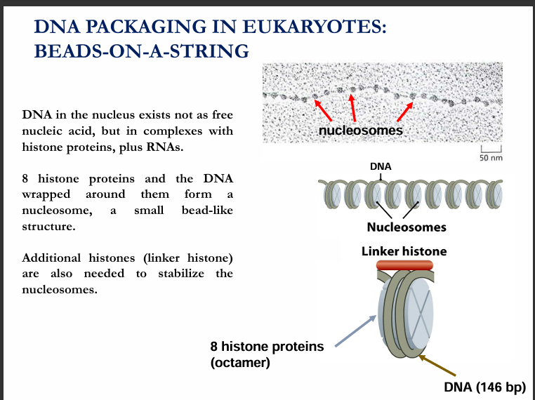
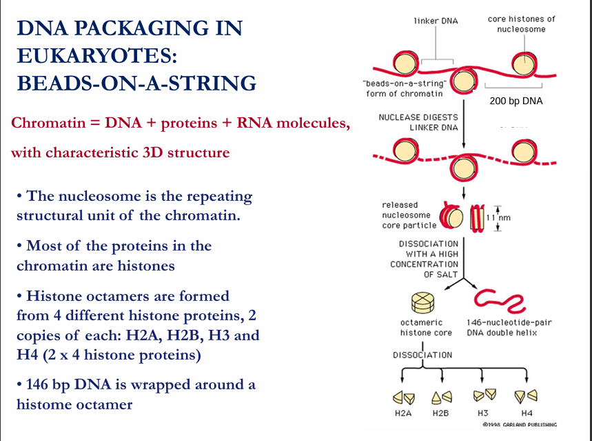
NOTE: A Nucleosome is the basic structural unit of chromatin.
They have a diameter of 11nm.
SOLENOID
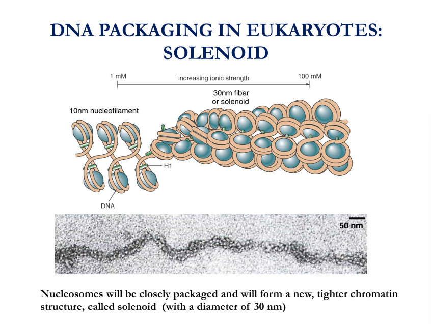
Solenoid (30 nm fibre)
These nucleosomes coil further into a helical structure called the solenoid (or zig-zag model, depending on the arrangement).
This packing gives a fibre of about 30 nm diameter.
Histone H1 helps stabilise this structure by binding the “linker DNA” between nucleosomes.
Levels of DNA packaging
Nucleosomes (11 nm fibre)
DNA wrapped around histone octamers (“beads on a string”).
Solenoid / 30 nm fibre
Nucleosomes coil into a tighter helix.
Histone H1 + linker DNA stabilises the structure.
Looped domains (~300 nm)
Additional proteins (non-histone scaffold proteins) anchor the 30 nm fibre into loops.
This creates more compact chromatin.
Higher-order folding (~700 nm)
Loops coil even further, forming the arms of chromatids.
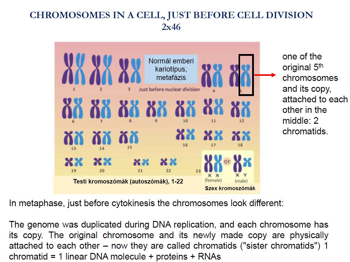
Metaphase chromosome (~1400 nm total width)
The most condensed state, only seen briefly during mitosis (metaphase).
Easily visible under a light microscope.
Chromosomes in non-dividing cells (cells in interphase) have much looser packaging, with alternating denser and looser regions, which makes them more difficult to study under the microscope, unlike the metaphase chromosomes, which are well visible on the microscope because the chromatin forms an extremely dense structure.
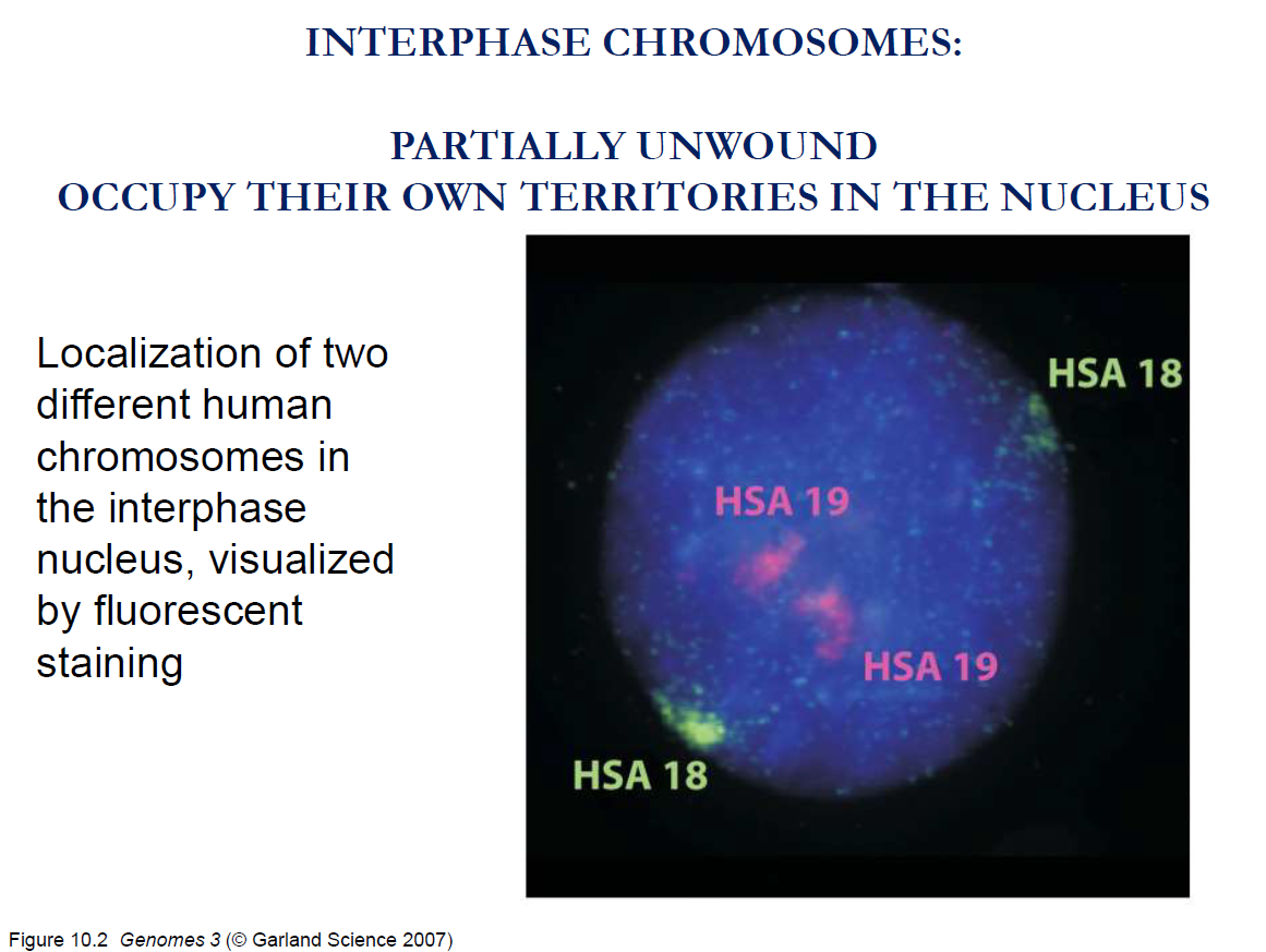
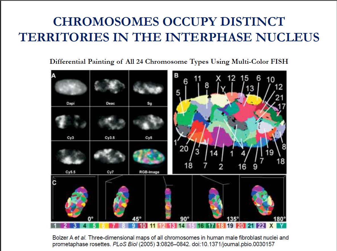
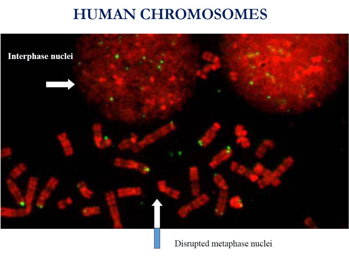
Key takeaway: Chromosomes occupy distinct territories in the nucleus of a cell in interphase.
Key takeaway: In non-dividing cells (interphase cells), the chromosomes do not have a regular shape. They are partially unwounded, with alternating denser and looser regions in their structures.
Key Terms to remember:
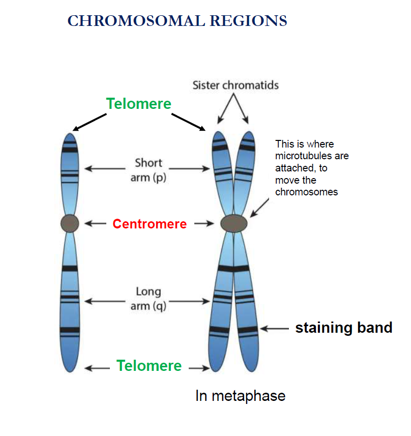
Telomeres
They are located at the end of chromosomes
They are made of repetitive DNA sequences (in humans: TTAGGG repeats).
Functions:
Protect chromosome ends from degradation.
Prevent chromosomes from fusing with each other.
Act like a "cap" (similar to plastic tips on shoelaces).
Shorten with each cell division → linked to ageing and genomic stability.
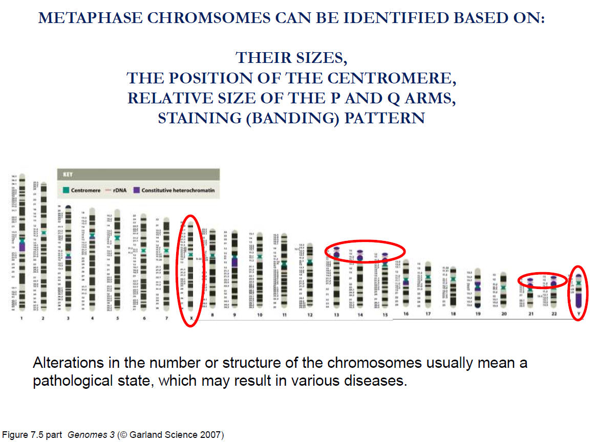
Centromeres:
This is where microtubules are attached to move the chromosomes.
Short arm: The shorter arm of the chromosome, located above the centromere. The short arm of a chromosome is also called the p arm.
Long arm: The longer arm of the chromosome, located below the centromere. The long arm of the chromosome is also called the q arm.
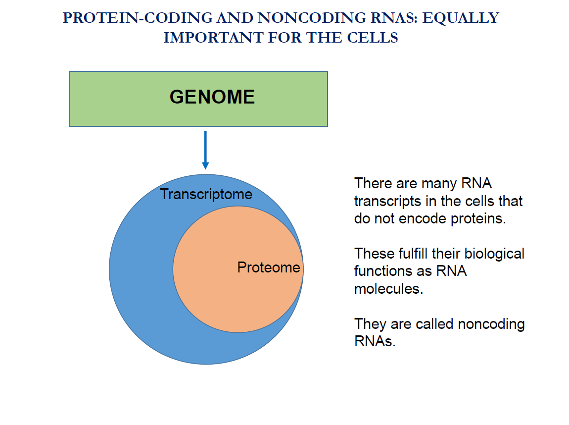
Non-coding RNAs (ncRNAs)
Definition: RNA molecules that are not translated into proteins.
Instead, they carry out functions directly as RNA (structural, regulatory, catalytic, etc.).
They make up a large fraction of the RNA in cells.
Note:
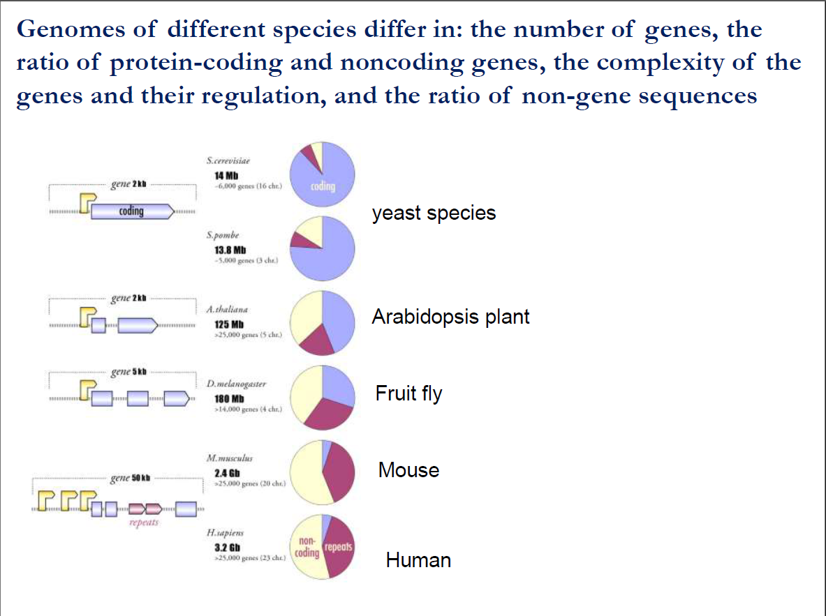
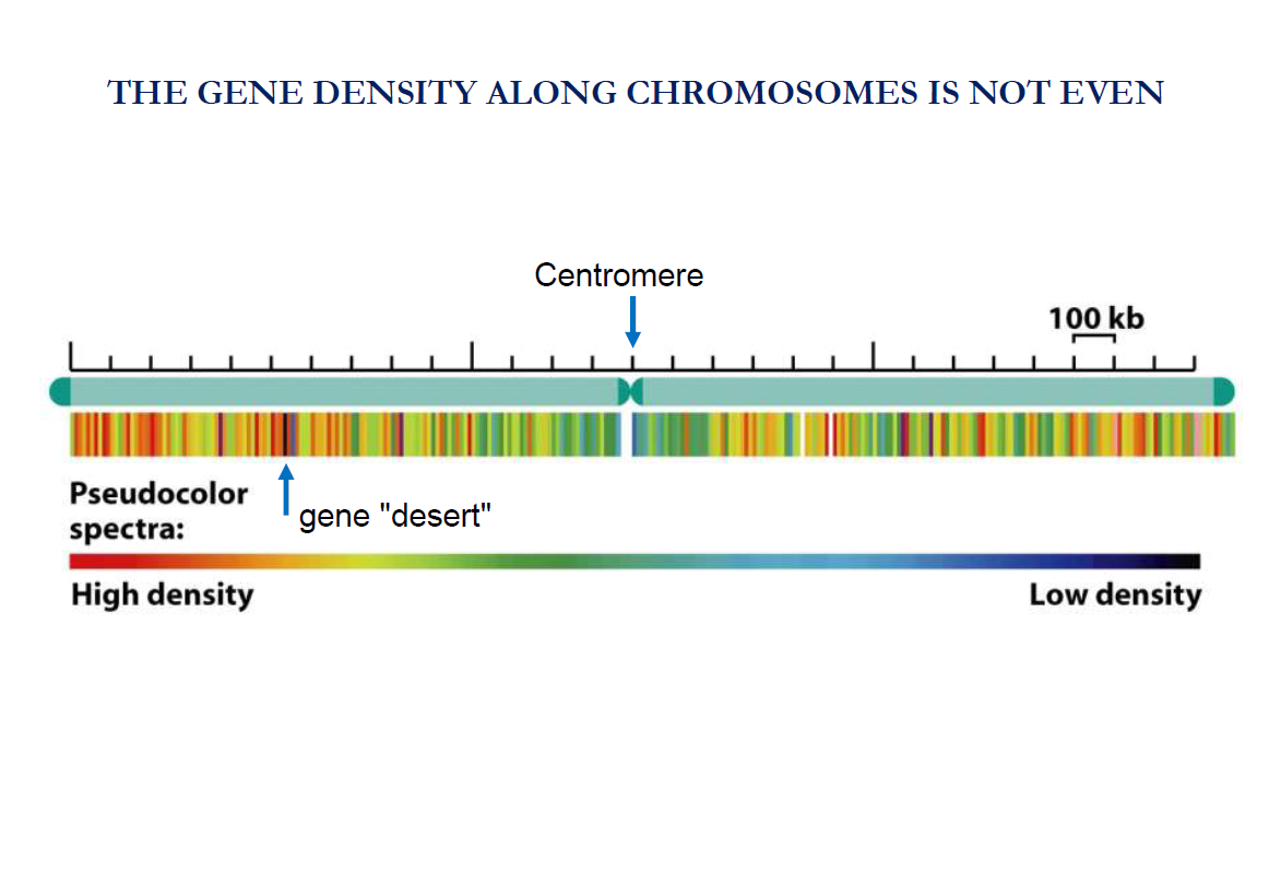
Genes are not evenly distributed along the chromosome.
Some regions of the chromosomes are gene-rich (lots of genes are packed together).
Other regions are gene-poor.
Take note:
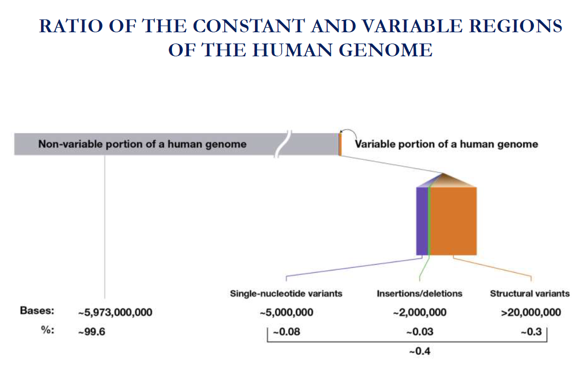
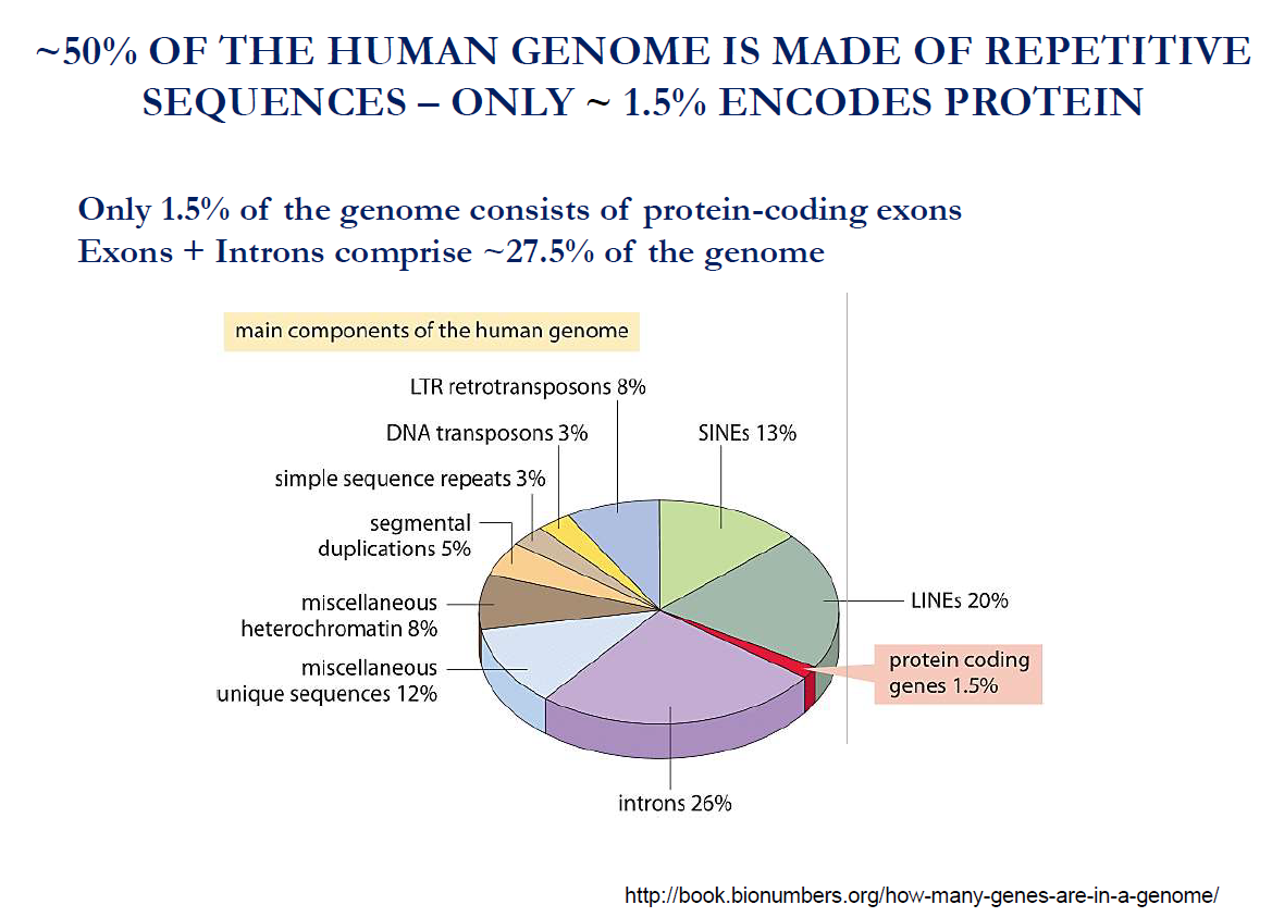
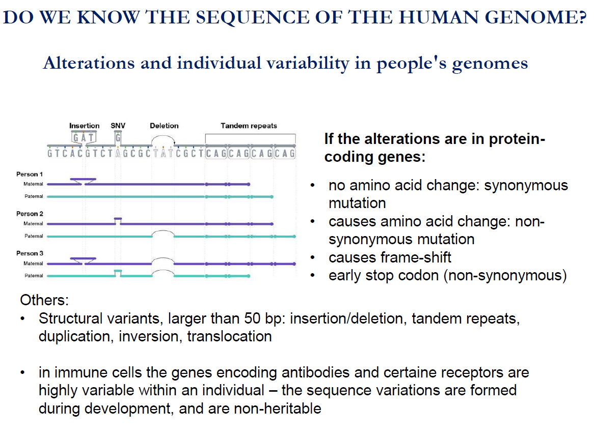
Approximately 50% of the human genome is made up of repetitive sequences
Only 1.5% encodes proteins, i.e. only 1.5% of the genome consists of protein-coding exons
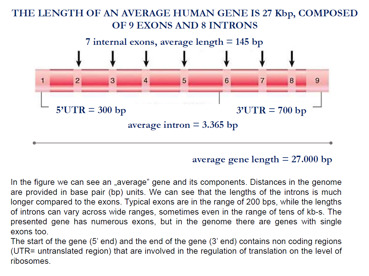
Exons+introns comprise approximately 27.5% of the genome.
TAKE NOTE:
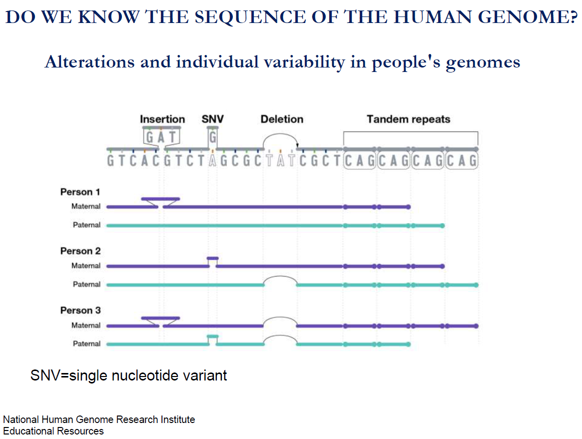
Unlike most genes, antibody and receptor genes are reshuffled like playing cards during immune cell development. This makes each immune cell unique, but these changes are not inherited — only your immune system has them.
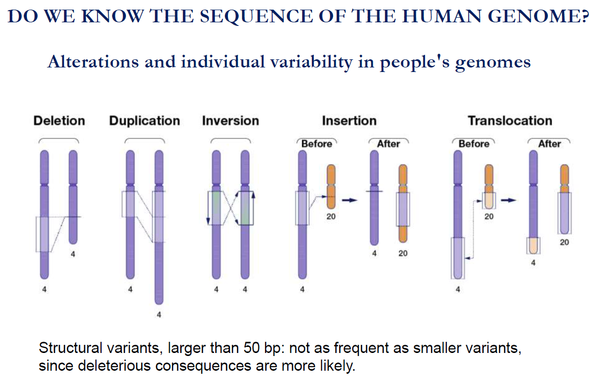
Mutations of the chromosome:
Deletion: In this type of mutation, a segment of DNA is lost/ removed.
Duplication: A DNA segment is copied and inserted again.
Inversion: A DNA segment is flipped (reversed) within the chromosome.
Insertion: A new segment of DNA is added into a chromosome.
Translocation: A segment of DNA is moved to a different chromosome or a different place on the same chromosome.
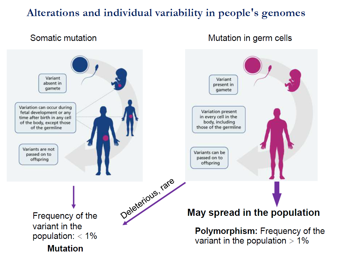
Take note:
A mutation in somatic cells is not transferred/ passed on to offspring.
But mutations in germ cells can spread to the offspring.
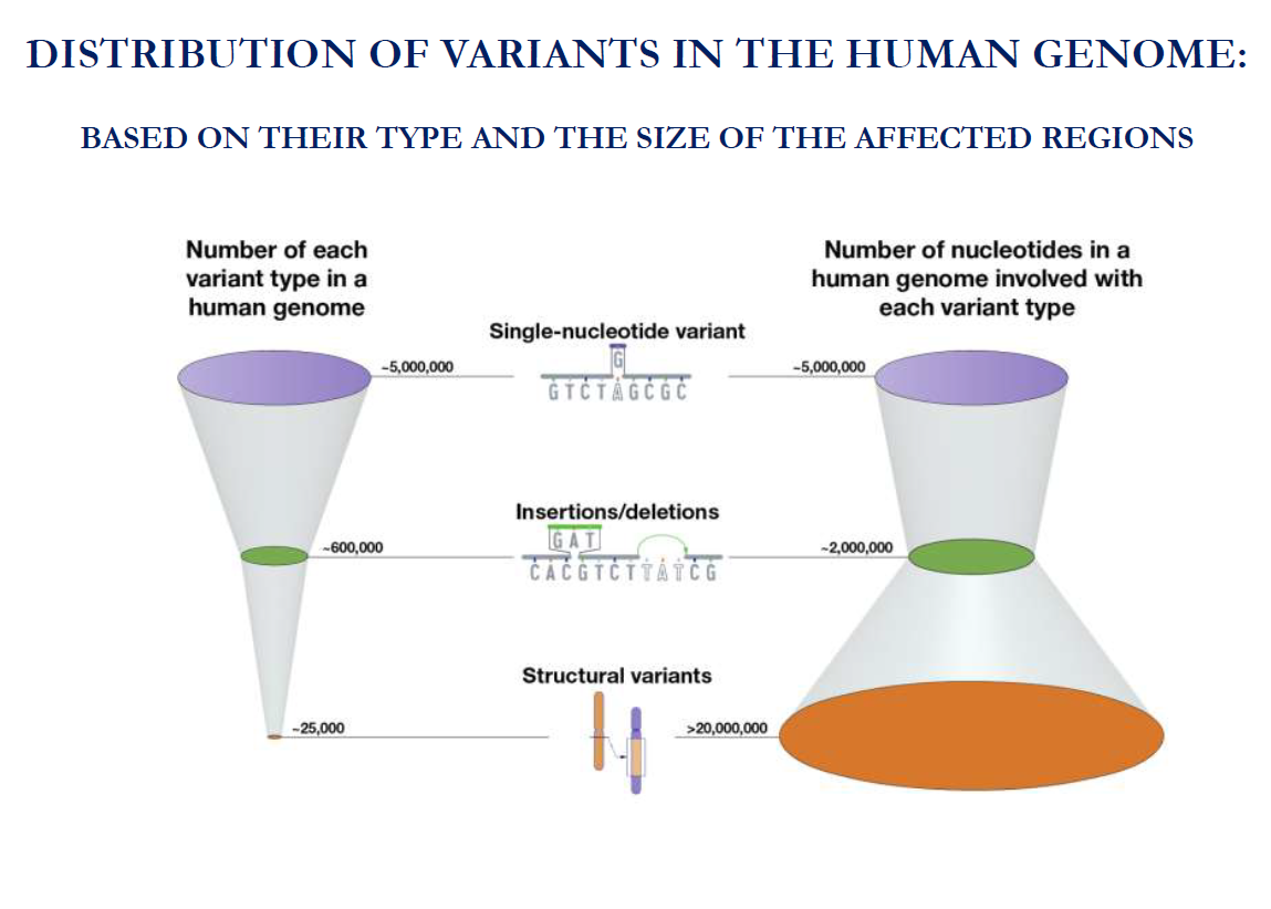
Mutation: Frequency os the variant in the population <1%.
Polymorphism: Frequency of the variant in the population >1%.