Comprehensive Genetics: Mapping, Chromosome Architecture, DNA Replication, Transcription, RNA Processing, Translation, and Sequencing Technologies
Genetic Linkage Mapping
Foundational Principles and Objectives:
Linkage mapping is a method utilized to identify the genomic position of causative genetic variation responsible for a phenotype of interest.
Historically, linkage mapping was employed to construct foundational de novo chromosome maps in model organisms and humans by measuring recombination frequencies between observable phenotypic traits.
Contemporary linkage mapping in model organisms and humans begins from near-complete reference genetic and physical genomic maps of all chromosomes. Rather than building maps from scratch, modern mapping locates mutations by determining their linkage to known molecular markers positioned throughout the genome.
Sturtevant originally mapped genes along the Drosophila X chromosome based on recombination frequencies, establishing a linear arrangement (y, w, v, m, r):
yellow (y): Sturtevant map position = ; contemporary map position =
white (w): Sturtevant map position = ; contemporary map position =
vermilion (v): Sturtevant map position = ; contemporary map position =
miniature (m): Sturtevant map position = ; contemporary map position =
rudimentary (r): Sturtevant map position = ; contemporary map position =

Limitations of Traditional Mapping in Humans:
Low number of progeny per family, which limits statistical power.
Ethical impossibility of performing controlled or directed experimental crosses.
Severe scarcity of discernible, non-deleterious visible phenotypes available to serve as genetic landmarks.
Molecular DNA Sequence Markers:
Modern mapping utilizes neutral DNA sequence markers—direct variations in DNA sequence at cataloged genomic coordinates—rather than morphological phenotypes.
Variable Number Tandem Repeats (VNTRs):
Short DNA sequence segments repeated in tandem head-to-tail arrays.
Alleles differ in the number of repeated units, producing variable length segments.
Genotyping is executed by designing locus-specific Polymerase Chain Reaction (PCR) primers flanking the repeat array, followed by separation using gel electrophoresis.
PCR exponentially amplifies the target DNA template: with initial template doubling each cycle, yielding target copies after cycles (e.g., ).
In gel electrophoresis, an electric field pulls negatively charged DNA molecules toward the positive anode through an agarose or polyacrylamide molecular sieve. Migration distance is inversely proportional to fragment length and compaction, resolving alleles by molecular weight.

Single Nucleotide Polymorphisms (SNPs):
A specific single nucleotide position where different base variants exist across individuals.
Abundance: In humans, SNPs occur approximately once every .
The vast majority of SNPs are biallelic (possessing two alternative alleles in a population, such as a G allele versus a C allele).
SNPs with high minor allele frequencies are the most informative for linkage studies.
Distribution: Located across coding exons, introns, and intergenic regions; the overwhelming majority are non-coding and not causative of any phenotype, functioning solely as physical positional landmarks.

Mathematical Framework of Linkage Analysis and LOD Scores:
When assessing linkage between a disease mutation and a marker locus with alleles (e.g., and ):
Recombination frequency is denoted as .
If unlinked (), parental and recombinant gametes each occur at a probability of .
If linked with recombination fraction :
Each parental class occurs at a probability of .
Each recombinant class occurs at a probability of .
Odds Ratio (OR): Compares the likelihood of the observed pedigree configuration under linkage versus independent assortment:
Log of the Odds (LOD) Score:
A indicates that the linkage hypothesis is more likely than independent assortment.
is the standard threshold for declaring statistically significant linkage ( odds in favor of linkage).
Evaluating LOD across multiple values of identifies the maximum LOD score; the value of corresponding to the maximum LOD represents the best estimate of recombination frequency between the loci.
Hypothetical pedigree result with 4 recombinants among 13 offspring ():
( greater likelihood than , maximum LOD estimate)
Familial Melanoma Case Study (TERT Promoter Mutation):
Study setup: Horn et al. (2013) evaluated a multi-generational pedigree afflicted with dominant malignant melanoma, genotyping 15 affected/unaffected individuals at approximately across all 23 human chromosomes.
Linkage mapping identified a peak maximum LOD score of on chromosome 5p at SNP locus rs2853669.
Downstream fine mapping: Sequencing the flanking genomic window revealed a causative mutation located within the core promoter of the TERT gene (which encodes the catalytic telomerase reverse transcriptase subunit).
The familial melanoma mutation was in complete allelic linkage with the G allele of marker rs2853669, situated exactly upstream from the ATG translation initiation site.
Follow-up screening established that identical and closely situated recurrent promoter mutations occur across multiple unrelated families with melanoma and in sporadic melanoma tumors.

Genome-Wide Association Studies (GWAS)
Linkage versus Linkage Disequilibrium:
Linkage: The failure of two loci to assort independently () due to physical proximity along a single chromosome during meiosis; detectable exclusively within familial pedigrees across recent generations.
Linkage Disequilibrium (LD):
The non-random association of alleles at different loci within an entire population, occurring when specific allele combinations are observed at higher frequencies than expected by chance.
LD is measured across broad cohorts of unrelated individuals.
Mechanism of LD decay: When a novel causative mutation arises, it occurs on a specific ancestral chromosome bearing a unique haplotype of linked SNPs. Over hundreds to thousands of generations (), meiotic crossing-over breaks down the association between the mutation and distant loci until they reach linkage equilibrium (random assortment). However, SNPs situated physically adjacent to the mutation rarely recombine away and remain in strong linkage disequilibrium.

Methodology and Experimental Design of GWAS:
The Common Disease, Common Variant Hypothesis: Proposes that common polygenic medical conditions are governed in part by relatively common ancestral genetic variants distributed throughout the human population.
Workflow:
Assemble a defined study cohort of unrelated individuals (e.g., case-control cohorts with and without a disease).
Record quantitative or qualitative clinical phenotypes.
Determine genotypes at hundreds of thousands to millions of SNPs distributed throughout the genome via high-density microarrays or sequencing.
Statistically evaluate the association between the allele frequencies of each SNP and the phenotypic trait using regression analyses, calculating corresponding -values.
Manhattan Plots:
A genomic scatter plot displaying association statistics.
X-axis: Chromosomal position ordered sequentially from chromosome 1 to 22, X, and Y.
Y-axis: Negative logarithm of the association -value (). Taller peaks indicate more significant statistical associations.
Significance thresholds:
Genome-wide significance threshold: (represented by a horizontal red dashed line, correcting for approximately independent hypothesis tests via Bonferroni correction).
Suggestive significance threshold: .

Case Study: Severe COVID-19 with Respiratory Failure:
A landmark GWAS (NEJM 2020) identified two major susceptibility loci reaching genome-wide significance () for severe COVID-19 with respiratory failure:
Chromosome 3p21.31 peak (): Flanks a multi-gene cluster containing SLC6A20, LZTFL1, FYCO1, CXCR6, XCR1, and CCR9.
Chromosome 9q34.2 peak (): Maps directly over the ABO blood group locus.
Applications and Limitations of GWAS:
Applications:
Unbiased discovery of biological pathways, physiological mechanisms, and candidate genes underlying complex polygenic traits.
Construction of Polygenic Risk Scores (PRS) to assess individual genetic susceptibility by summing risk alleles weighted by effect size.
Limitations:
Inability to detect rare causative variants () with modest effect sizes.
Requires phenotypic and genotypic variation within the population; cannot detect genes that are monomorphic across the cohort.
A statistically linked SNP does not establish causality; fine mapping and experimental perturbations are required to isolate functional variants.
The Ancestry Diversity Deficit:
GWAS publications have exhibited an extreme sampling bias toward populations of European ancestry.
As of July 8, 2021, approximately of all individuals included in the GWAS Catalog were of European descent, despite Europeans comprising only a minority fraction of the global population. East Asian participants represented the second largest slice, with African, Hispanic/Latino, South Asian, and Middle Eastern populations remaining severely under-sampled.
This structural imbalance restricts the clinical utility and generalizability of Polygenic Risk Scores across non-European cohorts.

DNA Structure and Chromosome Organization
Fundamental Properties of Genetic Material:
Must replicate accurately and completely once per cell cycle.
Must possess sufficient informational complexity to encode diverse phenotypes.
Must maintain biochemical stability to safeguard fidelity while accommodating mutations to facilitate evolutionary adaptation.
Chemical Architecture of DNA:
Composed of repeating deoxyribonucleotide monophosphates connected via covalent phosphodiester bonds.
Directional polarity: carries a terminal phosphate linked to the 5' carbon of deoxyribose; carries an unesterified hydroxyl () on the 3' carbon.
Sugar-phosphate backbone is invariant and polar, whereas nitrogenous base sequences (adenine, thymine, guanine, cytosine) are unrestricted.
Native DNA exists as an antiparallel double helix: one strand runs and the complement runs .
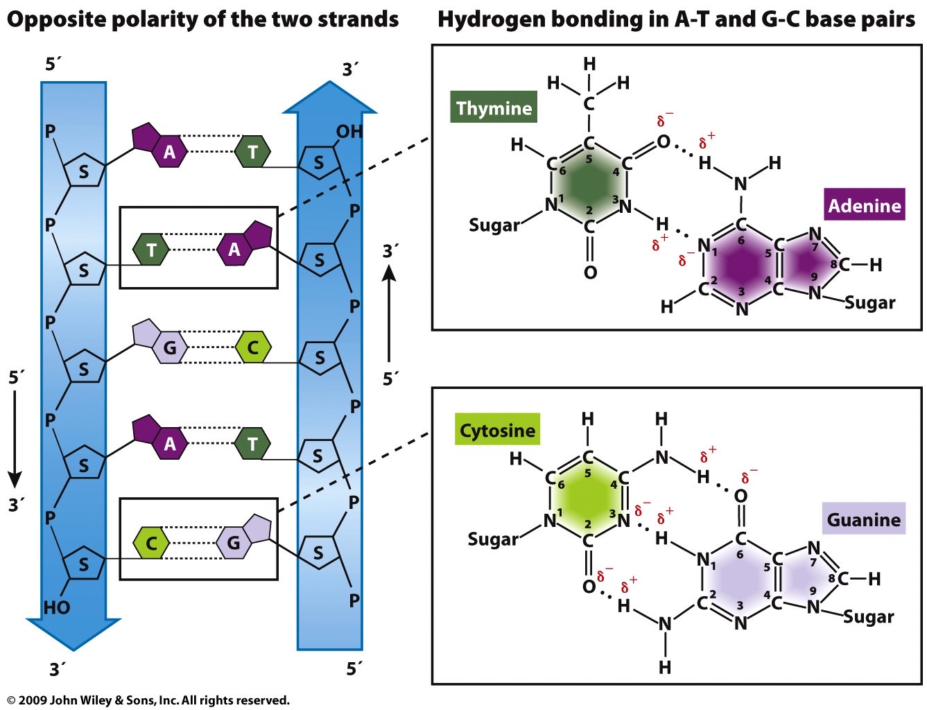
B-Form DNA Characteristics:
Right-handed double helix.
Helical pitch contains approximately .
Asymmetric glycosidic bonding relative to the helical axis generates alternating major grooves and minor grooves.
Base-Pairing Geometry:
Watson-Crick pairings: Adenine forms two hydrogen bonds with Thymine; Guanine forms three hydrogen bonds with Cytosine.
Complementary base pairs possess nearly identical spatial geometries:
pair: glycosidic bond angles of and , separation distance of .
pair: glycosidic bond angles of and , separation distance of .
Mismatched base pairs disrupt geometry:
mismatch: angles of and , separation distance of .
mismatch: angles of and , separation distance of .
Geometric distortions buckle the sugar-phosphate backbone, enabling scanning DNA repair enzymes to recognize lesions.
Information Content of the Major Groove:
Major grooves expose an array of chemical signatures from base pairs without unwinding the double helix:
Hydrogen bond acceptors (A)
Hydrogen bond donors (D)
Nonpolar hydrogens (H)
Methyl groups (M)
Sequence-specific DNA-binding proteins (e.g., transcription factors) recognize these patterns to dock into the major groove.

DNA Topology and Supercoiling:
Topoisomers: DNA molecules having identical primary sequences that differ in topological linking number (). Topoisomers can only exist in topologically constrained DNA (covalently closed circular DNA or linear DNA with ends fixed by scaffold proteins).
Relaxed DNA: The lowest-energy structural state, containing .
Supercoiled DNA:
Positively supercoiled DNA is overwound ().
Negatively supercoiled DNA is underwound ().
Biological systems universally store intracellular DNA in a negatively supercoiled state, storing torsional strain that facilitates strand separation during transcription and replication.
Topoisomerases: Enzymes that cleave, rotate, and religate DNA phosphodiester backbones to modulate linking number and resolve torsional strain.

Nucleic Acid Denaturation and In Situ Hybridization:
Complementary strands unwind and separate (denaturation) under thermal energy or alkaline conditions () by disrupting base-pair hydrogen bonds. When conditions return to neutral pH and physiological temperature, complementary single strands reanneal (renaturation/hybridization) sequence-specifically.
In Situ Hybridization (ISH): Utilizes single-stranded labeled nucleic acid probes (incorporating radioactive, biotinylated, digoxigenin, or fluorophore-tagged nucleotides) complementary to target sequences to visualize their precise spatial locations in preserved tissues or chromosome spreads.
Fluorescent In Situ Hybridization (FISH) and Chromosome Painting:
Multi-color FISH and chromosome painting employ chromosome-specific pools of fluorophore-conjugated probes to visualize whole chromosomes or specific cytogenetic regions.
Metaphase chromosomes appear condensed and individualized.
Interphase chromosomes decondense and occupy discrete non-random, three-dimensional nuclear volumes termed chromosome territories.

Eukaryotic Chromosome Anatomy and Cytogenetic Maps:
Centromere divides the chromosome into arms: shorter arm is designated p (petite); longer arm is designated q.
Centromere positions:
Metacentric: Centromere at the physical midpoint; arms of equal length.
Submetacentric: Centromere offset from center; distinct p and q arms.
Acrocentric: Centromere located near one end; very short p arms terminating in satellite repeats.
Telocentric: Centromere located at the extreme terminal end.
Cytogenetic G-banding (Giemsa staining) yields an invariant alternating pattern of dark bands (compact, gene-poor, late-replicating heterochromatin) and light bands (less condensed, gene-rich, early-replicating euchromatin).
Diploid chromosome counts () vary across species but remain invariant within normal members of a species:
Fruit fly (Drosophila melanogaster):
Frog (Rana pipiens):
Cat (Felis catus):
Mouse (Mus musculus):
Rat (Rattus norvegicus):
Rhesus monkey (Macaca mulatta):
Human (Homo sapiens):
Chimpanzee (Pan troglodytes):
Cow (Bos taurus):
Horse (Equus caballus):
Chicken (Gallus domesticus):
Dog (Canis familiaris):
Carp (Cyprinus carpio):
Chromatin Compaction and Histone Organization:
Chromatin consists of genomic DNA, structural proteins, and associated RNA.
Histone Proteins:
Small, highly conserved, basic proteins enriched in positively charged amino acids (lysine and arginine) that establish electrostatic interactions with the negatively charged sugar-phosphate backbone in the minor groove.
Histone H1: Basic-to-acidic amino acid ratio = ; Molecular weight = ; Residue count = ; Localized to linker DNA.
Histone H2A: Basic-to-acidic ratio = ; Molecular weight = ; Residue count = ; Core nucleosome component.
Histone H2B: Basic-to-acidic ratio = ; Molecular weight = ; Residue count = ; Core nucleosome component.
Histone H3: Basic-to-acidic ratio = ; Molecular weight = ; Residue count = ; Core nucleosome component.
Histone H4: Basic-to-acidic ratio = ; Molecular weight = ; Residue count = ; Core nucleosome component.
Levels of Chromatin Compaction:
10-nm Fiber ("Beads on a String"):
Core particle consists of an octamer of core histones: two molecules each of H2A, H2B, H3, and H4 ().
of double-stranded DNA wrap around the histone octamer.
Nucleosomes are separated by linker DNA spans ( in mammals).
Histone H1 functions as an external clamp on the exterior of the nucleosome core.
30-nm Solenoid Fiber:
The 10-nm nucleosomal strand coils into a spiral solenoid structure measuring across, requiring the presence of Histone H1.
300-nm Looped Chromatin Fiber:
The 30-nm solenoid loops out into large domains anchored along a non-histone protein scaffold.
Fully condensed metaphase chromosomes represent the highest compaction state ( chromatid width).

Position Effect Variegation (PEV):
The chromosomal location of a gene dictates its transcriptional accessibility through variations in local chromatin compaction.
Classic demonstration in Drosophila melanogaster: Chromosomal inversion of the X chromosome places the wild-type white gene () directly adjacent to pericentric heterochromatin.
Because heterochromatin spreading varies stochastically across individual cells during eye development, white expression is silenced in some cells and active in others, generating a variegated (mottled red and white) eye phenotype.
Large-Scale Chromosomal Mutations and Genome Evolution
Structural Chromosomal Abnormalities:
Terminal Deletions:
Involve a single double-strand break cleaving the terminal end of a chromosome, followed by loss of the acentric fragment (which lacks a centromere and cannot segregate at anaphase).
Manifests clinically in Cri-du-chat syndrome: Caused by a terminal deletion of the short arm of human chromosome 5 (). Cytogenetic breakpoint maps to band .

Interstitial Deletions: Two breaks occur within a single chromosome arm, excising an internal segment, followed by religation of the flanking segments.
Duplications: Amplification of an internal segment of DNA, altering gene dosage.
Inversions:
A segment of a chromosome is rotated and reinserted.
Paracentric Inversion: Does not span the centromere (both breaks reside in the same arm).
Pericentric Inversion: Spans the centromere (breaks reside in opposite arms).
Meiotic Consequences in Inversion Heterozygotes:
Homologous chromosomes align during prophase I by forming an inversion loop.
A crossover within a paracentric inversion loop produces an abnormal dicentric chromatid (possessing two centromeres) and an acentric fragment (possessing zero centromeres).
Anaphase I spindle tension creates a dicentric bridge that ruptures randomly, while the acentric fragment is lost. Meiosis yields one normal non-crossover chromatid, one inverted non-crossover chromatid, and two nonviable deletion-bearing recombinant gametes.

Translocations:
Transfer of chromosomal segments between non-homologous chromosomes.
Unbalanced Translocation: One-way movement of genetic material without reciprocal exchange.
Balanced Reciprocal Translocation: Mutual exchange of terminal segments between two non-homologs without net gain or loss of genetic material. During meiosis, pairing of translocation heterozygotes produces a cross-shaped quadrivalent figure; alternate segregation produces viable balanced gametes, whereas adjacent-1 and adjacent-2 segregations yield inviable duplications and deletions.
Robertsonian Translocations:
Fusion of the long (q) arms of two acrocentric chromosomes (human chromosomes 13, 14, 15, 21, and 22) with loss of the tiny short (p) arms.
Frequency: Occurs in approximately .
Carriers maintain 45 chromosomes and are phenotypically healthy because the lost p arms contain redundant repetitive ribosomal RNA (rDNA) gene clusters; however, they exhibit elevated risks of infertility, recurrent miscarriages, and unbalanced offspring (e.g., translocation Down syndrome).
Third-generation long-read sequencing revealed that the translocation breakpoints repeatedly localize to inverted repeat elements designated SST1 located within the pericentromeric regions between chromosomes 13 and 14, acting as non-allelic homologous recombination hotspots.
Synteny:
Conserved physical linkage and collinear gene order along chromosomes across divergent species.
Shared syntenic blocks between human and mouse genomes reflect conserved chromosome segments inherited from their last common mammalian ancestor, disrupted by lineage-specific translocations and inversions over evolutionary time.
Aneuploidy and Polyploidy:
Aneuploidy: Alteration in the baseline chromosome complement where individual chromosomes are lost or gained due to meiotic or mitotic nondisjunction.
Monosomy (); Trisomy ().
Highly deleterious or lethal in animals due to acute disruption of developmental gene dosage balances.
Polyploidy: Presence of three or more complete sets of chromosomes (, , , , etc.).
Common and well-tolerated in angiosperms; associated with increased cell volume and larger vegetative/fruit structures (e.g., octaploid commercial strawberries versus diploid wild species).
Autopolyploidy: Duplication of chromosome complements derived entirely from within a single species via complete meiotic nondisjunction.
Allopolyploidy: Chromosome doubling resulting from interspecific hybridization between two related species, followed by meiotic nondisjunction.
Evolution of Polyploid Wheat:
Around : Hybridization between diploid wild grass Triticum searsii (, BB genome) and wild einkorn wheat Triticum urartu (, AA genome) produced the cultivated allotetraploid emmer wheat Triticum dicoccum / Triticum dicoccoides (, AABB) and durum pasta wheat Triticum turgidum (, AABB).
Around : Hybridization between allotetraploid emmer wheat (, AABB) and wild goatgrass Triticum tauschii (, DD genome) generated modern allohexaploid bread wheat Triticum aestivum (, AABBDD) and spelt wheat Triticum spelta (, AABBDD).

DNA Replication
Semiconservative Mechanism and Essential Substrates:
Demonstrated by Meselson and Stahl: each parental DNA strand acts as a template for synthesizing one complementary daughter strand, yielding duplexes containing one conserved parental strand and one newly synthesized strand.
Substrates required for polymerization:
Deoxyribonucleoside triphosphates (dNTPs: dATP, dCTP, dGTP, dTTP).
Primed single-stranded DNA template providing a free 3'-hydroxyl () terminus.
Chemistry of phosphodiester bond formation: The free 3'-OH group of the primer carries out a nucleophilic attack on the inward-facing -phosphorus atom of the incoming dNTP, liberating the outer and phosphates as inorganic pyrophosphate ().

Enzymatic Properties of DNA Polymerases:
All DNA polymerases synthesize exclusively in the direction by adding nucleotides to a preexisting 3'-OH.
All DNA polymerases are template-directed.
Escherichia coli DNA Polymerases:
DNA Polymerase I: Single polypeptide; contains both exonuclease activity (for degrading double-stranded DNA and RNA primers) and exonuclease activity (proofreading); responsible for RNA primer excision and gap filling during replication and repair.
DNA Polymerase II: Single polypeptide; contains exonuclease activity; functions in DNA repair.
DNA Polymerase III: Multi-subunit holoenzyme complex; contains exonuclease activity (limited to single-stranded substrates) and exonuclease activity (proofreading); the primary replicative enzyme in E. coli.
DNA Polymerase IV: Single polypeptide; lacks exonuclease activity; error-prone translesion DNA repair.
DNA Polymerase V: Single polypeptide; lacks exonuclease activity; error-prone translesion DNA repair.
Prokaryotic Replication Initiation at OriC:
OriC is the single fixed origin of replication in E. coli, spanning .
Key functional sequence motifs:
13-mer tandem array: Three AT-rich repeating consensus sequences (
5'-GATCTATTTATTT-3'); site of initial strand separation.9-mer tandem array: Four binding sites (
5'-TTATCCACA-3') specific for the initiator protein.Initiation Pathway:
Initiator protein DnaA binds cooperatively to the 9-mer sites, forming a large oligomeric nucleoprotein complex that bends the DNA.
DNA wrapping introduces torsional strain that drives localized duplex unwinding at the AT-rich 13-mer repeats, opening a replication bubble.
DnaC (helicase loader) deposits DnaB (replicative hexameric helicase) onto the unwound single strands.
DnaB moves processively, unwinding the parental strands and advancing the replication forks bidirectionally.
Single-Stranded DNA-Binding Protein (SSB) coats exposed single strands to prevent reannealing or secondary structure formation.
DNA Primase (DnaG), a specialized RNA polymerase capable of de novo synthesis, synthesizes short RNA primers () onto the single-stranded template.

Fidelity and Kinetic Selectivity of DNA Replication:
Replication exhibits fidelity with net error rates below .
Kinetic Selectivity: The catalytic active site of DNA polymerase III geometrically accommodates only correctly paired Watson-Crick geometries. The rate of phosphodiester catalysis drops precipitously for mismatches. Initial base selection misincorporation rate is .
Proofreading ( Exonuclease): When an improper nucleotide is incorporated, the distorted terminal geometry stalls polymerization, shifting the 3' end into the exonuclease catalytic site where the aberrant base is cleaved. Proofreading failure occurs at an error rate of .
Total composite replication error rate before post-replicative repair:


Elongation Dynamics and Okazaki Fragment Maturation:
Processivity: Core DNA Polymerase III dissociates after synthesizing several hundred nucleotides. Association with the sliding clamp (-clamp dimer) encircles DNA, increasing processivity to over without dissociating.
Semidiscontinuous Synthesis:
Leading strand: Synthesized continuously in the direction toward the advancing replication fork.
Lagging strand: Synthesized discontinuously away from the fork in short segments termed Okazaki fragments ( in bacteria; in eukaryotes).
Fragment Maturation:
DNA Pol III terminates an Okazaki fragment upon reaching the 5' RNA primer of the preceding fragment.
DNA Polymerase I binds the single-stranded nick, utilizing its exonuclease activity to degrade the RNA primer ahead of it while simultaneously utilizing its polymerase activity to fill the gap with deoxyribonucleotides (nick translation).
DNA Ligase seals the remaining single-stranded nick, catalyzing phosphodiester bond formation between the adjacent 3'-OH and 5'-monophosphate groups.
DNA Topoisomerases (e.g., DNA Gyrase) travel ahead of the fork to relieve positive supercoils induced by helicase unwinding.

The End-Replication Problem and Telomeres:
Because DNA polymerases require a preexisting primer and synthesize exclusively , excision of the terminal RNA primer on the lagging strand leaves a gap that cannot be filled. Successive rounds of replication shorten linear eukaryotic chromosomes.
Telomeres: Repetitive, non-coding heterochromatic sequences located at linear chromosome terminals. In vertebrates, consists of tandem arrays of
5'-TTAGGG-3', spanning at birth.Telomerase:
A specialized ribonucleoprotein complex functioning as a reverse transcriptase.
Contains an internal RNA template (TERC) complementary to the telomeric repeat, alongside a catalytic protein reverse transcriptase subunit (TERT).
Binds the 3' single-stranded protrusion of the parental strand, synthesizing repetitive DNA extensions de novo . Primase and DNA polymerase then replicate the complementary lagging strand.
Shelterin and T-Loops:
The 3' single-stranded overhang loops back and invades duplex telomeric DNA, forming a protective T-loop.
Bound by the multiprotein Shelterin complex, shielding chromosome ends from being recognized as double-strand DNA breaks.
Senescence and Disease:
Telomerase is repressed in somatic cells. Somatic telomeres progressively shorten with cell division.
Hayflick Limit: Cultured human somatic cells undergo replicative senescence and arrest after approximately . Senescence triggers when telomeres erode below a critical threshold ().
Premature aging disorders (e.g., Progeria) correlate with accelerated telomere degradation.
Pathological telomerase activation occurs in of human cancers, driving cellular immortalization.
Transcription
Enzymology of Transcription:
Transcription produces a single-stranded RNA transcript complementary to the single-stranded antisense DNA template strand, matching the sequence of the sense (coding) strand (with uracil substituted for thymine).
Ribonucleoside triphosphates (rNTPs: ATP, CTP, GTP, UTP) serve as building blocks. Polymerization proceeds strictly via nucleophilic attack of the nascent RNA's 3'-OH on the incoming rNTP's -phosphate.

Prokaryotic Transcription Machinery and Promoters:
Bacterial RNA Polymerase Core Enzyme: Composed of subunits . Possesses catalytic activity but lacks promoter-recognition specificity.
Holoenzyme assembly: Addition of a sigma factor () to the core enzyme yields the functional holoenzyme (), conferring promoter-specific binding.
Prokaryotic Core Promoter Elements:
:
5'-TTGACA-3'(Pribnow Box):
5'-TATAAT-3'; site of localized duplex melting.Transcription start site (TSS) is designated .
Alternative Sigma Factors:
Direct RNA polymerase to distinct sets of genes sharing functional roles:
: Molecular weight = ; consensus =
TTGACA; consensus =TATAAT; directs expression of housekeeping genes.: Molecular weight = ; consensus =
TAAA; consensus =GCCGATAA; directs flagellar synthesis and chemotaxis.: Molecular weight = ; consensus =
CTTCAA; consensus =CCCCATTA; directs heat shock response.: Molecular weight = ; consensus =
CTGGPyAPyPu; consensus =TTGCA; directs nitrogen metabolism.
Stages of Prokaryotic Transcription:
Initiation:
Holoenzyme binds closed promoter complex ( and elements).
Unwinds approximately of DNA at the Pribnow box, generating an open promoter complex.
Synthesizes initial ribonucleotides de novo without an RNA primer.
Following promoter clearance, the subunit dissociates, and the core enzyme proceeds through elongation.
Elongation:
Unwinds DNA ahead and rewinds it behind, maintaining a moving transcription bubble.
Maintains an RNA-DNA hybrid zone of approximately .
Highly processive; transcript error rate is approximately .
Because bacteria lack a nuclear envelope, transcription and translation occur concurrently on the same nascent mRNA.

Termination Mechanisms:
Intrinsic (Rho-Independent) Termination:
Triggered by inverted repeat sequences followed by a run of adenines on the template strand.
As RNA polymerase transcribes the inverted repeats, the nascent single-stranded RNA folds into a stable GC-rich stem-loop hairpin.
The hairpin physically stalls RNA polymerase over the polyadenine stretch, aligning the transcript's terminal poly-U sequence with the template adenines.
The exceptionally weak thermodynamic stability of hydrogen bonding causes the transcript to dissociate from the template, collapsing the transcription bubble.
Rho-Dependent Termination:
Requires Rho, a hexameric ATP-dependent RNA helicase.
Rho binds to an unstructured cytosine-rich recognition sequence on the nascent RNA termed the rut (rho utilization) site.
Rho translocates along the transcript utilizing ATP hydrolysis.
RNA polymerase pauses upon encountering a terminator stem-loop structure downstream.
Rho catches up to the stalled polymerase, unwinds the RNA-DNA hybrid via its helicase activity, and releases both transcript and polymerase.

Eukaryotic RNA Polymerases and Promoter Architecture:
Eukaryotes deploy three nuclear RNA polymerases:
RNA Polymerase I: Synthesizes major ribosomal RNAs (precursor 45S transcript processing into 18S, 28S, and 5.8S rRNAs) within the nucleolus.
RNA Polymerase II: Transcribes protein-coding genes (pre-mRNAs) and most snRNAs/microRNAs.
RNA Polymerase III: Transcribes small non-coding RNAs including transfer RNAs (tRNAs), 5S rRNA, and the U6 snRNA.
RNA Polymerase II Promoters:
Modular combinations of conserved cis-acting core and proximal regulatory elements:
TATA Box (Goldberg-Hogness Box): Located at ; consensus
5'-TATAAA-3'.CAAT Box: Typically situated around .
GC-rich Box (SP1 binding site): Frequently positioned near ; consensus
5'-GGGCGG-3'.Octamer (OCT) Box: Frequently positioned near .
Promoters display variable arrangements of these elements:
Human -globin promoter contains a TATA box, a CAAT box, and a GC box.
Thymidine kinase promoter contains a TATA box, two GC boxes, and a CAAT box.
Histone H2B promoter contains an OCT box, a CAAT box, an OCT box, and a TATA box.
SV40 early promoter lacks a CAAT box, containing six tandem GC boxes upstream of a TATA element.

Assembly of the Pol II Pre-Initiation Complex (PIC):
Cis-regulatory elements are sequence motifs within the DNA, whereas trans-acting factors are freely diffusible molecules (proteins) that bind these sites.
Stepwise assembly of General Transcription Factors (GTFs) with RNA Polymerase II:
TFIID: Multiprotein complex containing TATA-Binding Protein (TBP) and multiple TBP-Associated Factors (TAFs). TBP binds the minor groove of the TATA box, inducing a severe DNA bend.
TFIIA and TFIIB: Bind to stabilize the initial TFIID-DNA binary complex.
TFIIF: Associates directly with unphosphorylated RNA Polymerase II, escorting the polymerase to the assembling platform.
TFIIE and TFIIH: Join sequentially, completing the closed pre-initiation complex.
TFIIH Multifunctionality: Functions as an ATP-dependent DNA helicase to melt promoter DNA (converting the closed complex to an open complex) and contains a cyclin-dependent kinase that phosphorylates Serine residues on the C-terminal domain (CTD) of RNA Polymerase II, releasing Pol II from the PIC to initiate elongation.
Electrophoretic Mobility Shift Assay (EMSA): A biochemical technique used to evaluate sequence-specific protein-DNA interactions in vitro. Labeled target DNA fragments incubated with regulatory proteins migrate slower through a native polyacrylamide gel than unbound naked DNA, generating a retarded band shift.
Post-Transcriptional RNA Processing
Anatomy of Mature Eukaryotic mRNA:
Eukaryotic primary transcripts (pre-mRNA) undergo three coupled processing events within the nucleus prior to nuclear export:
5' capping with 7-methylguanosine (
Removal of introns via splicing
3' end cleavage and polyadenylation
A mature mRNA consists of:
5' Cap: Required for mRNA stability, export, and ribosome recognition.
5' Untranslated Region (5' UTR): Non-coding leader segment regulating translation initiation.
Open Reading Frame (ORF / CDS): Protein-coding sequence extending from the initiator AUG start codon to an in-frame stop codon.
3' Untranslated Region (3' UTR): Contains regulatory motifs dictating mRNA stability, half-life, and localization.
3' Poly-A Tail: A non-templated homopolymeric tract of adenines promoting translation and protecting against degradation.
Capping and Polyadenylation Biochemistry:
5' Capping:
Initiated co-transcriptionally when the nascent pre-mRNA is long.
A terminal phosphate is removed, and guanylyltransferase adds a GMP residue in an unusual, inverted .
Guanine-7-methyltransferase adds a methyl group to the nitrogen-7 position of the terminal guanine ring, forming the mature 7-methylguanosine () cap.
3' Polyadenylation:
Pre-mRNA is cleaved downstream of a conserved polyadenylation signal sequence (
5'-AAUAAA-3').Downstream cleaved RNA fragments are degraded by exoribonucleases.
Poly-A Polymerase (PAP) synthesizes a poly-adenine tail ( long) onto the free 3'-OH in a template-independent manner.
This cleavage and polyadenylation reaction functions as the termination mechanism for RNA Polymerase II transcripts.
Poly-A tails shorten progressively in the cytoplasm; transcripts with longer tails are more stable and translated more efficiently.
The Spliceosome and Splicing Mechanism:
Pre-mRNA splicing removes non-coding internal intervening sequences (introns) and ligates the flanking conserved coding sequences (exons).
Consensus Splice Sequences:
5' Splice Site (Donor site): Invariant
5'-GU-3'at the 5' boundary of the intron.3' Splice Site (Acceptor site): Invariant
5'-AG-3'at the 3' boundary of the intron.Branch Point Sequence: Conserved sequence within the intron located upstream of the 3' splice site, containing a critical invariant adenine residue (branch point A).
Spliceosome Assembly and Catalysis:
The spliceosome is composed of five Small Nuclear Ribonucleoprotein complexes (snRNPs: U1, U2, U4, U5, and U6), each containing specialized snRNAs and proteins.
Step 1: U1 snRNP pairs with the 5' splice site via complementary base pairing. U2 snRNP binds the branch point sequence, sterically bulging out the un基paired branch point adenine.
Step 2: The pre-assembled U4/U6-U5 tri-snRNP complex enters the spliceosome, bringing the 5' splice site and branch point into physical proximity.
Step 3: U4 snRNP dissociates, triggering a conformational rearrangement that activates the catalytic center composed of U2 and U6 snRNAs.
Step 4 (First Transesterification Reaction): U1 snRNP cleaves the 5' splice site. The 2'-hydroxyl group () of the bulged branch point adenine carries out a nucleophilic attack on the phosphate at the 5' terminal guanine of the intron, forming a covalent . This generates a free 5' exon intermediate and an intron-exon lariat structure.
Step 5 (Second Transesterification Reaction): U5 snRNP aligns the exons. The free 3'-OH of the upstream exon carries out a nucleophilic attack on the phosphodiester bond at the 3' splice site, joining the exons through a standard phosphodiester linkage.
Step 6: The ligated mature mRNA is released, and the excised lariat intron is degraded.

Alternative Splicing and Proteomic Diversity:
The human genome contains approximately , yet encodes a proteome exceeding .
This discrepancy is driven by alternative pre-mRNA processing:
Alternative Promoters: Transcribe primary transcripts with alternative 5' terminal exons.
Alternative Polyadenylation Sites: Cleave pre-mRNAs at different positions, varying the 3' terminal sequence.
Alternative Splicing: Differential selection of splice sites results in exon skipping, mutually exclusive exons, or retained introns, generating multiple mature splice isoforms from a single gene.
Representative Examples:
Calcitonin/CGRP (CALCA) Gene: Undergoes alternative splicing to produce calcitonin (a thyroid calcium-regulating hormone comprising exons 1-4) or CGRP (calcitonin gene-related peptide, a neural vasodilator comprising exons 1-3, 5, and 6).
Tropomyosin Gene: Utilizes alternative promoters, alternative splicing, and alternative polyadenylation to yield diverse tissue-specific isoforms across skeletal, smooth, and non-muscle tissues.
Technologies for Detecting and Quantifying RNA:
Reverse Transcriptase PCR (RT-PCR):
Used to detect and quantify specific transcripts.
Total cellular RNA is extracted and reverse-transcribed into complementary DNA (cDNA) by viral reverse transcriptase (an RNA-dependent DNA polymerase) primed with oligo-dT or sequence-specific primers.
The resulting cDNA is amplified via PCR and resolved by gel electrophoresis. Quantitative RT-PCR product yields correspond to initial transcript abundance.

mRNA In Situ Hybridization:
Used to evaluate spatial and temporal patterns of transcript expression in whole organisms or histological sections.
Employs labeled antisense RNA probes complementary to the target transcript (e.g., mapping Pax-6 transcription during chick embryo neural tube development, or profiling distinct neural subpopulations in mouse retinas).
RNA Sequencing (RNA-Seq):
High-throughput platform for sequencing and quantifying the complete transcriptome of a sample.
Workflow:
Extract total cellular RNA.
Enrich for polyadenylated mRNA using oligo-dT magnetic beads.
Chemically or enzymatically fragment mRNA into short templates.
Synthesize double-stranded cDNA using random hexamer primers, followed by end repair and ligation of platform-specific adapters.
Perform high-throughput parallel sequencing (e.g., Illumina platforms), generating millions of short reads ().
Align and map sequencing reads to a reference genome.
Quantify transcript abundance per gene, normalized across samples as Transcripts Per Million (TPM).

Translation and Protein Structure
Amino Acid Classification and Side-Chain Chemistry:
Every amino acid features a central asymmetric -carbon bonded to four constituents: a basic amino group (), an acidic carboxyl group (), a hydrogen atom (), and a distinct side chain ( group).
Nonpolar / Hydrophobic Amino Acids:
Glycine (Gly, G): R group is a single hydrogen; non-chiral, provides conformational flexibility.
L-Alanine (Ala, A): Nonpolar methyl group ().
L-Valine (Val, V): Branched-chain isopropyl group.
L-Leucine (Leu, L): Branched-chain isobutyl group.
L-Isoleucine (Ile, I): Branched-chain sec-butyl group.
L-Proline (Pro, P): Cyclic pyrrolidine side chain covalently bonded back to the backbone amino nitrogen, forming a rigid imino ring that disrupts regular secondary structures.
L-Phenylalanine (Phe, F): Aromatic benzyl side chain.
L-Tryptophan (Trp, W): Bicyclic aromatic indole ring.
L-Methionine (Met, M): Nonpolar thioether chain ().

Polar / Hydrophilic Uncharged Amino Acids:
Serine (Ser, S), Threonine (Thr, T), and Tyrosine (Tyr, Y): Contain aliphatic or aromatic hydroxyl () groups that act as frequent substrates for kinase-mediated regulatory phosphorylation.
Asparagine (Asn, N) and Glutamine (Gln, Q): Contain uncharged amide side chains.
Cysteine (Cys, C): Contains a reactive sulfhydryl (thiol, ) group capable of forming covalent disulfide bonds () with other cysteines under oxidizing conditions.
Charged Hydrophilic Amino Acids:
Acidic (Negatively Charged at pH 7.4): Aspartate (Asp, D) and Glutamate (Glu, E) carry carboxylate groups ().
Basic (Positively Charged at pH 7.4): Lysine (Lys, K) carries an amino group (); Arginine (Arg, R) carries a guanidinium group.
Histidine (His, H): Features an imidazole ring with an ionization dissociation constant of . Because this value is near physiological pH, small pH shifts toggle histidine between protonated (positively charged) and deprotonated (neutral) states, allowing it to function as a biological pH sensor.
Peptide Bond Formation and Structural Hierarchy:
Amino acids are joined through a condensation reaction between the -carboxyl group of one amino acid and the -amino group of the next, eliminating water to form a covalent peptide bond.
Polypeptides have a directional polarity () that aligns collinearly with the polarity of the mRNA transcript.
Levels of Protein Structure:
Primary Structure: The linear amino acid sequence.
Secondary Structure: Local folding patterns stabilized exclusively by hydrogen bonds between the backbone carbonyl oxygen () and amide nitrogen () atoms.
-Helix: Right-handed helical coil containing . Intramolecular hydrogen bonds connect each carbonyl oxygen to the amide proton four residues ahead (). Side chains project outward from the core. Amphipathic helices segregate hydrophobic side chains to one face and hydrophilic side chains to the opposite face.
-Pleated Sheet: Extended polypeptide strands aligned parallel or antiparallel, stabilized by inter-strand backbone hydrogen bonds. Side chains project alternately above and below the plane.
Regions lacking defined repeating geometry form flexible random coils.

Tertiary Structure: The complete three-dimensional folded conformation of a single polypeptide chain, mediated by non-covalent interactions (hydrophobic collapse, hydrogen bonding, ionic salt bridges, van der Waals interactions) and one covalent interaction: disulfide bridges between cysteine residues. Assisted in vivo by molecular chaperone proteins.

* **Quaternary Structure**: The spatial arrangement and assembly of multiple polypeptide subunits into multimeric protein complexes (e.g., functional hemoglobin forming a heterotetramer of two -globin and two -globin chains bound to four heme prosthetic groups).
Transfer RNAs (tRNAs) and the Adaptor Hypothesis:
Formulated by Francis Crick: There is no chemical complementarity between mRNA codons and their corresponding amino acids; instead, translation relies on an intermediate adaptor molecule: transfer RNA (tRNA).
tRNA Structure: Folded into a cloverleaf secondary structure that packs into an L-shaped tertiary configuration. Includes an anticodon loop (displaying a three-nucleotide triplet complementary to the mRNA codon) and a 3' terminal single-stranded acceptor arm ending in the invariant sequence
5'-CCA-3'.Aminoacyl-tRNA Synthetases (aaRS):
A family of 20 distinct enzymes, each dedicated to linking one amino acid to its cognate tRNAs through an ester bond at the 3'-OH of the terminal adenosine, driven by ATP hydrolysis.
The ribosome does not verify whether an amino acid matches its carrier tRNA; accuracy is established exclusively during the initial charging reaction.
The Chapeville Experiment:
Verified the adaptor hypothesis by charging cysteine onto its cognate using cysteinyl-tRNA synthetase, forming cysteinyl-.
Treated the complex with a strong reducing agent (Raney nickel) to chemically convert the bound cysteine into alanine without altering the tRNA anticodon, generating alanyl-.
Tested in two translation systems:
Synthetic repeating
poly(UG)messenger RNA: Alanine was incorporated into the growing polypeptide despite alanine codons beingGCU,GCC,GCA, andGCG, because theUGUcodon was recognized by the anticodon.Native rabbit reticulocyte system: Alanine was incorporated into positions in rabbit globin chains normally occupied by cysteine.
Proved that the translational machinery responds exclusively to codon-anticodon base pairing, irrespective of the amino acid attached to the tRNA.

Ribosome Architecture and Translational Catalysis:
Ribosomes are ribonucleoprotein complexes containing a small subunit (mediating mRNA-tRNA codon-anticodon decoding) and a large subunit (catalyzing peptide bond synthesis via its peptidyl transferase center).
Ribosome Composition:
Prokaryotes (70S Ribosome):
Small 30S subunit: Contains 16S ribosomal RNA () and 21 ribosomal proteins.
Large 50S subunit: Contains 23S ribosomal RNA (), 5S ribosomal RNA (), and 31 proteins.
Eukaryotes (80S Ribosome):
Small 40S subunit: Contains 18S ribosomal RNA and approximately 33 proteins.
Large 60S subunit: Contains 28S, 5.8S, and 5S ribosomal RNAs and approximately 46 proteins.
In eukaryotes, rRNA genes are present in high copy numbers ( in humans) and transcribed in the nucleolus by RNA Polymerase I (with the exception of 5S rRNA, transcribed by RNA Polymerase III).
tRNA Binding Sites:
A (Aminoacyl) Site: Binds incoming charged aminoacyl-tRNA molecules.
P (Peptidyl) Site: Carries the tRNA linked to the growing nascent polypeptide chain.
E (Exit) Site: Accommodates uncharged tRNAs prior to dissociation.

Steps of Protein Synthesis:
Initiation in Prokaryotes:
The 30S subunit binds Initiation Factor 3 (IF-3), preventing premature association with the 50S subunit.
The 16S rRNA aligns with the mRNA by base-pairing with the Shine-Dalgarno consensus sequence (
5'-AGGAGG-3'), located upstream of the AUG start codon.A dedicated initiator tRNA charged with -formylmethionine () complexes with IF-2 and GTP.
This complex docks alongside IF-1 onto the 30S subunit, placing directly into the future P site.
IF-3 dissociates, allowing the 50S large subunit to bind; GTP is hydrolyzed, and IF-1 and IF-2 dissociate, forming the active 70S initiation complex.

Initiation in Eukaryotes:
Initiation factors (eIFs) bind simultaneously to the 5' 7mG cap and the 3' poly-A tail, circularizing the mRNA to verify transcript integrity.
The small 40S ribosomal subunit, charged initiator tRNA (), and eIFs assemble into a pre-initiation complex at the 5' cap and scan through the 5' UTR until encountering the Kozak consensus sequence (
5'-ACCAUGG-3').The 60S large subunit docks, initiation factors dissociate, and translation initiates.
Prokaryotic Elongation Cycle:
Decoding: An incoming aminoacyl-tRNA is delivered to the vacant A site as a ternary complex with Elongation Factor Tu (EF-Tu) and GTP. Codon-anticodon base pairing triggers EF-Tu GTP hydrolysis, releasing EF-Tu-GDP.
Peptide Bond Formation: Catalyzed by the peptidyl transferase activity of the 23S rRNA ribozyme within the 50S subunit. The ester bond linking the polypeptide to the P-site tRNA is cleaved, and the chain is transferred to the amino acid of the A-site tRNA.
Translocation: Elongation Factor G (EF-G) binds the ribosome; driven by GTP hydrolysis, EF-G translocates the ribosome by three nucleotides along the mRNA. The uncharged tRNA moves from P to E site and dissociates, the peptidyl-tRNA shifts from A to P site, and the A site clears to receive the next aminoacyl-tRNA.
Prokaryotic Termination:
Occurs when an in-frame stop codon (
UAA,UAG, orUGA) enters the ribosomal A site. There are no complementary tRNAs for these codons.Protein Release Factors enter the A site: Release Factor 1 (RF-1) recognizes
UAAandUAG; Release Factor 2 (RF-2) recognizesUAAandUGA.Binding of the release factor alters the peptidyl transferase center, transferring the nascent polypeptide's carboxyl terminus to a water molecule () rather than an amino acid, cleaving the completed protein from the P-site tRNA.
Ribosome recycling factors promote dissociation of the mRNA-tRNA-ribosome complex.

Antibiotic Mechanisms on Bacterial Ribosomes:
Clinically utilized antibiotics exploit structural divergences between prokaryotic 70S and eukaryotic 80S ribosomes to inhibit bacterial protein synthesis:
Tetracyclines: Bind the 30S subunit, sterically blocking aminoacyl-tRNA entry into the A site.
Aminoglycosides (Streptomycin, Spectinomycin): Bind 30S decoding center, inducing codon misreading.
Chloramphenicol: Binds the 50S subunit, inhibiting peptidyl transferase peptide bond formation.
Macrolides (Erythromycin): Bind the 50S peptide exit tunnel, arresting translocation.
Oxazolidinones: Inhibit formation of the active 70S initiation complex.
Properties of the Genetic Code and the Wobble Hypothesis:
Properties of the Genetic Code:
Triplet Code: Three consecutive nucleotides constitute a codon; .
Non-Overlapping: Successive codons are read sequentially without sharing nucleotides.
Unpunctuated: Lacks internal commas or spacers; read continuously between defined start and stop codons.
Degenerate: Redundant; 61 sense codons encode 20 standard amino acids (e.g., leucine is specified by 6 codons). Only methionine (
AUG) and tryptophan (UGG) are specified by single codons.Ordered: Codons specifying the same or chemically related amino acids cluster together within the codon table, minimizing the phenotypic impact of point mutations.
Nearly Universal: Operates identically across archaea, bacteria, and eukaryotes, with limited exceptions (such as minor modifications in mitochondrial genomes).
The Wobble Hypothesis:
Formulated by Francis Crick: Base-pairing between the first two positions of an mRNA codon and the last two positions of a tRNA anticodon follows strict Watson-Crick rules. However, spatial constraints are relaxed at the third position of the codon (the "wobble" position, corresponding to the 5' position of the tRNA anticodon).
Enables a single tRNA species bearing a single anticodon to recognize multiple synonymous mRNA codons, explaining why organisms require fewer than 61 distinct tRNAs to decode the genetic code.
DNA Sequencing Technologies
Mechanistic Overview of DNA Sequencing:
All DNA sequencing technologies adapt the template-directed enzymatic synthesis reaction carried out by DNA polymerase.
Three sequencing generations are recognized based on methodology and scale:
First-Generation (Sanger Dideoxy Sequencing): Low-throughput, high accuracy, chain termination chemistry.
Second-Generation (Next-Generation Sequencing / NGS): Massively parallel sequencing-by-synthesis, high-throughput, short read lengths.
Third-Generation (Long-Read Single-Molecule Sequencing): Massively parallel, ultra-long read lengths, real-time single-molecule detection.
Sanger Dideoxy Sequencing Chemistry:
Chain-Terminating Substrates: Relies on 2',3'-dideoxynucleoside triphosphates (ddNTPs: ddATP, ddTTP, ddGTP, ddCTP). These synthetic analogs lack the essential 3'-hydroxyl group () present on natural dNTPs. Once a ddNTP is incorporated into a growing chain, no phosphodiester bond can form with subsequent nucleotides, terminating elongation.
Reaction Components:
Target DNA template (e.g., PCR product or plasmid)
Single sequence-specific oligonucleotide primer
DNA polymerase I enzyme
High concentration of standard deoxyribonucleotide triphosphates (dATP, dTTP, dCTP, dGTP)
Low, limiting concentration of four chain-terminating ddNTPs, each tagged with a distinct fluorescent dye
Reaction Dynamics:
Because ddNTPs are present in low concentrations, polymerase inserts a normal dNTP most of the time, allowing chain extension to proceed.
Random, occasional insertion of a dye-labeled ddNTP produces a nested array of terminated DNA fragments representing every single nucleotide position along the template strand.
Resolution and Detection:
Synthesis products are resolved by capillary gel electrophoresis, which separates single-stranded DNA fragments with single-nucleotide resolution based on molecular weight.
As fragments pass a laser detector at the exit of the capillary, fluorescent emissions are recorded, producing a four-color electropherogram/chromatogram representing the sequence.
Characteristics: Generates a single sequence read per reaction (), highly accurate, slow, low throughput, and expensive per base pair.

Next-Generation Sequencing (NGS / Illumina):
Massively parallel sequencing platforms that process millions to billions of clonal DNA clusters simultaneously on a solid-state glass flow cell.
Library Preparation and Clonal Amplification:
High-molecular-weight genomic DNA is randomly sheared into short fragments ().
Synthetic double-stranded oligonucleotide adapters are ligated onto the ends of all fragments.
Fragments are hybridized onto the surface of a flow cell decorated with complementary lawn oligonucleotides.
Bridge amplification PCR generates spatially localized, dense monoclonal clusters, with each cluster containing thousands of identical copies of a specific template.
Reversible Chain-Terminator Chemistry:
Sequencing-by-synthesis utilizes four modified dNTP substrates, each tagged with a distinct fluorescent label and a chemically removable 3'-O-blocking group.
Step 1 (Incorporation): DNA polymerase incorporates a single modified dNTP into the growing strand. Elongation arrests immediately because the 3'-OH is blocked.
Step 2 (Imaging): Unincorporated reagents are washed away. Lasers excite the flow cell, and a high-resolution camera records fluorescent signals across all clusters simultaneously.
Step 3 (Cleavage): Chemical reagents cleave the fluorophore and remove the 3'-O-blocking group, regenerating a free 3'-OH.
Step 4 (Cycle Repeat): The flow cell is washed, and the cycle is repeated sequentially ().
Characteristics and Computational Assembly:
Produces massive volumes of short reads ().
Inexpensive per base pair and highly accurate.
Assembly Limitations: Short read lengths can cause sequence gaps during de novo genome assembly, because reads cannot span large repetitive DNA arrays, segmental duplications, or complex structural rearrangement breakpoints.

Third-Generation Sequencing (Long-Read Technologies):
Massively parallel sequencing of unamplified single DNA molecules without prior PCR amplification.
Technological Methodologies:
Single-Molecule Real-Time (SMRT) Sequencing (PacBio): An engineered DNA polymerase is immobilized at the bottom of a zero-mode waveguide (ZMW) nanostructure. Polymerase incorporates dNTPs tagged with fluorescent dyes on their terminal phosphate, emitting real-time light pulses prior to phosphate cleavage.
Nanopore Sequencing (Oxford Nanopore): A motor protein feeds an intact single-stranded native DNA molecule through a synthetic protein nanopore set within an electrically resistant synthetic membrane. As individual bases pass through the constriction, they disrupt the local ionic current in a sequence-specific manner, reading the strand directly.
Characteristics:
Produces continuous reads spanning thousands to hundreds of thousands of nucleotides ().
Displays a higher raw error rate and higher per-run operational costs than second-generation sequencing.
Key Applications:
Bridges gaps across repetitive genomic regions (e.g., centromeric heterochromatin, telomeres, ribosomal RNA repeat arrays).
Accurately maps structural breakpoints associated with large chromosomal inversions and translocations (e.g., identifying SST1 recombination hotspots underlying Robertsonian fusions).
Sequences complete genomes of rapidly mutating RNA viruses in single uninterrupted reads, phasing distinct viral quasispecies variants within an infected patient without computational assembly artifacts.