#Class 4 - The Muscular System
Jan 15, 2025
Objectives
• Mention the three types and the four key functions of muscle tissues
• Describe the structure and functions of the skeletal muscle tissue
• Describe the organization of skeletal muscle and its connective tissue
coverings
• Describe the muscle attachment sites: origin and insertion
• Define the lever and fulcrum and compare the three types of levers
• Identify the types of fascicle arrangements of muscles
• Describe the coordination among muscles for functioning
• Name the skeletal muscles by their features
• TD Chapter 10, 11
3 Types of Muscle Tissue
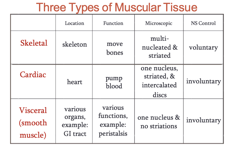
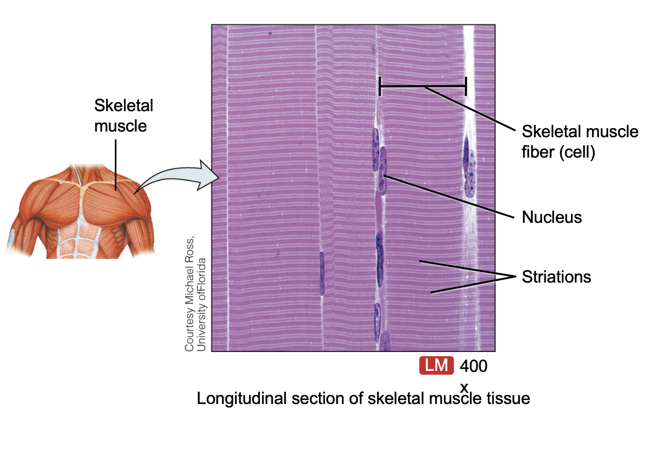
Skeletal Muscle
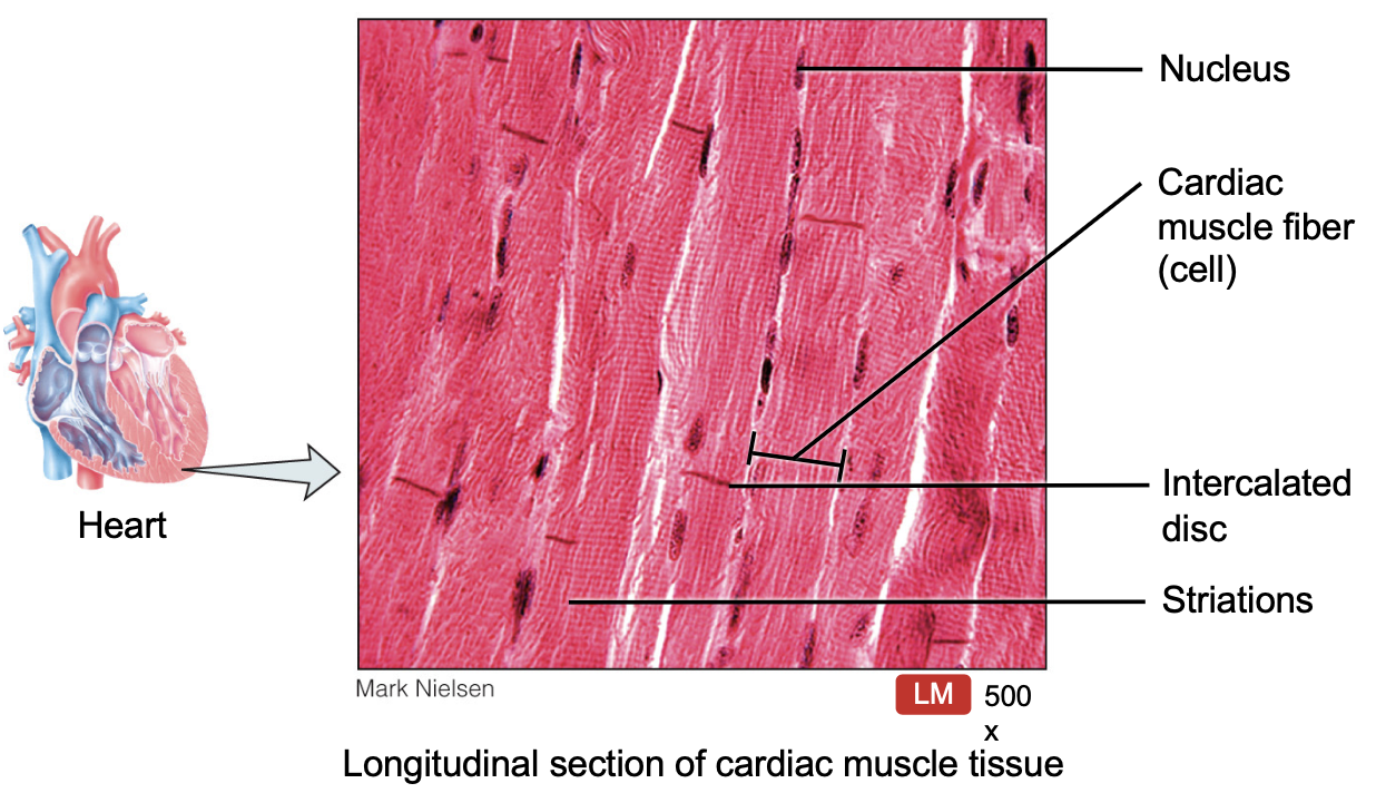
Cardiac muscle tissue
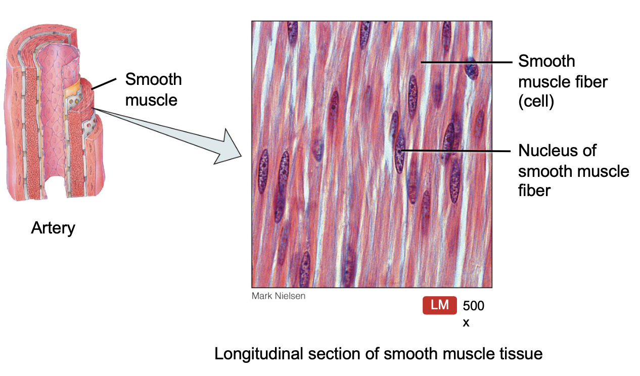
Smooth Muscle
Functions of Muscles
Producing Body Movements
Stabilizing body positions
Storing & moving substances within the body
Generating heat, thermogenesis
Anatomy of the Skeletal Muscles
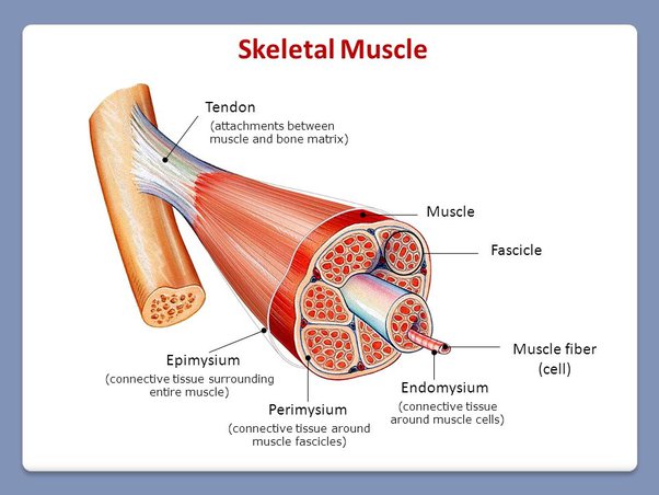
Each muscle cell is covered by connective tissue called endomysium
The muscle cells are arranged in small bundle (fasicles) covered by connective tissue around the bundles called the perimysium
The entire muscle is then encased in a thick layer of connective tissue known as the epimysium.
Perimysium covering the fascicle
3 Layers (Outside to Inside)
Epimysium - Around entire muscle
Perimysium - Around fascicles (bundles)
Endomysium - Around individual muscle fibers
Mnemonic
"EPI-PERI-ENDO = Entire-Part-Individual
EPImysium = Entire muscle
PERImysium = Part of muscle (fascicles/bundles)
ENDOmysium = Individual fibers
Tendons: Tendons are extensions of the three coverings attaching the muscle to the periosteum of the bone
youtube: anatoli
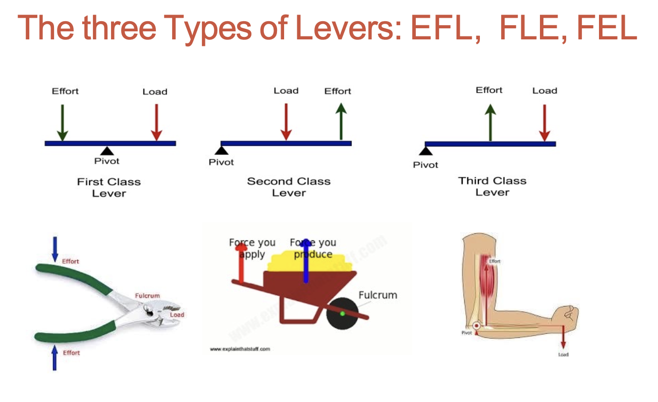
3 Types of levers: EFL, FLE, FEL
Mnemonic “123 FRE”
Fulcrom
Resistance
Effort
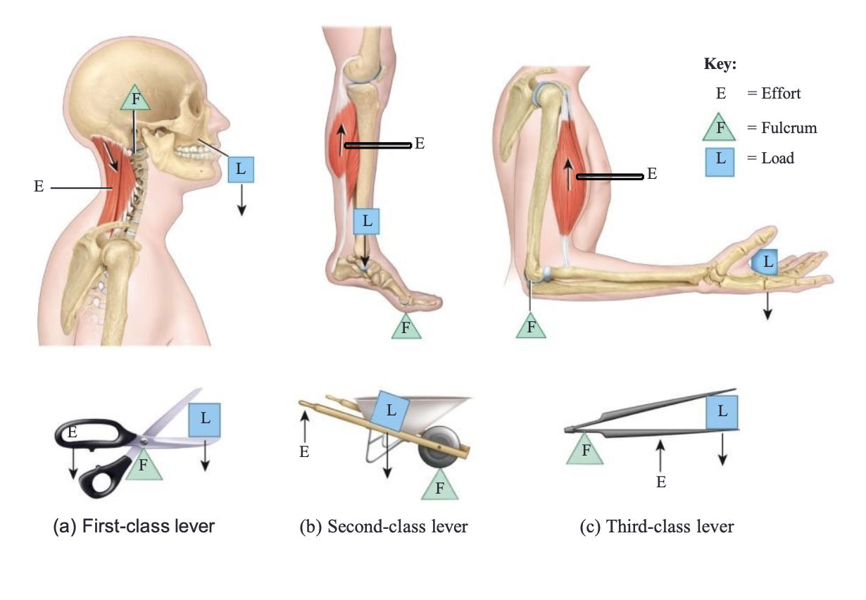
1st class - Fulcrom in middle = mechanical advantage / mechanical disadvantage
2nd class - Resistance in middle, fulcrom on one end, effort on other = mechanical advantage
3 class - Effort in middle, fulcrom on me end, resistance on the other = mechanical disadvantage
Effects of Fascicle Arrangement
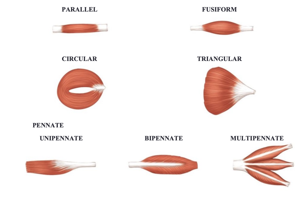
The muscle fascicles, muscle fibers are all arranged parallel to each other
The 5 pattern with respect to tendons:
Parallel
Fusiform (spindle-shaped) - hamstrings, bicep
Circular - esophogal sphincter, eyes
Triangular - pectoralis major
Pennate (feather shaped) - extensor digitorum
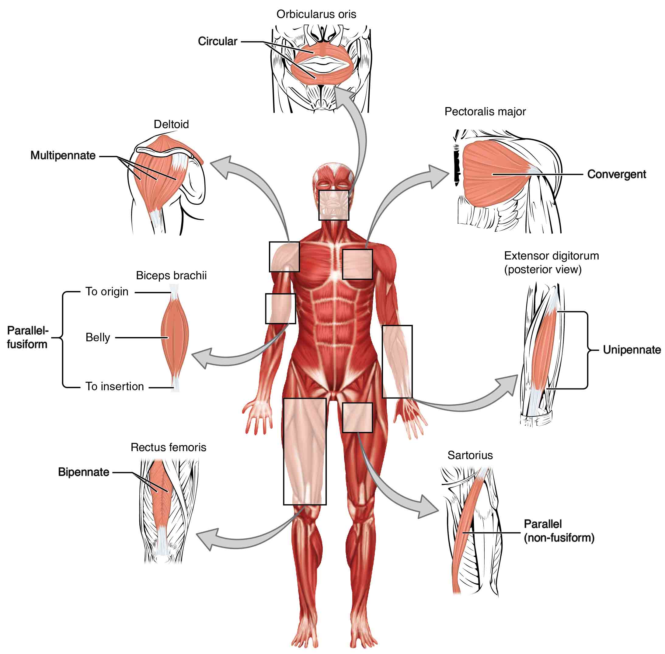
Parallel - stretch
Pennate - deltoid
Arrangement of muscles for movement
Agonist - prime mover
Antagonist - stretches / yields to effects of prime mover
Synergist - aids prime mover
Fixator - stabilizes