endocrine system pt 2 extra notes
Posterior Pituitary Gland Physiology
Hypothalamic Control of Posterior Pituitary
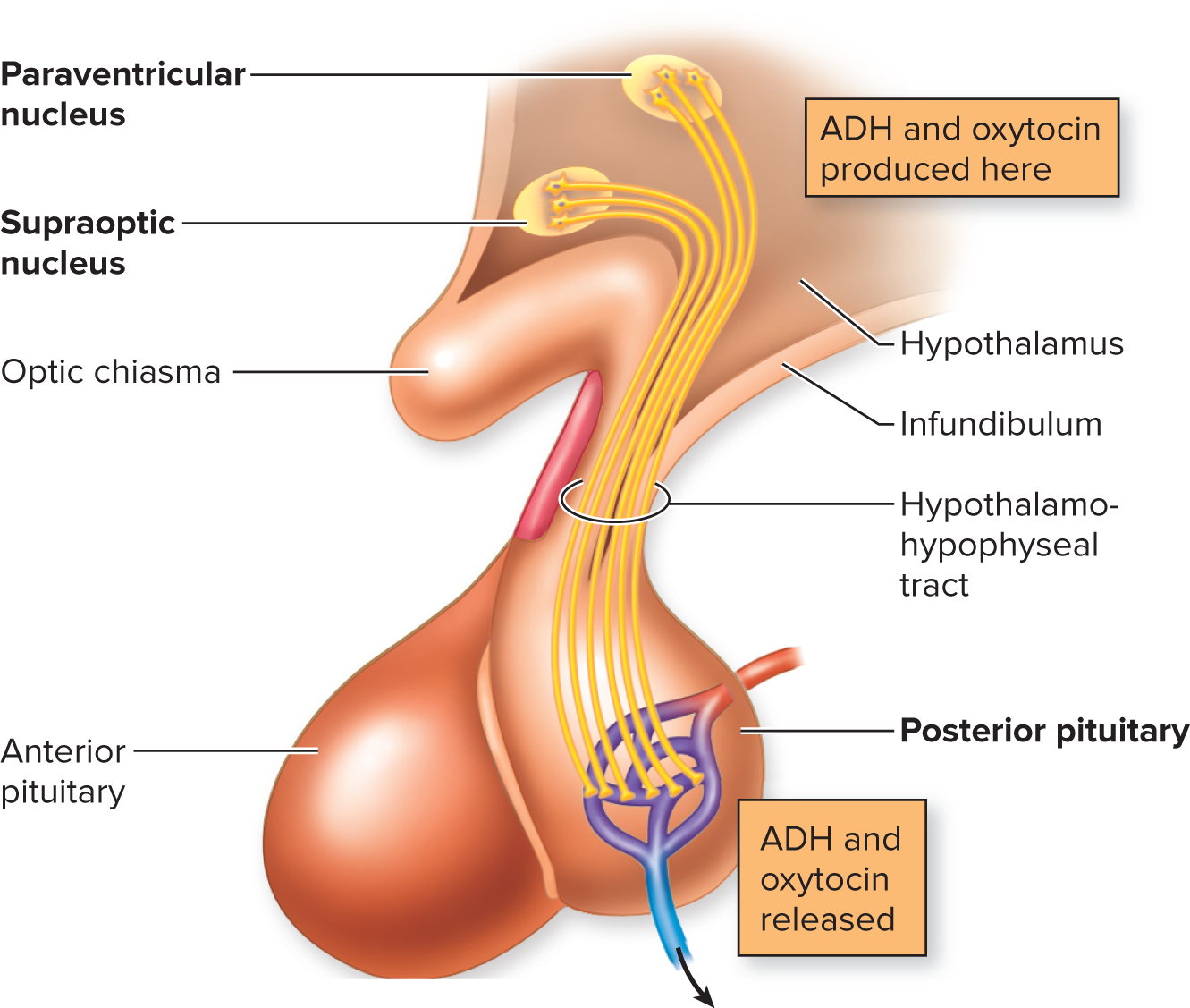
hormones are produced in neurons that are in the hypothalamus
hypothalamic nuclei are located in the hypothalamus
hormones are transported along axon of hypothalamo-hypophyseal tract
hypothalamo-hypophyseal tract: tract of nerve fibers (axons) that transports antidiuretic hormone and oxytocin from the hypothalamus to the posterior pituitary.
hormones are released from synaptic knob into capillary bed, entering the blood stream in the posterior pituitary
neurons axons extend into posterior pituitary gland, releasing hormones directly into capillary bed
although the messenger signal is made in a neuron, it is called hormones (instead of neurotransmitter) because released directly into blood
would be called neurotransmitter if it was released directly on synapse
2 Hormones Released by the Posterior Pituitary Lobe:
1. Antidiuretic Hormone (ADH)
Target: Kidneys and blood vessels.
Effects:
stimulation of water retention in kidneys
less water is secreted in the urine
what gives the hormone its name
vasoconstriction of blood vessels.
rise in blood pressure due to vasoconstriction
Secreted when hydration levels are low (dehydration).
Regulation:
secretion of ADH is stimulated by hypothalamic osmosensors and angiotensin II
osmoreceptors neurons: A sensory neuron in hypothalamus that responds to changes in the osmotic pressure of the surrounding fluid.
secretion of ADH is inhibited by Atrial baroreceptors
baroreceptors is a type of mechanoreceptor that is a stretch receptor
located in left atrium of the heart
stimulated when there is a rise in blood volume, causing inhibition
2. Oxytocin
Target: Mammary glands and uterus.
Effects:
Triggers milk ejection during lactation
stimulates contraction of mammary gland alveoli and ducts
stimulates uterine contractions during childbirth.
Regulation: More complex regulation, particularly in reproductive scenarios.
Anterior Pituitary Gland Physiology
Hypothalamic Control of Anterior Pituitary
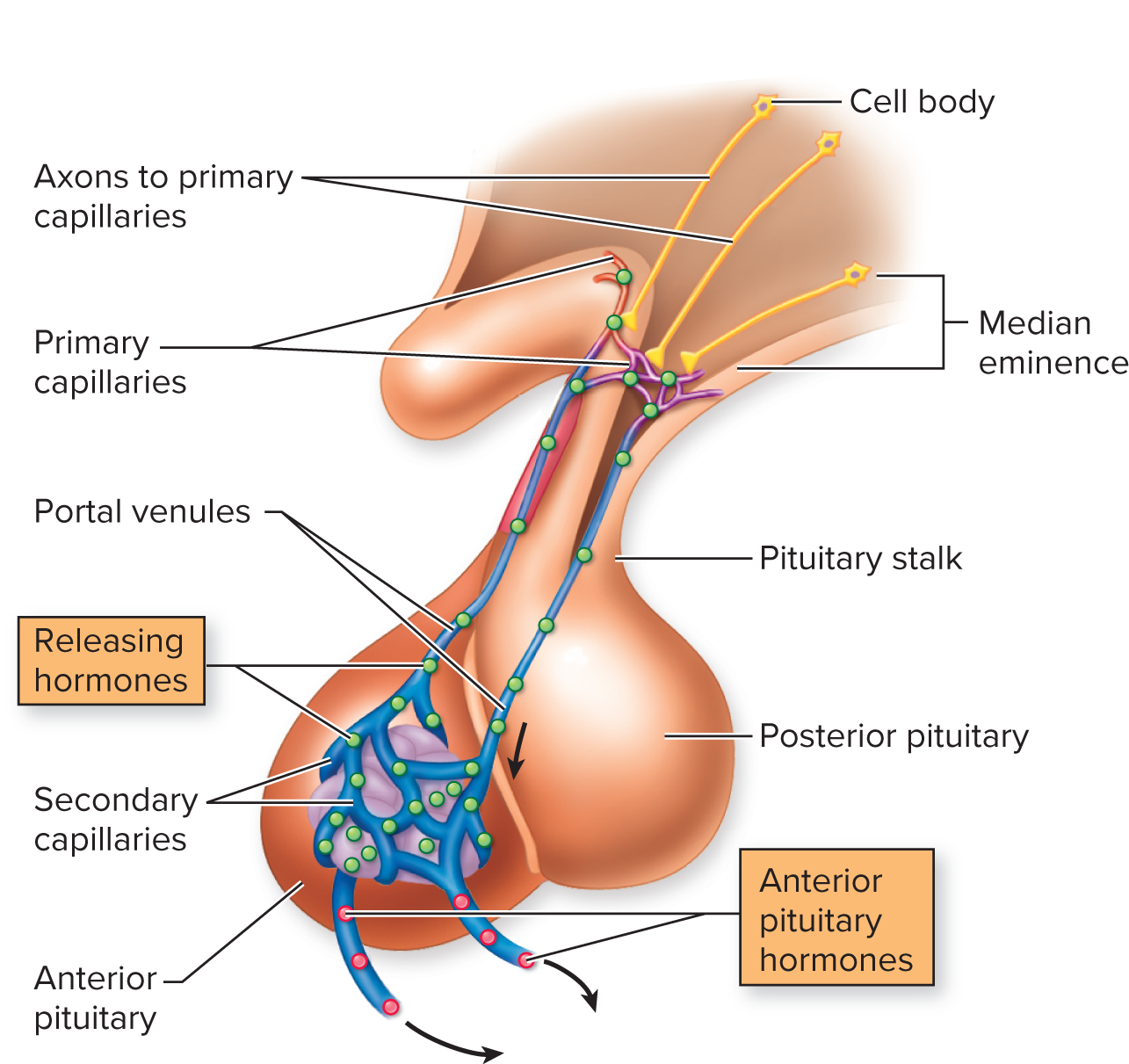
Axons are only in hypothalamus, do not enter anterior pituitary lobe
Neurons secrete regulatory hormones into hypophyseal portal system
hypophyseal portal system: vascular system with contains two capillary beds, transporting hormones from the hypothalamus to the anterior pituitary
regulatory hormones are secreted into first capillary bed at the base of the hypothalamus
then delivered to another capillary bed in anterior pituitary gland
Regulatory hormones either stimulate or inhibit release of hormones
Negative feedback loop can either work at the top in the hypothalamus or at the bottom in the anterior anterior pituitary lobe
Hypothalamic Regulatory Hormones: hormones released in the hypothalamus
Growth Hormone-Releasing Hormone (GHRH)
Growth Hormone-Inhibiting Hormone (GHIH)
Thyroid-Releasing Hormone (TRH)
Corticotropin-Releasing Hormone (ACTH)
Prolactin-Inhibiting Hormone (PIH)
Gonadotropin-Releasing Hormone (GRH)
Anterior Pituitary Hormones: hormones released at anterior pituitary gland
Growth Hormone (GH)
Thyroid-stimulating Hormone (TSH)
Adrenocorticotropic Hormone (ACTH)
Follicle-stimulating Hormone (FSH)
Luteinizing Hormone (LH)
Prolactin (PRL)

1. Growth Hormone (GH): released at anterior pituitary gland
Targets: Bone, muscle, adipose tissue, cartilage, and many others
Effects:
Stimulates growth in bone, cartilage, and muscle
increase lipolysis and lipid immobilization
break down of lipids (fats) and mobilizes it
Hypothalamic Regulatory Hormones:
Growth hormone-releasing hormone (GHRH): stimulates release of growth hormone
Growth hormone-inhibiting hormone (GHRH): inhibits release of growth hormone
Hypothalamic-Pituitary-Somatotropic Axis - Hypothalamic Regulation of Growth Hormone:
Negative feedback loop
Pituitary gland secretes growth hormone (GH)
increase of growth hormone (GH) causes increase in somatomedins from the liver
increase of somatomedins levels blocks growth hormone-release hormone AND stimulates growth hormone-inhibiting hormone in the hypothalamus
NOT negative product inhibition

2. Adrenocorticotropic Hormone (ACTH): released at the pituitary gland
adreno: target of hormone
cortico: cortisol
tropic: ACTH is a tropic hormone
Tropic Hormone: target is another endocrine gland
ACTH is released from pituitary gland (endocrine gland) and targets adrenal gland (another endocrine gland)
Trophic Hormone: causes growth in target
Target: Adrenal cortex.
(adrenal medulla is stimulated by Sympathetic Nervous System from hypothalamus)
Effects: Stimulates the secretion of glucocorticoids (e.g., cortisol)
glucocorticoids: hormones whose effects include glucose metabolism
also stress hormones
increases gluconeogenesis
gluconeogenesis: production of glucose from a non-carbohydrate
increases lipolysis
increase protein degeneration
protein degeneration: protein digestion
depresses immune/inflammatory response
all stress hormones have same functions on immune system
Hypothalamic Regulatory Hormones:
Corticotropin-Releasing Hormone (CRH)
tropic hormone
targets pituitary gland to stimulate release of Adrenocorticotropic Hormone (ACTH), causing release of cortisol
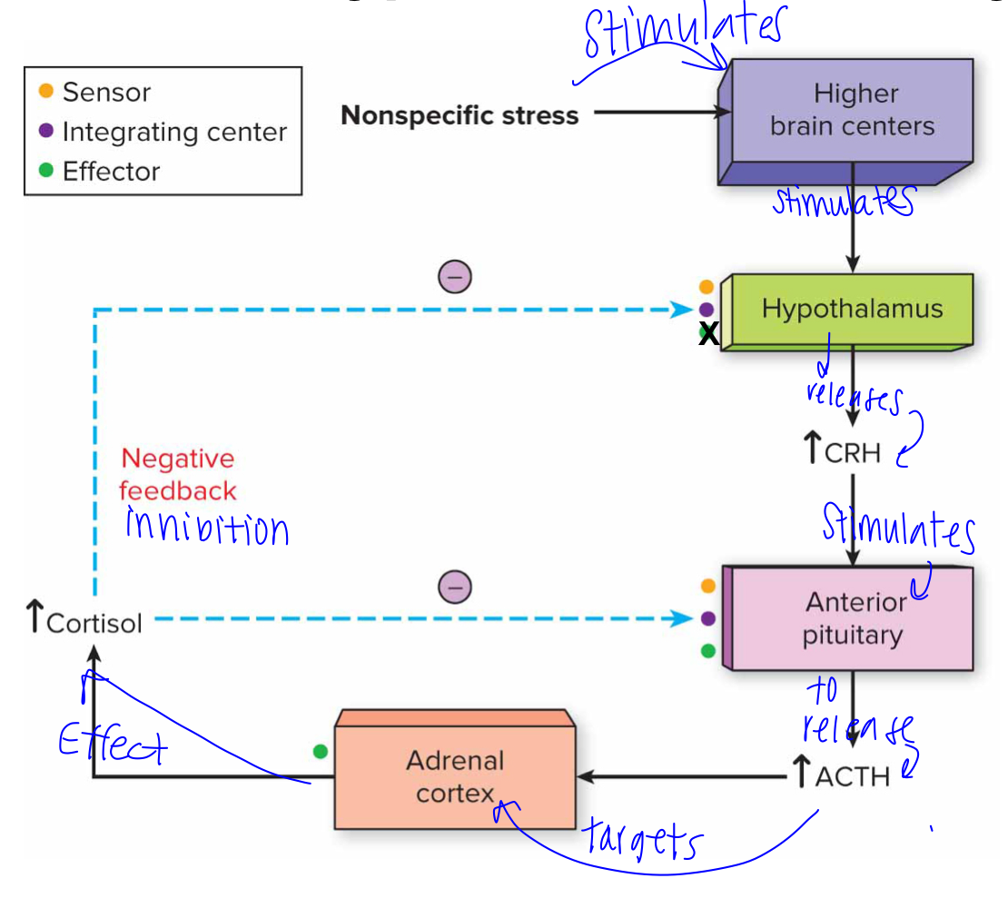
stress stimulates higher brain centers higher brain centers stimulate hypothalamus to release CRH CRH stimulates anterior pituitary gland to release ACTH ACTH targets adrenal cortex adrenal cortex increases cortisol levels
Hypothalamic-Pituitary-Adrenal Axis:
anterior pituitary often has negative feedback inhibitions at two levels:
hypothalamus
increase of cortisol inhibits hypothalamus’ secretion of Corticotropin Releasing Hormone (CRH)
hypothalamus has cortisol receptors that detect elevated cortisol levels
hypothalamus acts as sensor and integrating center
leads to a decrease in CRH production
adrenal cortex acts as effector, decreasing amount of cortisol being secreted
Pituitary
increase in cortisol inhibits anterior pituitary’s responsiveness of Corticotropin Releasing Hormone (CRH)
anterior pituitary has cortisol receptors detect elevated cortisol levels, decreasing responsiveness to CRH, leading to a reduction in Adrenocorticotropic Hormone (ACTH) secretion and thus lower cortisol production from the adrenal glands.
anterior pituitary acts as sensor, integrating center and effector
Adrenal Insufficiency Disease: disease involving hypo-secretion (not enough secretion) of glucocorticoids
Two Types:
Primary Adrenal Insufficiency
disease of adrenal cortex
adrenal cortex is stimulated by ACTH but is unable to produce cortisol
most common type: Addison’s disease
autoimmune disease of the adrenal cortex
other causes include congenital, drug-related, infections
common symptoms: hair loss, blurred vision, abdominal pain, decreased appetite, darkening of skin, shaking or tremors, depression
Secondary Adrenal Insufficiency
disease at the anterior pituitary where it does not produce enough ACTH, leading to inadequate stimulation of the adrenal cortex and resulting in decreased cortisol production.

Testing to diagnose primary or secondary adrenal insufficiency: injection of ACTH
if nothing happens after injection, diagnosis is primary adrenal insufficiency
problem is in the adrenal cortex because adrenal cortex does not increase levels of cortisol
if injection causes an production of cortisol, diagnosis is secondary adrenal insufficiency
problem is in the anterior pituitary because it fails to produce adequate amounts of adrenocorticotropic hormone (ACTH), which stimulates the adrenal cortex to release cortisol.
Cushing's Syndrome: hyper-secretion (excessive secretion) of glucocorticoids causing increased lipolysis and redistribution of fat
Two types:
Primary Cushing Syndrome: typically from tumor in adrenal cortex
excessive cell division of adrenal cortex cells, causing more cells to secrete cortisol
Secondary Cushing Syndrome: typically from tumor in pituitary gland
excessive cell division of pituitary gland cells, causing more cells to secrete ACTH, increasing cortisol levels

3. Thyroid-Stimulating Hormone (TSH): released at anterior pituitary gland
Target: Thyroid gland.
tropic hormone AND Trophic hormone
tropic: released by anterior pituitary gland
trophic: increase size of thyroid
Effects: Stimulates the secretion of thyroid hormones, Triiodothyronine (T3) and Thyroxine (T4)
increases basal metabolic rate and body temperature
increases catabolism of carbohydrates and proteins
Hypothalamic Regulatory Hormones:
Thyrotropin Releasing Hormone (TRH)
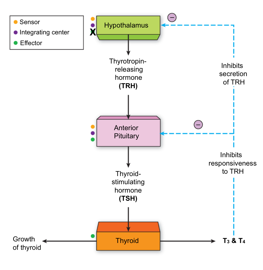
hypothalamus secretes Thyrotropin Releasing Hormone (TRH) TRH triggers anterior pituitary gland to produce thyroid-stimulating hormone (TSH) TSH causes thyroid to secrete Triiodothyronine (T3) and Thyroxine (T4) AND increases growth of thyroid
Hypothalamic-Pituitary- Thyroid Axis:
Negative Feedback Inhibition at two levels:
hypothalamus
increase of T3 and T4 inhibits hypothalamus’ secretion of Thyrotropin Releasing Hormone (TRH)
hypothalamus has receptors that detect elevated T3 and T4 levels
hypothalamus acts as sensor and integrating center
leads to a decrease in TRH production
thyroid acts as effector, decreasing amount of T3 and T4 being secreted
Pituitary
increase in T3 and T4 inhibits anterior pituitary’s responsiveness of Thyrotropin Releasing Hormone (TRH)
anterior pituitary has T3 and T4 receptors detect elevated T3 and T4 levels, decreasing responsiveness to TRH, leading to a reduction in TSH secretion and thus lower T3 and T4 production from the thyroid glands
anterior pituitary acts as sensor, integrating center and effector
Synthesis of thyroid hormone requires iodide (iodine)
iodide must be in diet
if there is not enough iodide in diet, there is no production of Triiodothyronine (T3) and Thyroxine (T4)
Goiter Disease: insufficient dietary iodide leads to hypertrophy of thyroid gland
excessive growth of thyroid gland
if not enough iodide, leads to low levels of T3 and T4
results in lack of negative feedback inhibition causing anterior pituitary gland to secrete excessive TSH
excessive TSH causes abnormal growth of thyroid

Graves’ Disease: an autoimmune disease, caused by auto-antibodies binding to TSH receptors, which stimulates overproduction of thyroid hormones

Hypothalamic Regulation of Adrenal Medulla
Sympathetic Nervous System Control of Adrenal Medulla
adrenal medulla is stimulated by hypothalamus through sympathetic innervation
Effects: stimulate secretion of epinephrine and norepinephrine, which target other organs:
increase heart rate
dilate bronchioles
vasoconstriction in skin
vasodilation in muscle
“Fight or Flight” Response

Hormonal Regulation of Blood Calcium Levels
Calcitonin and Parathyroid Hormone (PTH)
Calcitonin: produced by thyroid glands, inhibits dissolution of Ca2+ from bone and stimulates Ca2+ excretion
if blood calcium is too high, thyroid gland produces calcitonin
Lowers blood calcium by causing calcium deposition in bone (inhibiting osteoclast activity) and increasing renal excretion of calcium
leads to blood calcium levels decreasing
Parathyroid Hormone. (PTH): Produced by parathyroid glands, promotes dissolution of Ca2+ from bone and inhibits Ca2+ excretion
if blood calcium is low, parathyroid gland produces parathyroid hormone
raises blood calcium by releasing stored calcium from bone (stimulating osteoclasts and inhibiting osteoblast activity) and enhancing reabsorption of calcium in kidneys (decrease calcium excretion in kidneys)
increases blood calcium levels
Calcitonin and Parathyroid Hormone work in antagonist effects
Therapeutic Use of Cortisol Mimics
Cortisol Mimics: Synthetic molecules that act like cortisol, binding to cortisol receptors, causing same physiological response as cortisol
most often used for immunosuppressive effects
depresses immune’s systems response and reduces inflammation
Common Medications:
Prednisone
Prednisolone
Dexamethasone
Caution: Long-term use can lead to adrenal suppression
careful dosing and tapering are necessary to prevent withdrawal symptoms.