Cardiovascular System PP #1
THE HEART
- The heart is a hollow, muscular organ.
- It is located between the lungs, behind the sternum.
- Its exact location is in the pericardial cavity, which is in turn located in the thoracic cavity.
- The apex, or the lower tip of the heart, is on the left side.
- The human heart is about the size of a loosely clenched fist.
- The function of the heart is to pump blood throughout the body, delivering oxygen and nutrients.
THE PERICARDIUM
- The heart is located in the pericardial cavity.
- Within the pericardial cavity is the pericardial sac, which is a sac that surrounds the heart.
- The pericardial sac is a double-membraned sac.
- A membrane is a thin, pliable tissue layer that surrounds or covers a body part.
- The outer layer of the pericardium is called the parietal pericardium.
- The inner layer of the pericardium is called the visceral pericardium, or the epicardium. Viscer/o means “internal organ,” and –al means “pertaining to.” Epi- means “above,” cardi/o means “heart” and –um is a noun ending.
- Pericardial fluid is located between the parietal and visceral layers of the pericardium. This fluid acts as a lubricant during the beating of the heart, as well as a cushion against physical trauma.
WALLS OF THE HEART
The heart is a muscle composed of three layers:
- The epicardium is the outermost layer of heart muscle and the internal layer of the pericardium.
- When referring to the heart muscle, the term “epicardium” is used. When referring to the pericardium, the term “visceral pericardium” is used.
- The myocardium (my-oh-KAR-dee-um) is the muscular portion of the heart, and is the thickest of the three layers. My/o means “muscle,” cardi/o means “heart,” and –um is a noun ending.
- The myocardium is composed of cardiac muscle, which is also called myocardial muscle. This is a unique muscle type that maintains a constant contraction and relaxation, without conscious effort.
- The endocardium is the innermost layer of the heart, and is composed of squamous epithelial tissue.
- It is continuous with the blood vessels, and has direct contact with blood.
BLOOD SUPPLY TO THE HEART
- The heart requires a continuous blood supply, and it has its own independent supply, called coronary circulation.
- Coronary circulation consists of the coronary arteries and coronary veins.
- The coronary arteries start at the base of the aorta, which is the large artery that carries oxygenated blood to the heart.
- Because the coronary arteries are located at the starting point of the aorta, they provide a constant supply of oxygenated blood to the cardiac muscle.
- The coronary veins carry wastes away from the cardiac muscle.
THE CHAMBERS OF THE HEART AND BLOOD FLOW
- There are four distinct chambers of the heart, which are separated by walls called septa (SEP-tah) (singular = septum).
- The atria (atrium) are the upper two chambers of the heart, separated by the interatrial septum. All blood enters the heart through the right atrium.
- The ventricles are the lower two chambers of the heart, separated by the interventricular septum.
- The flow of blood through the heart follows a regular pattern.
- The right atrium gets deoxygenated blood (meaning that the oxygen in that blood has been distributed to the body tissues) from the superior and inferior vena cavae, which are the large veins that collect blood from the body
- The blood then moves to the right ventricle
- Then pulmonary arteries, deliver the blood to the lungs for oxygenation.
- Once the blood has been oxygenated, it returns to the heart via the pulmonary veins.
- Then it goes to the left atrium
- The blood then moves to the left ventricle,
- Lastly, it goes to the aorta and the rest of the body
- The left ventricle is the strongest of the two; it has to contract hard enough to send blood through the entire body, while the right ventricle only has to get the blood through the pulmonary artery.
VALVES OF THE HEART
- Just as there are four major chambers of the heart, there are four major valves in the heart which help control blood flow.
- These valves also prevent blood from flowing backwards through the heart.
- The tricuspid valve, also known as the right atrioventricular valve, controls the opening between the right atrium and the right ventricle.
- The term “tricuspid” refers to the shape of the valve, which has three cusps.
- The pulmonary semilunar valve is shaped like a half-moon and controls the opening between the right ventricle and the pulmonary artery.
- The mitral valve is also known as the bicuspid valve, and controls the opening between the left atrium and the left ventricle.
- It is referred to as the “bicuspid” valve because it has two cusps.
- The aortic semilunar valve is similar to the pulmonary semilunar valve, and controls the blood flow between the left ventricle and the aorta.
THE HEARTBEAT
- A heartbeat is the coordinated contraction and relaxation of the chambers of the heart to pump blood efficiently.
- Each heartbeat is regulated through electrical impulses from the nervous system, which stimulate the myocardium.
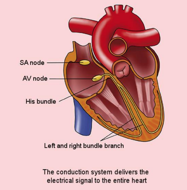
- The electrical impulses are controlled by the conduction system of the heart, which consist of three electrical nodes:
- The sinoatrial (SA) node
- The atrioventricular (AV) node
- The bundle of HIS (HISS)
THE CONDUCTION SYSTEM
- The sinoatrial (SA) node is located on the back wall of the right atrium, near the superior vena cava.
- This node establishes the rhythm and rate of the heart’s beats, so it is also referred to as the natural pacemaker.
- It starts each wave of muscle contraction. The impulse of the right atrium spreads to both atria, stimulating contraction to force the blood flow to the ventricles.
- The atrioventricular (AV) node is located on the floor of the right atrium, near the interatrial septum.
- The AV node transmits the electrical impulse to the Bundle of His.
- The bundle of His is a group of fibers in the interventricular septum, and travels to the right and left ventricles.
- The bundle of His ensures the sequence of contractions remains steady through its fibers, called Purkinje fibers.
- The Purkinje fibers run throughout the ventricles, causing them to contract.
- This contraction forces the blood out of the heart to either the pulmonary artery (from the right ventricle) or the aorta (from the left ventricle).
ELECTRICAL WAVES OF THE HEART
- The actions of the electrical conduction system can be visualized as electrical waves, which can help determine whether a heartbeat is normal or abnormal.
- A normal heartbeat is described as a sinus rhythm.
- The image on this slide shows a sinus rhythm.
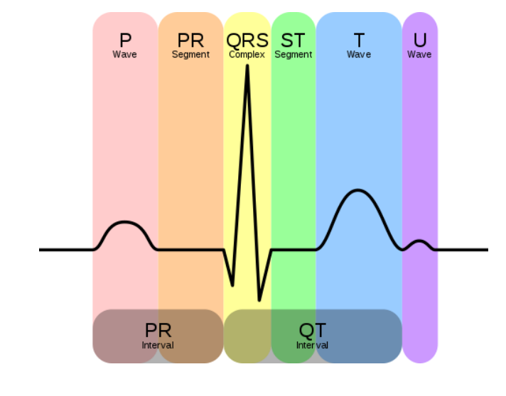
- The P wave correlates with the stimulation of the atria by the SA node.
- The QRS complex shows the simulation of the ventricles.
- The T wave shows the relaxation of the ventricles.