Lecture 1- levels of organizations, feedback loops, Radiology, Sonograms
Levels of Organization of the Human Body
Levels (as listed on the slide):
Chemical components – Hydrogen, oxygen atoms and water
A cell is the smallest independently functioning unit of a living organism. Includes cell membrane, cytoplasm and organelles
A tissue is a group of many similar cells (though sometimes composed of a few related types) that work together to perform a specific function.
Skeletal muscles are voluntary, controlled by your brain impulses.
Smooth muscles are involuntary, controlled automatically
An organ is an anatomically distinct structure of the body composed of two or more tissue types. Each organ performs one or more specific physiological functions.
An organ system is a group of organs that work together to perform major functions or meet physiological needs of the body.
Organ Systems of the Human Body
1. Integumentary System
Skin and layers of skin, includes hair, nails, and glands which protect inside layers from outside environments
2. Skeletal System
3. Muscular System
4. Nervous System
5. Endocrine System
6. Cardiovascular System
7. Lymphatic System
8. Respiratory System
9. Digestive System
Urinary System
Reproductive System
Metabolism
Basic function of metabolism: to consume energy and molecules from food, convert some into fuel for movement, sustain body functions, and build/maintain body structures.
Two types of reactions: anabolism and catabolism
Anabolic reactions: building reactions into bigger compounds, requires energy.
Recovery, new growth
Example: Tissue injury
Catabolic reactions: break materials down into smaller compounds, release energy.
Example: Digestion
Metabolism includes both anabolic and catabolic reactions
ATP (adenosine triphosphate) is the chemical compound used by every cell to store and release energy
Homeostasis
Homeostasis: the state of steady internal conditions maintained by living things.
Set point - Normal range.
Example: Body temperature; normal set point at 37C (which equals 98.6F).
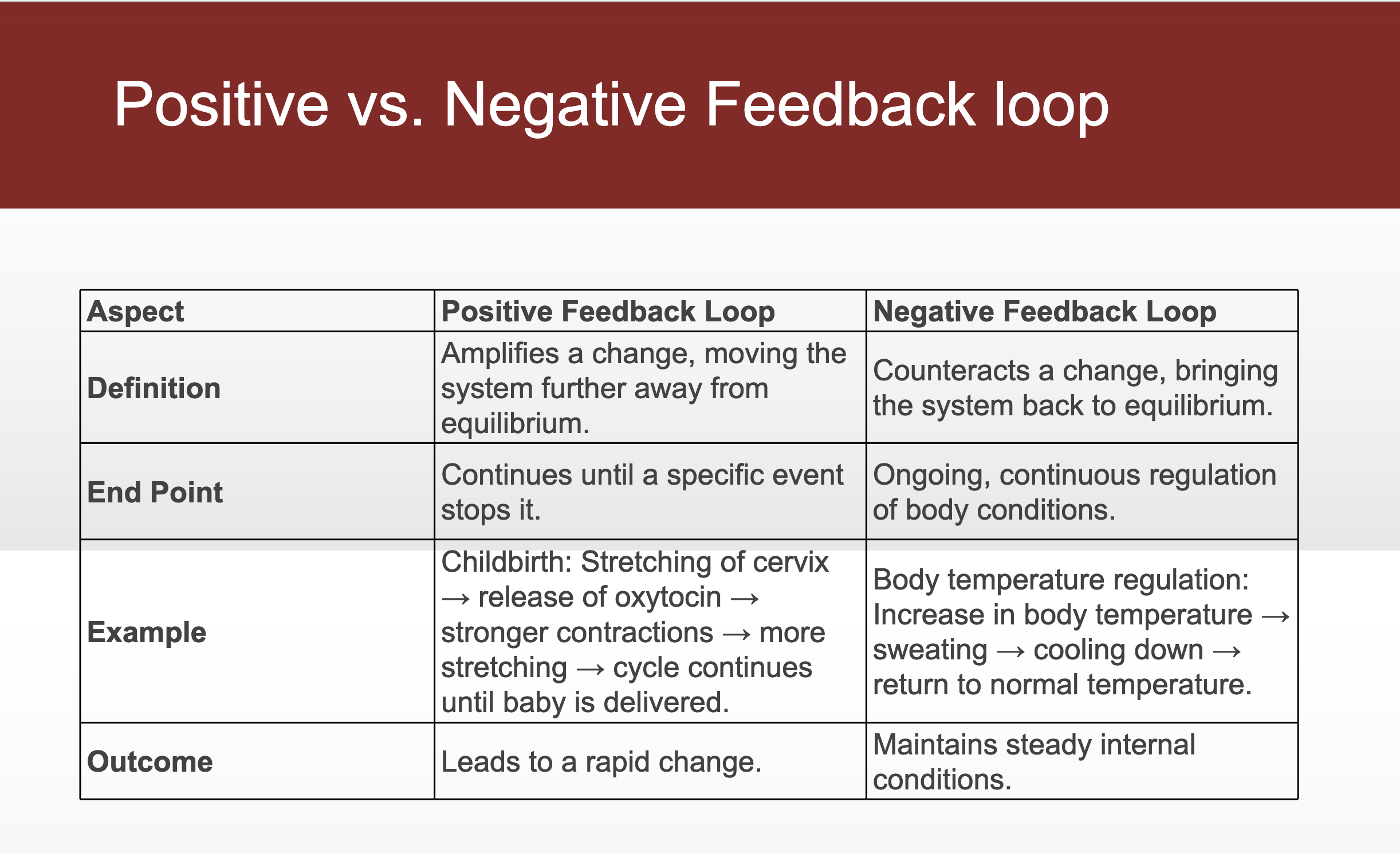
Negative feedback loop: When deviation is detected form set point, the body activates mechanisms to counteract the change and restore balance
Negative feedback loop maintains homeostasis with three parts:
Sensor (receptor): monitors deviations from the set point
Control: compares deviations to the set point; if deviations are too high, it activates the effector
Effector: helps return the body system to its set point
Body temperature regulation
Body temp exceeds 37C
Sensor: Nerve cells in skin and brain
Control: temp regulatory center in brain
Effector: sweat glands throughout body
Risk of developing seizures when there’s high temperature: when the brain is unable to regulate the temperature, especially in infants, this can lead to an overload in the brain, causing a seizure.
Positive Feedback Loop:A positive feedback loop results in a rapid change in the body’s status.
No continuous stimulus
Example: Childbirth
Stimulus - first contractions of labor push the baby towards the cervix
Sensors - stretch-sensitive nerve cells in the cervix, that monitor the degree of stretching
Effectors - nerve cells send signals to the brain causing the pituitary gland to release the hormone Oxytocin into the blood
Right after the child is born, Oxytocin is no longer secreted, because there is no longer a stimulus, therefore a positive feedback loop.
another example: blood clotting, lactation
Medical Imaging (Non-surgical Methods to Study the Living Body)
X-ray
High energy electromagnetic radiation with a short wavelength capable of penetrating solids and ionizing gases
Soft tissues appear gray; bones appear white
Good for visualizing teeth and bones
Drawback/risk: Radiation damage
Computed Tomography (CT)
Uses computers to analyze several cross-sectional X-rays to reveal minute details about structures
Magnetic Resonance Imaging (MRI)
Patient is exposed to magnetic fields and radio waves; the resulting radio signals are analyzed
Cancer cells may have a different signal pattern than normal tissue
Drawbacks: High cost, noise, and discomfort to the patient during imaging
Positron Emission Tomography (PET)
Uses radiopharmaceuticals that emit short-lived radiation
Advantage: studies physiologic activity (e.g., blood flow)
Ultrasonography
Uses high-frequency sound waves to generate an echo signal interpreted as real-time images of anatomy and physiology
Drawbacks: Cannot penetrate bone or gas