B2 : Structure Function and Main Disorders of the Cardiovascular System
pathway of blood in the body
- heart
- arteries
- arterioles
- capillaries
- venules
- veins
- back to the heart
Structure and Function of the Cardiovascular System
Structures:
- heart
- veins
- arteries
- capillaries
Two circulatory systems: Systemic and Pulmonary.
Cardiac cycle: Two phases of systole and diastole
Function:
- Circulates oxygen and nutrients to every cell in the body. (systemic circulation)
- Removes waste such as CO2 (gaseous exchange, pulmonary circulation)
- Protects body against disease and infection (white blood cells)
- Provides clotting after injury (platelets)
Blood vessels
Overview: where blood cells flow through to transport its materials to the entire body. Three types include Arteries Capillaries and Veins.
Keywords
==Lumen== - Cavity in which blood flows through
==Pressure== - force or rate in which blood cells flow
==Valves== - prevents backflow of blood. Only in veins (artery pressure is high enough to ensure one direction of blood)
==Walls== - thick as to maintain pressure and flow in arteries, thin as pressure is low in veins and capillaries
==arterioles== - small branch of artery connects to capillaries, venules - small branch of vein connecting to capillaries
A(away) - arteries: Vessel that flows blood away from the heart to all the cells in the body.
- High pressure + Has a pulse
- Oxygenated
- Bright red
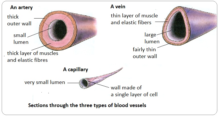
Lumen is small
Walls are thick and elasticated. Contains no valves.
Veins - flows blood towards the heart from the body. (IN)
- No pulse - low pressure
- Deoxygenated
- Purply red
Lumen is large
Walls are thin and do not stretch. Contains Valves
Capillaries
connects ARTERIES to VEINS
- Artery delivers oxygen rich blood to capillaries
- Exchange of oxygen and carbon dioxide occurs to all cells in the body
- Capillary delivers deoxygenated blood to Veins to be taken back to heart and lungs for oxygenation.
Allows diffusion of gases and nutrients from blood into the body cells, organs and tissues.
- Both deoxygenated blood + Oxygenated blood
- Decreasing pressure as blood transfers to veins from arteries - No pulse
Lumen is small. One red blood cell passes at a time.
One cell thick and permeable
No valves.
Structure and Function of Blood
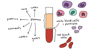
- Red blood cells (erythrocytes) 44% of Blood.
Structure: No nucleus. Doughnout shaped (maximises efficiency in transport) Filled with haemoglobin. Produced in bone marrow. Large surface area. Survives up to 120 days.
Function: To hold haemoglobin - the iron rich protein responsible for carrying oxygen to all cells.
- Plasma 55% of Blood.
Structure: Colourless fluid. Water, ions, proteins, nutrients, wastes, gases.
Function: The liquid in which blood cells float in. Aids in helping blood pressure to come up.
- Platelets
Structure: No colour. No nucleus
Function: Helps to clot cuts and wounds after an injury. Prevents excessive blood loss.
- Leukocytes (White Blood Cells)
Two types - Neutrophils (most common type and most essential part of immune system)
and Monocytes ( influences process of adaptive immunisation e.g when given immunisation jabs and vaccines )
Function: Mostly helps to protect from developing infection. Forms the immune system.
Engulfs and destroys bacteria fungi and virus’s (but not all)
Pulmonary and Systemic Circulation - Double Circulation Cycle
double circulatory system - blood passes through the heart twice each time it goes round the body.
Pulmonary circulation
the movement of blood from the heart to the lungs and back to the heart again.
- Deoxygenated blood leaves through the right ventricle and through the pulmonary artery.
- Deoxygenated blood goes to the lungs where blood absorbs oxygen and releases CO2.
- Oxygenated blood returns to the heart via the pulmonary vein.
gas exchange - movement of gas from area of high concentration to area of low concentration.
Systemic Circulation
movement of blood between heart and the rest of the body and back to the heart again.
- Heart pumps oxygenated blood out of the left ventricle and into the aorta.
- Blood supplies cells throughout the body with oxygen and nutrients
- Returns as deoxygenated blood to the superior vena cava.
Cardiac Cycle
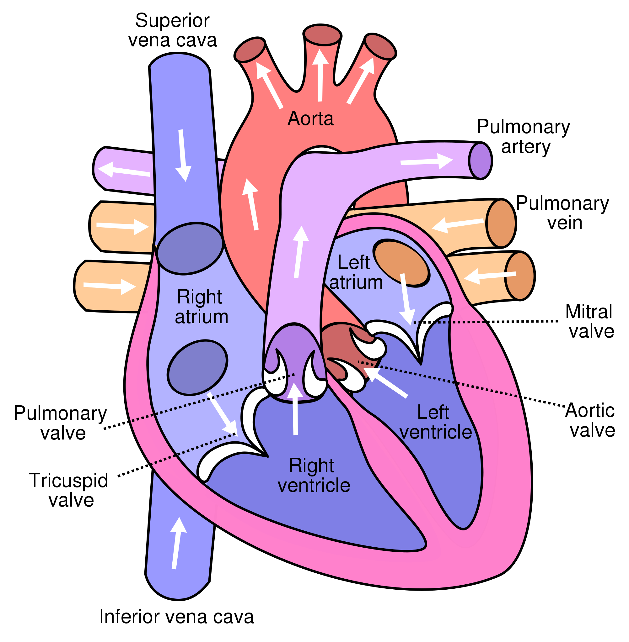
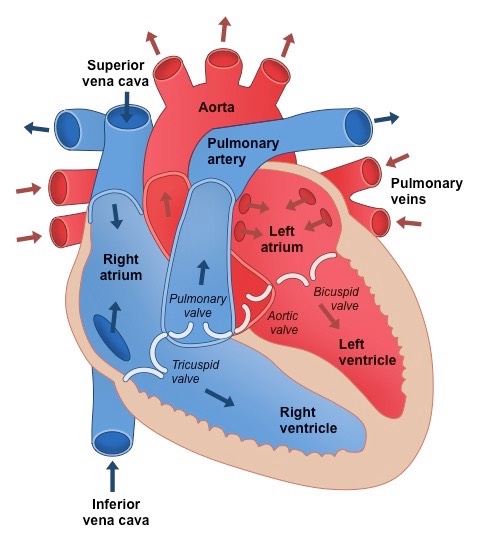
Deoxygenated blood enters the superior and inferior vena cava and into the right atrium
Deoxygenated blood then travels through the tricuspid valve into the right ventricle
Deoxygenated blood via the pulmonary valve then travels into the pulmonary artery out of the heart towards the lungs for oxygenation and gaseous exchange.
Blood releases CO2 and absorbs oxygen.
Oxygenated blood returns to the heart via the pulmonary vein.
Blood enters the left atrium and through the mitral valve enters the left ventricle.
Blood then travels through the aortic valve and out the aorta to the rest of the body.
Blood delivers its oxygen and becomes deoxygenated again and returns back to the vena cava.
Heart structure
There are 4 chambers of the heart
Lower 2 chambers - Ventricles
Top 2 chambers - atria (atrium).
Septum - the wall separating left and ride sides of the heart.
Valves in order - 1) Tricuspid Valve - right atrium to right ventricle
- Pulmonary Valve - right ventricle to pulmonary artery
- Mitral valve - left atrium to left ventricle
- Aortic Valve - left ventricle to aorta
semilunar valves - aortic valve and the pulmonary valve. Blood flows up.
atrioventricular valves - mitral valve and tricuspid valve. Blood flows down.
Movement of the Heart
Systole - heart muscles contract and pumps blood out of its chambers.
==Atrial Systole== - Muscles in atria walls contract
Blood is pushed into the ventricles
Semi Lunar Valves (pulmonary and aortic valves) are closed. AV valves (tricuspid and mitral valves) are open.
==Ventricular Systole== - Muscles in ventricle walls contract
Blood is pushed upwards. AV valves close and Semi Lunar valves open.
Blood flows into arteries (aorta and pulmonary artery)
==Diastole== - Heart muscles are relaxed.
Blood trickles into the heart from veins (superior and inferior vena cava)
Semi lunar valves are closed. AV valves are open.
Heart Rate & Regulation
sympathetic nervous system, (part of the autonomic nervous system)
speeds up the heart and causes blood vessels to contract.
Regulates sweat glands and during stress ] heart rate increases
parasympathetic nervous system - conserves energy by slowing heart rate.
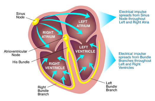
sinoatrial node (SA NODE) specialised cells - pacemaker of the heart. controls rhythym of the heart. LOCATED IN RIGHT ATRIUM.
atrial ventricular node - cells between atria and ventricles.
process
SA node - generates impulse and contracts the atria. Blood is forced into ventricles
AV node slows impulse before entering ventricles. Delay of impulse enables atria to contract before ventricles.
Bundle of His & Purkinje network - pathway of fibers sends impulse to muscular walls of ventricles where they then contract.
Blood is then forced out of the heart - to lungs and body.
SA node fires another impulses and cycle starts again.