
A&P: Cellular Biology
FALL SEMESTER 2024
Instructor: Anthony Uzwiak
uzwiak@dls.rutgers.edu

Cellular biology digs deeper into the system and function of the cell.
Describe the structure and function of the cell membrane
Describe types of non-active and active transport through the plasma membrane
Describe the contents of the cytoplasm and its organelles
Describe the structure and function of the endomembrane system
Describe gene expression
Describe the components and functions of the cytoskeleton

Cells are the basic units of all living things. In humans, cells can vary in size, structure, and function, but all share several common characteristics.
Cells as Units: Cells are the fundamental functional and structural units of living organisms.
All life forms are made up of cells.
Activity Dependence: The overall activity of an organism relies on both the individual and collective activities of its cells.
Biochemical Activities: The biochemical activities of cells are determined by their subcellular structures, reflecting the principle of complementarity.
The structure of a cell influences its function.
Continuity of Life: The continuity of life is fundamentally based on cellular processes.
All living things come from pre-existing cells.
Major Parts:
Nucleus: Contains genetic material— directs cell activity
Located near the center of the cell
Consists of nucleoplasm surrounded by a nuclear envelope.
Nuclear envelope is composed of two membranes separated by a space
Nuclear envelope has nuclear pores, openings where molecules move between the nucleus and cytoplasm
Cytoplasm: Houses organelles and is the site of most cellular activities.
Contains organelles
Plasma Membrane: The outer boundary of the cell that regulates the entry and exit of substances.
Organelles:
Ribosomes: Located on the (rough) endoplasmic reticulum, serves as site of protein synthesis.
Free ribosomes float freely into the cytoplasm and synthesize proteins used in the cell
Endoplasmic reticulum: Network of membranous tubules and sacs (cisternae) within the cytoplasm
Continuous with nuclear envelope
Two regions— smooth ER and rough ER
Smooth ER: Lacks ribosomes
Calcium storage
Synthesis of lipids, phospholipids, and steroids
Carbohydrate metabolism
Detoxifies drugs
Rough ER: protein synthesis (has ribosomes)
Ribosomes attached to ER synthesize secretory proteins
Growing polypeptide is threaded through ER membrane into cisternal space
Protein folds into native conformation
Protein departs in a transport vesicle pinched off from the ER
Golgi apparatus: Modifies, packages, and distributes proteins from the rough ER (packaging system)
Organelle of stacked, flattened membranous sacs
Has polarity
Cis face— receives transport vesicles from rough ER
Trans face— pinches off vesicles
Rough ER products are modified as they mouth through golgi apparatus
Lysosome: Contains digestive enzymes and breaks things down
Digest all major classes of macromolecules
Digest bacteria
Digest organelles that are no longer functioning (autophagy)
Pompe’s Disease and Tay-Sachs disease are genetic disorders caused by enzyme deficiencies. Pompe’s disease is caused by the lack of acid alpha-glucosidase (acid maltase), causing glycogen to build up in the lysosomes. This causes muscle weakness, respiratory issues, and cardiomyopathy. Tay-Sachs disease is the deficiency of hexosaminidase A, which causes fat to accumulate in the nerve cells of the brain and spinal cord. This can result in neurological deterioration, loss of motor skills, and seizures. |
Peroxisome: Breaks down hydrogen peroxide using the enzyme catalase
Proteasomes: Large protein complexes containing enzymes that break down and recycle other proteins in the cell
Mitochondria: Converts oxygen into ATP (energy)
The powerhouse of the cell
The endomembrane system is the network of membranes within a cell that synthesize, modify, and transport the degradation of biomolecules. The system facilitates the transport of materials within the cell via vesicles.
The several key structures of the system are:
Nuclear Envelope
Endoplasmic Reticulum
Golgi Apparatus
Lysosomes
Vacuoles
The mitochondria has a double membrane (similarly to the plasma membrane.)
The outer membrane is permeable to small solutes.
The inner membrane contains embedded proteins involved in cellular work.
The material within the inner membrane is called the matrix.
The cristae are folds of the inner membrane.

The cytoplasm is the cellular material outside of the nucleus but inside the plasma membrane. It is made up of cytosol, organelles, and inclusions.
Cytosol is the fluid portion of the cytoplasm. It is a viscous solution.
Inclusions are non-functioning chemicals substances that are unique to cell types.
Within the cell also contains the cytoskeleton, which supports the cell and holds the nucleus + other organelles into place. It is a network of fibers throughout the cytoplasm that form a framework for support, movement, and regulation.
Functions of the cytoskeleton are:
Mechanical support to maintain shape
Allows cell to change shape when environment changes
Associated with motility
Regulatory role in transmitting signals from cell’s surface to its interior
The components of the cytoskeleton are responsible for changes in cell shape and movement of cell organelles. It is constructed from three types of fibers.
Microtubules (thickest)
Hollow tubes composed primarily of protein units called tubulin
Radiate from cell’s center
Determine cell shape
Provide tracks for organelle movement
Involved in separation of chromosomes during cell division
Actin Filaments/Microfilaments (thinnest)
Made up of contractile protein (actin)
Attach to cytoplasmic side of plasma membrane
Participate in muscle contraction
Localized cell contraction
Intermediate Filaments
Most stable and permanent cytoskeletal element
Act as guy wires to resist pulling forces on the cell
Fix organelle position

The plasma membrane serves five different important functions to the life/regulation of a cell.
Serves a boundary separating the cytoplasmic (intracellular) substances from outside substances (extracellular)
Enclose and supports the cell
Attaches cells to extracellular environment, or to other cells
Cells communicate and recognize each other through plasma membranes
Regulates what comes in and out of the cell
The plasma membrane is composed of a bilayer of phospholipids. The (polar, charged) hydrophobic tails are directed towards each other while the (nonpolar, uncharged) hydrophilic heads are directed toward the cytoplasm or extracellular fluid.
This structure of the plasma membrane is described as the fluid mosaic membrane. This is because the plasma membrane is neither rigid nor static in structure. It is highly flexible and can change shape and composition.
This fluidity allows:
Important means of distributing molecules within plasma membrane
If slight damage occurs, membrane can be repaired as phospholipids reassemble around damaged sites
Fluid nature enables membranes to fuse with one another
There are little carbohydrates in the plasma membrane. However, the carbs can combine with lipids to form glycolipids— and combine with proteins to form glycoproteins.
Glycocalyx is a combination of glycolipids, glycoproteins, and carbohydrates on the plasma membrane. It is involved in cell adhesion, protection, and signaling, allowing cells to recognize and interact with each other.
Basic structure and some functions of the plasma membrane are determined by lipids, however many other functions are determined by proteins— depending on location or attachment, proteins can be integral or peripheral.
Integral membrane proteins penetrate deeply into the lipid bilayer—extending from one surface to the other.
Includes channels and carriers
Peripheral membrane proteins are attached to the membrane surface.
Involved in enzymatic activity and structural support
Functions of membrane proteins include:
Transport
Enzymatic activity
Receptor sites
Intercellular junctions
Cell-cell recognition
Cytoskeletal and extracellular matrix attachment
In order to communicate between cells, cell membranes have membrane junctions. The three types of junctions are tight junctions, desmosomes, and gap junctions.
Tight junctions create an impermeable barrier between adjacent cells. Substances are prevented from passing through. They are barriers.
Desmosomes are anchoring junctions that provide mechanical stability to tissues. They provide structural support.
Gap junctions allow the passage of small molecules and ions. They allow passage.
The cell membrane is selectively permeable. This means that the membrane can allow certain substances to pass while blocking others.
There are multiple ways in which substances move through the membrane. These three transport methods are called passive, active, and bulk transport.
Passive Transport: Does not require energy
Passive Transport (Filtration): When water and solutes are forced through membrane or capillary by pressure
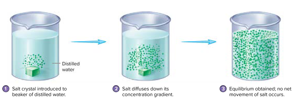
Diffusion— movement down a concentration gradient; movement of solutes from an a area of high concentration to lower concentration
A concentration gradient changes over a distance in a particular direction
Results from intrinsic kinetic energy
Affected by temperature and molecular size
Movement is random
Continues until dynamic equilibrium is reached
 |
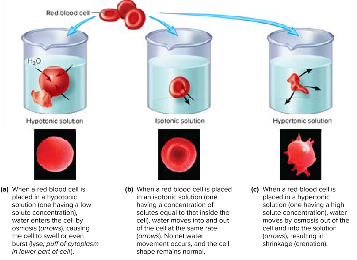
Osmosis— diffusion of a water (solvent) through a selectively permeable membrane; water movement
Hypertonic solution: solution with greater solute concentration than inside a cell
Hypotonic solution: solution with a lower solute concentration than inside a cell
Isotonic solution: equal parts solute in and out of the cell
Osmolarity— total concentration of all solutes in a solution
Osmotic pressure—amount of pressure required to prevent net movement of water into a solution
 |
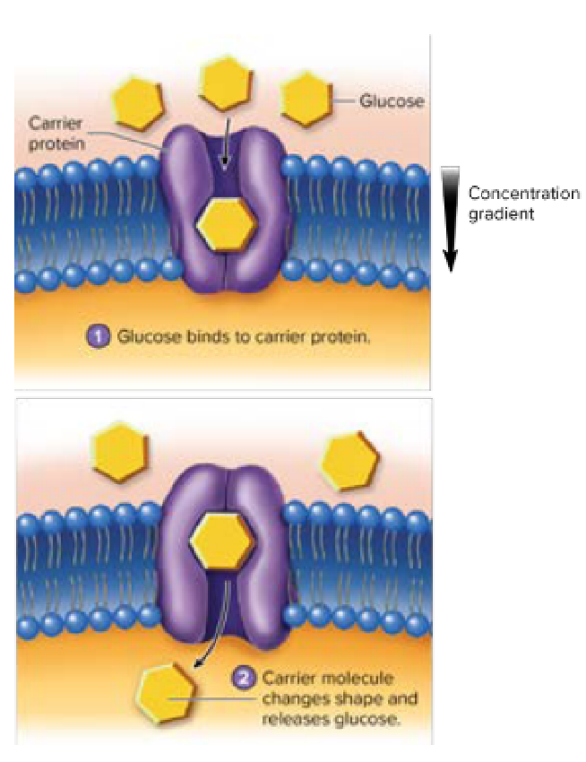
Facilitated diffusion— carrier proteins move substances across membrane
Occurs when molecules are too large to diffuse through membrane pores
Selective— not just size and lipid solubility (specific)
Limited by number of carrier molecules present (saturated)
 |
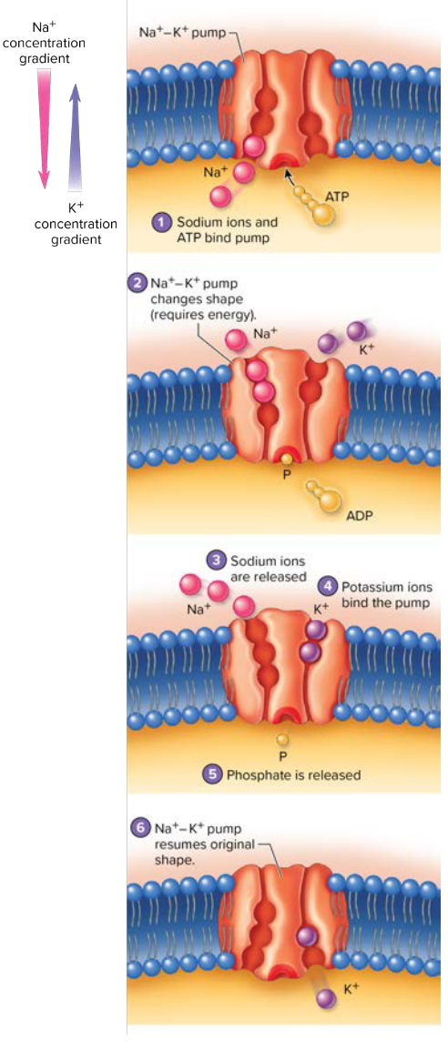
Active Transport: Transport process that requires energy provided by ATP
Cell uses energy to move substances across membrane
Transport molecules harvest energy from ATP to pump molecules against concentration gradients
Symport— same direction
Aniport— opposite direction
Sodium-potassium (Na+ K+ pump) exchange one substance for another using active transport
 |
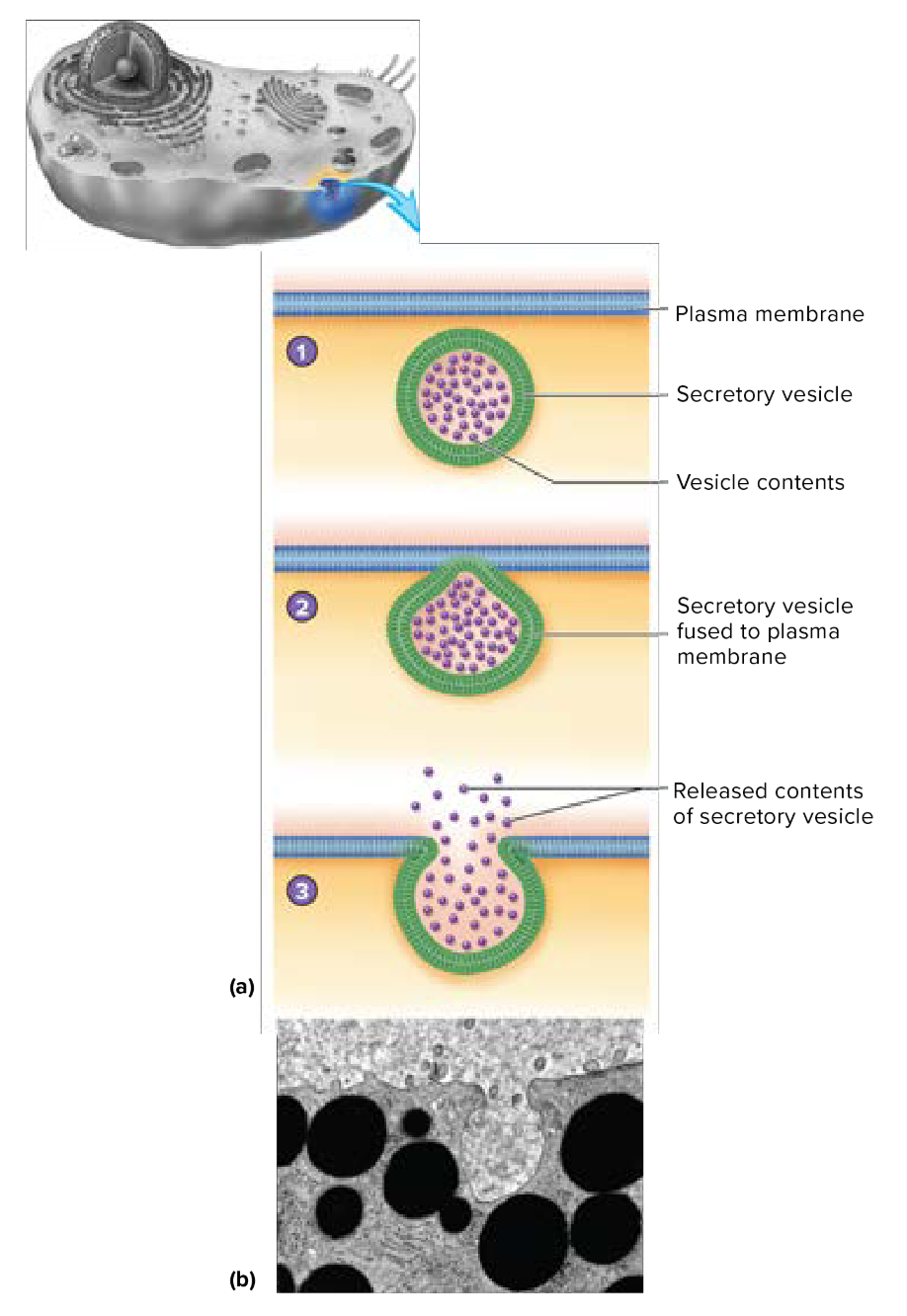
Bulk Transport (aka Vesicular Transport): Movement of large quantities of substances into or out of the cell using vesicles (membrane-bound sacs).
Exocytosis: Substance is released from vesicle
Fuses with membrane and releases contents to outside
 |
Endocytosis: Large substances enclosed by the membrane and taken into cell
Phagocytosis— “cell eating” of large particles
Solid particles are ingested and phagocytic vesicles are formed
Important in eliminating harmful substances from the body
Pinocytosis— “cell drinking” of fluids
Similar to phagocytosis, except that vesicles are smaller and material inside the vesicle is liquid
A coated pit are indentations in the plasma membrane which form when the membrane indents to capture extracellular material. Clathrin (a protein) helps stabilize and shape these pits to facilitate the formation of vesicles that transport materials into the cell. |
Endocytosis can exhibit specificity. The plasma membranes contain specific receptor molecules that recognize certain substances and allow them to be transported in or out of the cell via phagocytosis or pinocytosis. Receptor-mediated endocytosis is a term used to describe the specific uptake of molecules via receptors.
FALL SEMESTER 2024
Instructor: Anthony Uzwiak
uzwiak@dls.rutgers.edu
Cellular biology digs deeper into the system and function of the cell.
Describe the structure and function of the cell membrane
Describe types of non-active and active transport through the plasma membrane
Describe the contents of the cytoplasm and its organelles
Describe the structure and function of the endomembrane system
Describe gene expression
Describe the components and functions of the cytoskeleton
Cells are the basic units of all living things. In humans, cells can vary in size, structure, and function, but all share several common characteristics.
Cells as Units: Cells are the fundamental functional and structural units of living organisms.
All life forms are made up of cells.
Activity Dependence: The overall activity of an organism relies on both the individual and collective activities of its cells.
Biochemical Activities: The biochemical activities of cells are determined by their subcellular structures, reflecting the principle of complementarity.
The structure of a cell influences its function.
Continuity of Life: The continuity of life is fundamentally based on cellular processes.
All living things come from pre-existing cells.
Major Parts:
Nucleus: Contains genetic material— directs cell activity
Located near the center of the cell
Consists of nucleoplasm surrounded by a nuclear envelope.
Nuclear envelope is composed of two membranes separated by a space
Nuclear envelope has nuclear pores, openings where molecules move between the nucleus and cytoplasm
Cytoplasm: Houses organelles and is the site of most cellular activities.
Contains organelles
Plasma Membrane: The outer boundary of the cell that regulates the entry and exit of substances.
Organelles:
Ribosomes: Located on the (rough) endoplasmic reticulum, serves as site of protein synthesis.
Free ribosomes float freely into the cytoplasm and synthesize proteins used in the cell
Endoplasmic reticulum: Network of membranous tubules and sacs (cisternae) within the cytoplasm
Continuous with nuclear envelope
Two regions— smooth ER and rough ER
Smooth ER: Lacks ribosomes
Calcium storage
Synthesis of lipids, phospholipids, and steroids
Carbohydrate metabolism
Detoxifies drugs
Rough ER: protein synthesis (has ribosomes)
Ribosomes attached to ER synthesize secretory proteins
Growing polypeptide is threaded through ER membrane into cisternal space
Protein folds into native conformation
Protein departs in a transport vesicle pinched off from the ER
Golgi apparatus: Modifies, packages, and distributes proteins from the rough ER (packaging system)
Organelle of stacked, flattened membranous sacs
Has polarity
Cis face— receives transport vesicles from rough ER
Trans face— pinches off vesicles
Rough ER products are modified as they mouth through golgi apparatus
Lysosome: Contains digestive enzymes and breaks things down
Digest all major classes of macromolecules
Digest bacteria
Digest organelles that are no longer functioning (autophagy)
Pompe’s Disease and Tay-Sachs disease are genetic disorders caused by enzyme deficiencies. Pompe’s disease is caused by the lack of acid alpha-glucosidase (acid maltase), causing glycogen to build up in the lysosomes. This causes muscle weakness, respiratory issues, and cardiomyopathy. Tay-Sachs disease is the deficiency of hexosaminidase A, which causes fat to accumulate in the nerve cells of the brain and spinal cord. This can result in neurological deterioration, loss of motor skills, and seizures. |
Peroxisome: Breaks down hydrogen peroxide using the enzyme catalase
Proteasomes: Large protein complexes containing enzymes that break down and recycle other proteins in the cell
Mitochondria: Converts oxygen into ATP (energy)
The powerhouse of the cell
The endomembrane system is the network of membranes within a cell that synthesize, modify, and transport the degradation of biomolecules. The system facilitates the transport of materials within the cell via vesicles.
The several key structures of the system are:
Nuclear Envelope
Endoplasmic Reticulum
Golgi Apparatus
Lysosomes
Vacuoles
The mitochondria has a double membrane (similarly to the plasma membrane.)
The outer membrane is permeable to small solutes.
The inner membrane contains embedded proteins involved in cellular work.
The material within the inner membrane is called the matrix.
The cristae are folds of the inner membrane.
The cytoplasm is the cellular material outside of the nucleus but inside the plasma membrane. It is made up of cytosol, organelles, and inclusions.
Cytosol is the fluid portion of the cytoplasm. It is a viscous solution.
Inclusions are non-functioning chemicals substances that are unique to cell types.
Within the cell also contains the cytoskeleton, which supports the cell and holds the nucleus + other organelles into place. It is a network of fibers throughout the cytoplasm that form a framework for support, movement, and regulation.
Functions of the cytoskeleton are:
Mechanical support to maintain shape
Allows cell to change shape when environment changes
Associated with motility
Regulatory role in transmitting signals from cell’s surface to its interior
The components of the cytoskeleton are responsible for changes in cell shape and movement of cell organelles. It is constructed from three types of fibers.
Microtubules (thickest)
Hollow tubes composed primarily of protein units called tubulin
Radiate from cell’s center
Determine cell shape
Provide tracks for organelle movement
Involved in separation of chromosomes during cell division
Actin Filaments/Microfilaments (thinnest)
Made up of contractile protein (actin)
Attach to cytoplasmic side of plasma membrane
Participate in muscle contraction
Localized cell contraction
Intermediate Filaments
Most stable and permanent cytoskeletal element
Act as guy wires to resist pulling forces on the cell
Fix organelle position
The plasma membrane serves five different important functions to the life/regulation of a cell.
Serves a boundary separating the cytoplasmic (intracellular) substances from outside substances (extracellular)
Enclose and supports the cell
Attaches cells to extracellular environment, or to other cells
Cells communicate and recognize each other through plasma membranes
Regulates what comes in and out of the cell
The plasma membrane is composed of a bilayer of phospholipids. The (polar, charged) hydrophobic tails are directed towards each other while the (nonpolar, uncharged) hydrophilic heads are directed toward the cytoplasm or extracellular fluid.
This structure of the plasma membrane is described as the fluid mosaic membrane. This is because the plasma membrane is neither rigid nor static in structure. It is highly flexible and can change shape and composition.
This fluidity allows:
Important means of distributing molecules within plasma membrane
If slight damage occurs, membrane can be repaired as phospholipids reassemble around damaged sites
Fluid nature enables membranes to fuse with one another
There are little carbohydrates in the plasma membrane. However, the carbs can combine with lipids to form glycolipids— and combine with proteins to form glycoproteins.
Glycocalyx is a combination of glycolipids, glycoproteins, and carbohydrates on the plasma membrane. It is involved in cell adhesion, protection, and signaling, allowing cells to recognize and interact with each other.
Basic structure and some functions of the plasma membrane are determined by lipids, however many other functions are determined by proteins— depending on location or attachment, proteins can be integral or peripheral.
Integral membrane proteins penetrate deeply into the lipid bilayer—extending from one surface to the other.
Includes channels and carriers
Peripheral membrane proteins are attached to the membrane surface.
Involved in enzymatic activity and structural support
Functions of membrane proteins include:
Transport
Enzymatic activity
Receptor sites
Intercellular junctions
Cell-cell recognition
Cytoskeletal and extracellular matrix attachment
In order to communicate between cells, cell membranes have membrane junctions. The three types of junctions are tight junctions, desmosomes, and gap junctions.
Tight junctions create an impermeable barrier between adjacent cells. Substances are prevented from passing through. They are barriers.
Desmosomes are anchoring junctions that provide mechanical stability to tissues. They provide structural support.
Gap junctions allow the passage of small molecules and ions. They allow passage.
The cell membrane is selectively permeable. This means that the membrane can allow certain substances to pass while blocking others.
There are multiple ways in which substances move through the membrane. These three transport methods are called passive, active, and bulk transport.
Passive Transport: Does not require energy
Passive Transport (Filtration): When water and solutes are forced through membrane or capillary by pressure
Diffusion— movement down a concentration gradient; movement of solutes from an a area of high concentration to lower concentration
A concentration gradient changes over a distance in a particular direction
Results from intrinsic kinetic energy
Affected by temperature and molecular size
Movement is random
Continues until dynamic equilibrium is reached
|
Osmosis— diffusion of a water (solvent) through a selectively permeable membrane; water movement
Hypertonic solution: solution with greater solute concentration than inside a cell
Hypotonic solution: solution with a lower solute concentration than inside a cell
Isotonic solution: equal parts solute in and out of the cell
Osmolarity— total concentration of all solutes in a solution
Osmotic pressure—amount of pressure required to prevent net movement of water into a solution
|
Facilitated diffusion— carrier proteins move substances across membrane
Occurs when molecules are too large to diffuse through membrane pores
Selective— not just size and lipid solubility (specific)
Limited by number of carrier molecules present (saturated)
|
Active Transport: Transport process that requires energy provided by ATP
Cell uses energy to move substances across membrane
Transport molecules harvest energy from ATP to pump molecules against concentration gradients
Symport— same direction
Aniport— opposite direction
Sodium-potassium (Na+ K+ pump) exchange one substance for another using active transport
|
Bulk Transport (aka Vesicular Transport): Movement of large quantities of substances into or out of the cell using vesicles (membrane-bound sacs).
Exocytosis: Substance is released from vesicle
Fuses with membrane and releases contents to outside
|
Endocytosis: Large substances enclosed by the membrane and taken into cell
Phagocytosis— “cell eating” of large particles
Solid particles are ingested and phagocytic vesicles are formed
Important in eliminating harmful substances from the body
Pinocytosis— “cell drinking” of fluids
Similar to phagocytosis, except that vesicles are smaller and material inside the vesicle is liquid
A coated pit are indentations in the plasma membrane which form when the membrane indents to capture extracellular material. Clathrin (a protein) helps stabilize and shape these pits to facilitate the formation of vesicles that transport materials into the cell. |
Endocytosis can exhibit specificity. The plasma membranes contain specific receptor molecules that recognize certain substances and allow them to be transported in or out of the cell via phagocytosis or pinocytosis. Receptor-mediated endocytosis is a term used to describe the specific uptake of molecules via receptors.

