9 Ultrasound Imaging
Introduction
- Pros of ultrasound imaging
- Inexpensive
- Simple
- Fast
- Portable
- Non-ionizing
- True: True or false, ultrasound imaging is non-ionizing.
- Excellent depth resolution
- Anatomical & functional info
- True: True or false, ultrasound imaging shows anatomical and functional info.
- Blood flow is the best example of the functional information that an ultrasound can show.
- Cons of ultrasound imaging
- Poor angular resolution
- Fan beams from ultrasounds don’t help show the angular rays, because it uses mostly parallel beams.
- Depth limited
- Some beams are immediately reflected at the skin, so beams that pass through are already low energy and then they still have to travel back to the receptor
- Material specific limitations
- False: True or false, ultrasounds can handle a big difference in density (i.e. between bone and air) and can see beyond bone.
- Think, why would ultrasound imaging be bad for looking at the lungs? Because it has to pass through the ribcage and you have to deal with air reflection
Common Imaging Modes
- A-mode: An imaging mode of ultrasound, where the amplitude of returning signal is returned and plotted; measures one line at a time
- X axis is time and y axis is amplitude
- A mode is a 1-D plot
- B-mode: An imaging mode of ultrasound, where A-line the a line plot of amplitude is shown as brightness over a certain distance
- B mode is a 2-D plot
- M-mode: An imaging mode of ultrasound where a plot of A-line, converted to brightness, and then repeated over time; shows motion; the plot is position vs time
- M mode is a 2-D plot
- Doppler: An imaging mode of ultrasound; if the object is moving then the returning wave will change, and that change in frequency is measured as velocity; color overlays are used to show velocity of flow and direction
- Continuous doppler requires two transducers
- Pulse doppler works like a speed trap meter
- Doppler mode is a 2-D plot with an overlay
Ultrasound Physics
What is Sound?
- Sound is a mechanical pressure wave
- Audible waves have a frequency of 20 Hz to 20 kHz
- Ultrasound waves operate with frequencies greater than 20 kHz
- Speed of sound = frequency * wavelength
Ultrasound Physics
- Sound needs a medium to travel through
- Sound is a longitudinal mechanical wave, which means the wave goes out and comes back along the same line
- Transverse waves can scatter, but aren’t used by ultrasounds
- Particles vibrate back-and-forth with a “zero” net movement in ultrasound
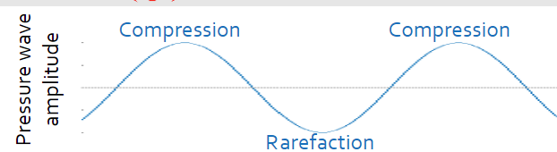
- Represent compression (particles bump down the line like a spring) and rarefaction (particles move away from each other) of travel medium particles
Wave Propagation
- Particle velocity: How fast a particle is moving back and forth, not wave speed
- u = 𝝏𝒛/𝝏𝒛
- u = 𝝏𝒛/𝝏𝒛
Wave Equations

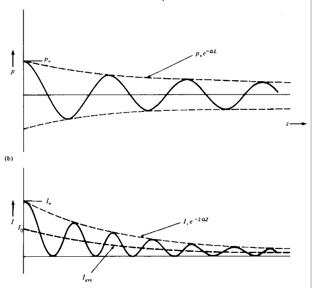
- Exponential sinosoid decay in pressure variation
- ρ is the average material density (kg/m^3)
- K is the compressibility constant of the material (ms^2 / kg)
- Pm is the maximum pressure intensity and amplitude (kPa)
- α is the linear attenuation coefficient (1/cm)
- ω is the frequency (rad/s) or 2pif where f is frequency (Hz)
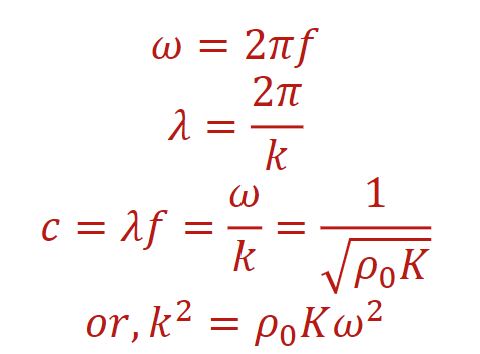
- k is the wave number or the propagation constant (1/m)
- The average speed of sound through soft tissue is 1540 m/s
- Intensity is the square of the pressure
Ultrasound Imaging
- The gel that goes on the skin where the transducer applies decreases the amount of air between transducer and skin, so you get less uneccessary reflection
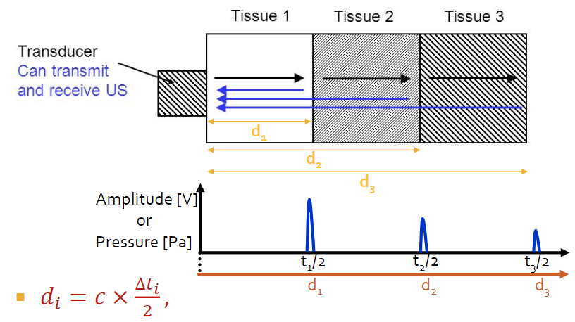
- A-Mode
- Distance = c * ∆t * 1/2
- Smaller peaks in the [V] vs time mean that the wave has less energy when it reaches the transducer
- B-mode imaging is formed by combining multiple A-mode lines to form a frame
- Frame time = number of lines x (time of a line + time of a pulse) *for individually pulsed
- Frame time = time of line + time of pulse *for simultaneously pulsed
- Frame time determines the maximum depth of return echos in ultrasound.
- Refresh rate: Numbe of frames drawn per second (1/time of frame)
- Refresh rate being higher means it’s closer to real-time
Transducers
- Sector: Ultrasound probe best for large structures that are deep in the tissue
- Sector transducers allow imaging through a narrow sonographic window in ultrasounds
- Sector transducers have an image shaped like a pie slice
- Linear: Ultrasound probe best for imaging small structures that works best for structures just beneath the skin
- Curved: An array ultrasound probe that combines sector and linear formats, best for a broad sonographic window
Acoustic Impedance
- Acoustic impedance is denoted by the letter Z
- Z = 𝜌0 c = 𝜌0 x (𝜌0 * k)^-1/2 = (𝜌0 / k)^1/2
- Z of soft tissue is 1.63 x 10^6 kg / m^2 *s
- Z of water is 1.52 x 10^6 kg / m^2 *s
- Z of the skull is 7.8 x 10^6 kg / m^2 *s
- Units of acoustic impedance are kg / m^2 *s
Material & Wave Interaction
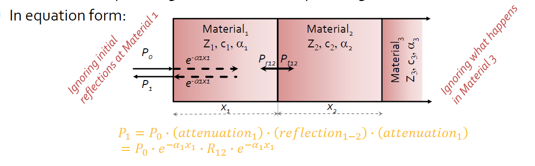
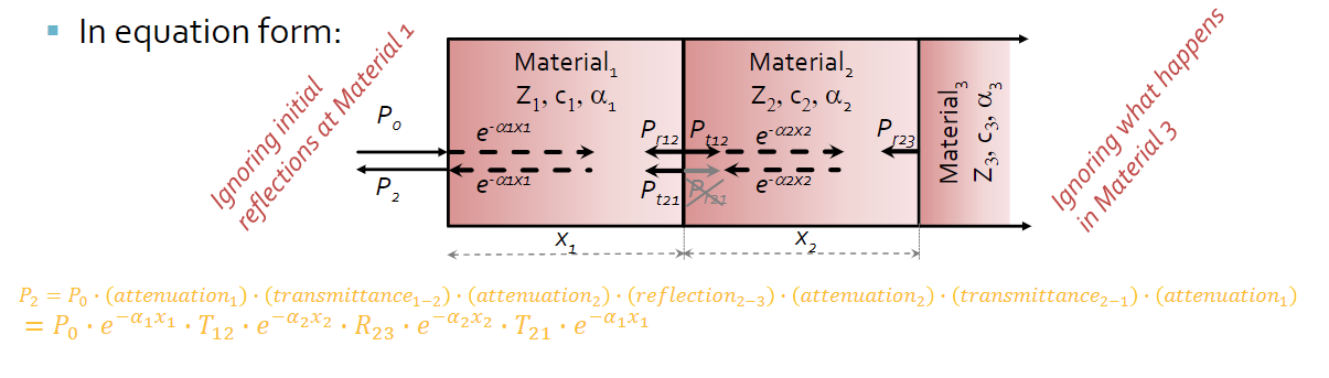
- There are two types of acoustic interaction, which is at interfaces between different materials (boundaries & transmission) and within the material itself (attenuation)
- At a new interface, some energy is reflected back and some is transmitted (refracted)
- The reflecting and refracting at a new interface during ultrasound is due to the acoustic impedance
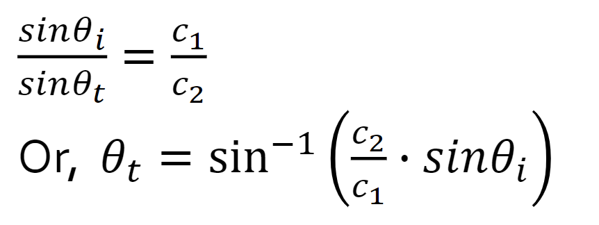
- Snell’s Law
- At the critical angle, we will have total reflection of the incident wave and no transmission into second medium
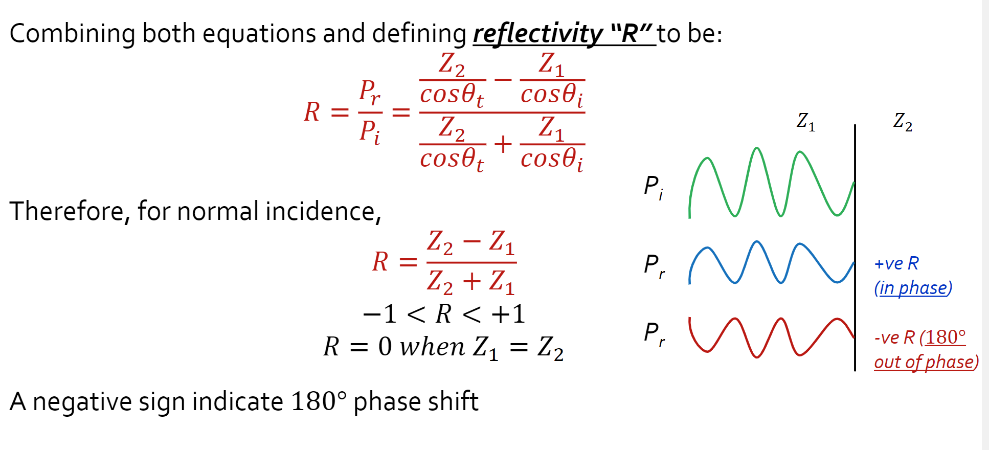
- A negative value for R indicates a 180 degree phase shift (flip over x-axis); this affects phase shift, but not amplitude or anything else
- ==Reflectivity is zero when the acoustic impendences are equal to one another==
- Transmittivity (T) is transmitted pressure over initial presssure
- Transmittivity is between zero and positive 2
- 2 occurs when the reflected energy is the same as the initial, so it doubles (reflects on the same path)
- Transmission = 1 + R
- 1 + (Z2 - Z1) / (Z2 + Z1)
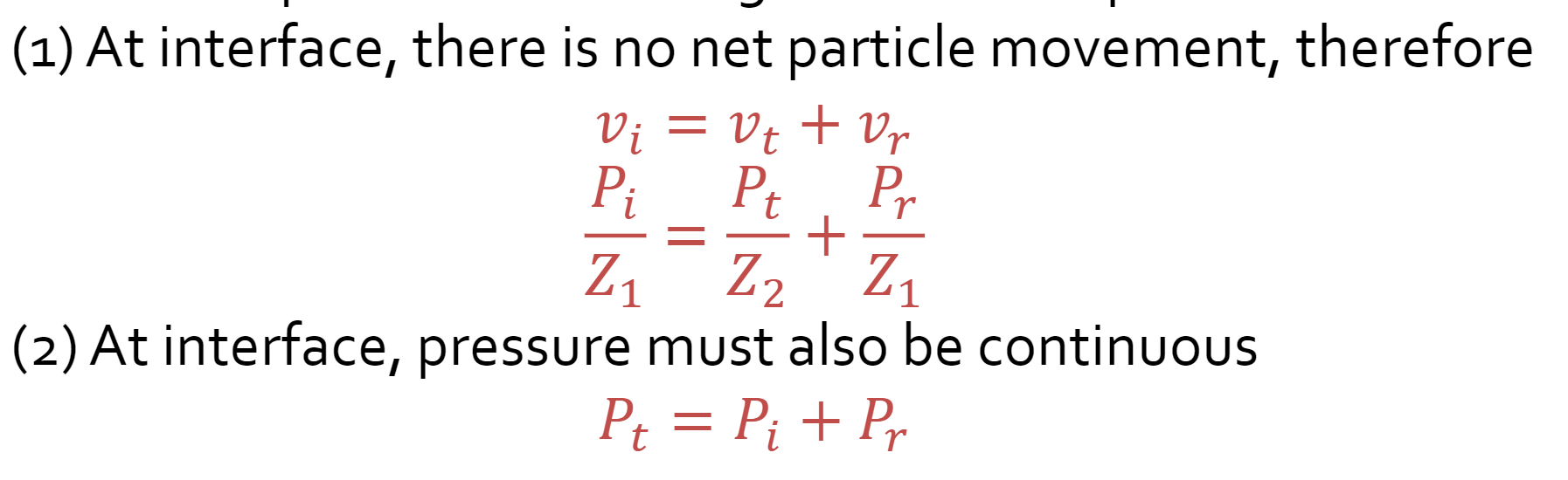
- At interface
- At interface, there is no particle movement.
- At interface, pressure must be continuous
- The linear attenuation coefficient in ultrasound imaging is a linear function of frequency
- As frequency increases in an ultrasound, the linear attenuation also increases
- Pressure vs Intensity
- Acoustic wave intensity is used to measure the power in the wave
- I = Pressure^2 / Z
- P = Pm e ^ - 1 alpha *** frequency * distance
- Alpha needs to be in the inverse unit of the distance
- To increase distance, you need to decrease the energy
- Combined effects of interaction through multiple materials are multiplicative