Bio 301 - Ch. 2 Book Notes
Chapter 2 - Observing the Microbial Cell
fluorescence microscopy captures single molecules within a living cell
the fluorophores can label specific kinds of DNA or proteins
electron microscopy explores the cell’s interior
cryo-electron microscopy reveals the cells the 3D & models the shape of viruses
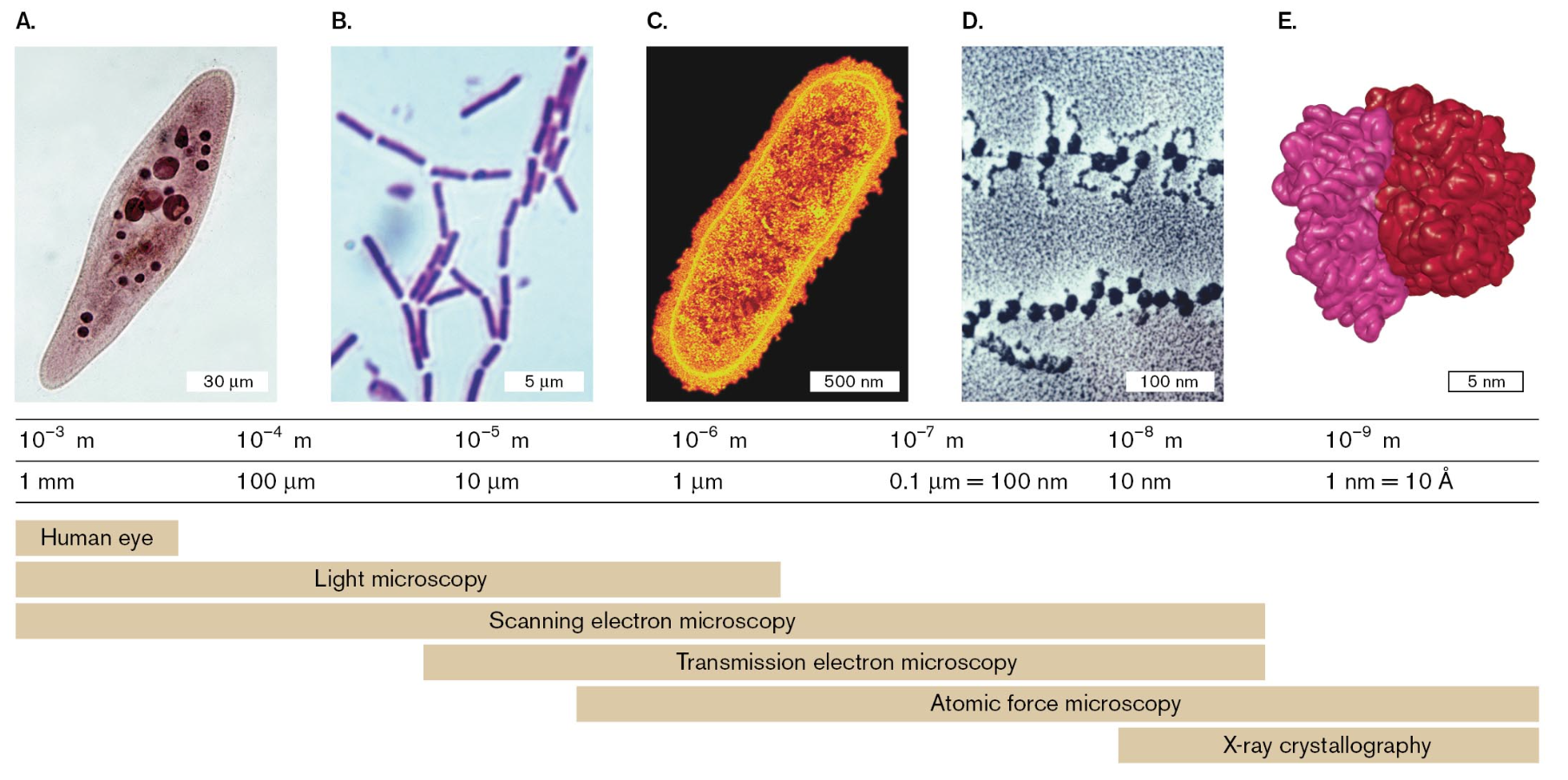
Example of how the technology has advanced since Leeuwenhoek’s first invention
spiral-shaped methane oxidizing bacterium were isolated in a peat bog in northern Russia
these bacteria are significant for curbing the release of methane from the archaea that produce it
these bacteria were visualized as a whole by phase-contrast microscopy
phase contrast utilizes the wave property of light to bend “out of sync” at the edge between a microbe and its surrounding medium to generate high contrast
electron transmission microscopy
the sample is sliced into very thin sections, stained with electron-dense metal atoms and bombarded with a beam of electrons
upon viewing the specimen, the electron beam reveals layers of membranes stacked like pancakes
these intracellular membranes are packed with proteins that transfer electrons from methane into oxygen
much of the methane is trapped in the cell’s biomass which prevents an escape of a greenhouse gas into the atmosphere
resolution of objects by our eyes
animals’ eyes observe an object by focusing its image on a retina packed with light-absorbing photoreceptor cells
the image appears sharp if the eye’s lens and cornea bend all the light rays from each point of the object to converge at one point on the retina
in the human eye, the finest resolution of two separate points is perceived by the fovea, the portion of the retina where the photoreceptors are packed at the highest density
the foveal photoreceptors are cone cells which detect primary colors and finely resolved detail
the distance between two foveal cones limits our resolution to 100-200 um
our eyes can detect a large population of microbes
examples include: a spot of mold or a cloudy tube of bacteria
observing these examples means the eye can detect the presence of mold or bacteria but it cannot resolve distinct cells
magnification is needed to resolve microbial cells individually to their natural shapes
as the distance increases between points of detail, our eyes can now resolve the object’s shape as a magnified image
microbial size & shape
eukaryotic microbes have a range of cell sizes to which a light microscope can resolve intracellular compartments such as the nucleus and vacuoles
protists show complex shapes and appendages
example includes an ameba from a freshwater ecosystem that has a large nucleus and pseudopods to engulf prey
another example includes Trypanosoma brucei which is an insect-borne blood parasite that causes African sleeping sickness; a nucleus and flagellum can be observed under a light microscope
certain shapes of bacteria are common to many taxonomic groups
both bacteria & archaea form similarly shaped rods, bacilli and cocci
it can be noted rods and spherical shapes evolved independently within different taxa
another shape distinction is a spirochete in which species of this shape cause diseases such as Lyme borreliosis and syphilis; the spiral form is maintained by internal axial filaments, flagella, and an outer sheath
an unrelated spiral form is known as spirillum which is a wide, rigid spiral cell that is similar to a rod-shaped bacillus
microscopy at different size scales
light microscopy (LM) - resolves images of individual bacteria by their absorption of light
bright-field microscopy - the specimen is commonly viewed as a dark object against a light-filled field
advanced techniques based on special properties of light include phase-contrast and fluorescence microscopy
electron microscopy (EM) - uses beams of electrons to resolve details several orders of magnitude smaller than those seen under LM
scanning electron microscopy (SEM) - uses a beam of electrons that is scattered from a metal-coated surface of an object which can generate an appearance of 3D depth
transmission electron microscopy (TEM) - a type of EM microscope where the electron beam travels through the object and the electrons are absorbed by an electron-dense metal stain
chemical imaging microscopy - uses spectrometry to map the chemical contents of a specimen like distribution of nitrogen and carbon compounds
X-ray crystallography - detects the interference pattern of X-rays entering the crystal lattice of a molecule; from this pattern a computational model of the structure can be built of the individual molecule
optics and properties of light
regions of the electromagnetic spectrum are defined by wavelength
visible spectrum ranges from 400-750 nm
radiation of longer wavelengths includes infrared and radio waves
radiation of shorter wavelengths includes ultraviolet rays and x-rays
information carried by radiation can be used to detect objects
all electromagnetic radiation travels through a vacuum at 3 × 10^8 m/s (speed of light)
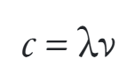
c is constant; the longer the wavelength, the lower the frequency of v (Hz)
wavelength limits the size of objects that can be resolved as separate from neighboring objects
requirements for resolution
contrast between the object and its surroundings
if an object and its surroundings absorb or reflect radiation equally, then the object will be undetectable
wavelength smaller than the object
the radiation must be equal to or smaller than the size of the object
if the wavelength of the radiation is larger than the object, then most of the wave’s energy will simply pass through the object
magnification
the smallest distance our retina can resolve is 150 um which means we are virtually unable to access all the information contained in the light than enters our eyes
we must adapt and spread the light rays far enough apart for our retina to perceive the resolved image
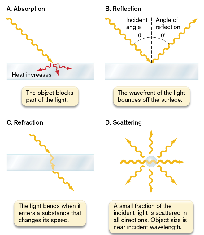
light interacts with an object
the physical behavior of light resembles a beam of particles, photons, and a waveform
each photon has an associated wavelength that determines how the photon will interact with a given object
the combined properties of particle and wave enable light to interact with an object in several different ways
absorption: the absorbing object gains the photon’s energy which is converted to a different form of energy; usually heat
when a microbial specimen absorbs light, it can be observed as a dark spot against a bright field (bright-field microscopy)
some molecules exhibit fluorescence which is when the absorbed light of a specific wavelength reemits energy as light with a longer wavelength
reflection: the wavefront redirects from the surface of an object at an angle equal to its incident angle
refraction: the light bends as it enters a substance / medium that slows its speed
a substance that exhibits this type of property is said to be refractive and has a higher refractive index than air
scattering: a portion of the wavefront is converted to a spherical wave originating from the object
if a large # of particles simultaneously scatter light, we observe a haze
magnification by a lens
refraction magnifies an image when light passes through a refractive material shaped so as to spread its rays
a parabolic curve is a type of shape that would spread light rays
when light rays enter a lens of refractive material with a parabolic surface, parallel rays each bend at an angle so that all the rays meet at a focal point
from this focal point, the light rays continue to spread out with an expanding wavefront
this expansion magnifies the image carried by the wave
focal distance is determined by the degree of curvature of the lens and by the refractive index of its material
resolution of detail
empty magnification occurs when there is magnification without increasing detail
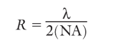
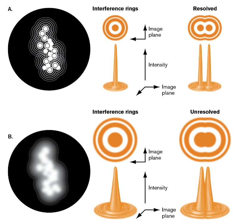
the resolution of detail in microscopy in limited by the wave nature of light
interference occurs because the width of the lens is infinite but light rays form wavefront of infinite extent so only part of the wavefront enters
the converging edges of the waves interfere with each other to form alternation regions of light and dark
when viewing a specimen with LM, each point source generates a central peak of intensity to which the width of this central peak will define the resolution between any two points of the object
this resolution determines the degree of detail that can be observed
factors that limit the resolution of an image
the wavelength of light limits the sharpness of the peak of intensity of a point of detail
the finite width of the wavefront captured by the lens leads to interference and widens the peak intensity
bright-field microscopy resolves only details that are greater than half the wavelength of light (about 200 nm)
super-resolution imaging can allow for the tracking of cellular molecules at a precision of 20-40 nm
magnification
wavelength & resolution
the greatest magnification that can improve our perception of what is usually 100-200 um is about 1,000x
any greater magnification expands the image size but the peaks expand without resolution between them (empty magnification)
light and contrast
balanced amount of light yields the highest contrast between the dark specimen and the light background
high contrast is needed to perceive the full resolution at a given magnification
lens quality
all lens possess aberrations that detract from perfect curvature
optical properties limit the perfection of a single lens but manufactures can construct microscopes with a series of lens to multiply each other’s magnification as a correction
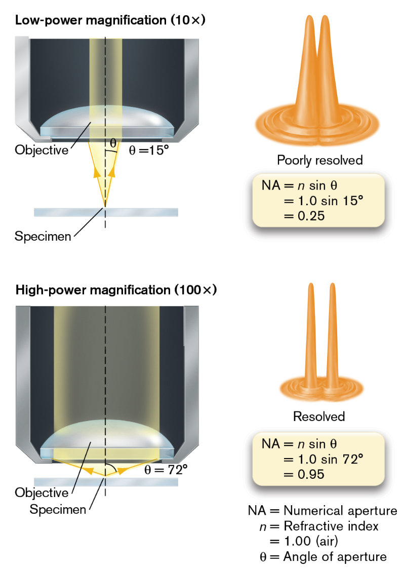
an object at the focal point of a lens sits at the tip of a cone of light formed by rays from the lens converging at the object
the angle of the light cone is determined by the curvature and refractive index of the lens
as theta increases, the horizontal width of the light cone increase giving way to a wider cone of light to pass through the specimen
the wider the cone of light rays, the less the interference between the wavefronts and the narrower the peak intensities in the image
a wider light cone enables us to resolve smaller details
the greater the angle of aperture of the lens will give better resolution
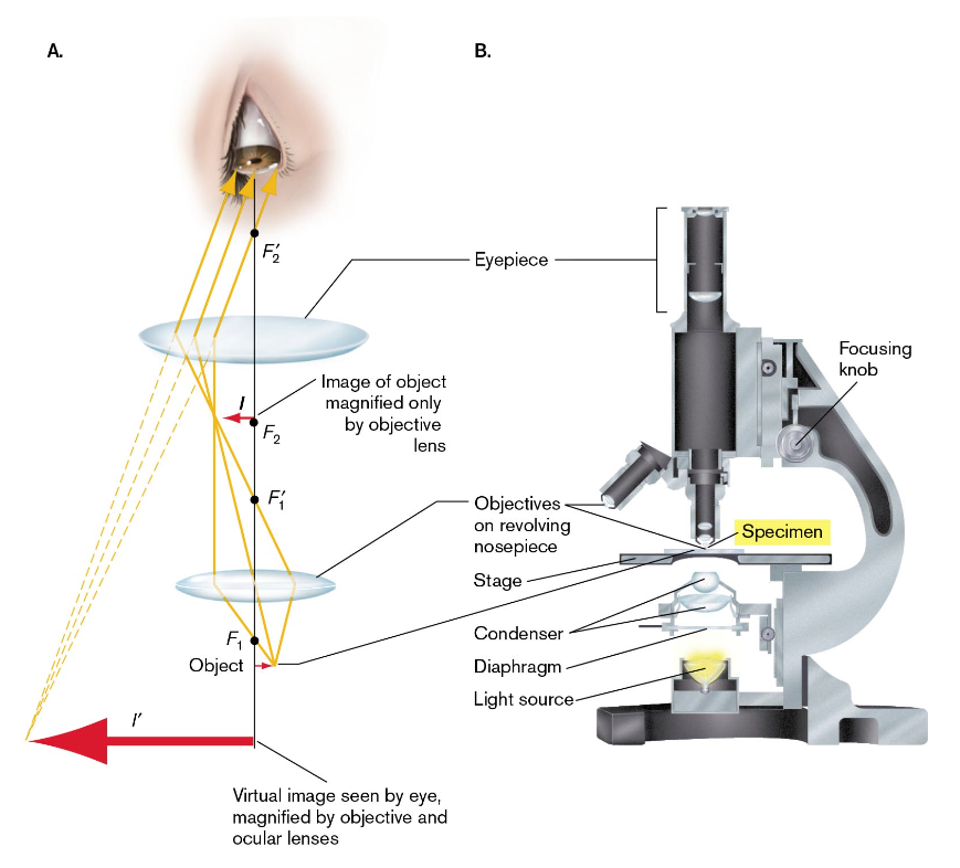
the compound microscope
a series of lower-power lens is used to multiply their magnification with minimal aberration (condenser & ocular lens)
lower-power lens require lower light levels because the excess light makes it impossible to observe the darkening effect of specimen absorbance
for higher-power lenses, they spread the light rays further and require an open diaphragm to collect sufficient light for contrast between the dark specimen and the bright field
condenser lenses increase light available for contrast but do not participate in magnification
in a parfocal system, when a specimen is viewed from one lens and the lens are rotated, it remains in focus
steps for observing a specimen under a compound microscope
position the specimen centrally in the optical column
optimize the amount of light to produce the optimal amount of contrast
focus the objective lens - typically start with a low-power objective which generates a greater depth of field
preparing a specimen for microscopy
wet mount; to observe microbes by placing a drop of water on a slide with a coverslip
advantages of wet mounting include: the organism being viewed is in it’s most natural state without artifacts resulting from chemical treatment
disadvantages of wet mounting includes: there is little contrast with the external medium and the specimen of interest (detection and resolution are minimal) & the sample can get easily dried out
the microbe observed must adhere to a specially coated slide within the temperature-controlled flow cell to avoid overheating
focusing the objects
the shape of the dark object is defined by points of light surrounding its edge when in focus
at higher power, these points of light are only partially resolved (limit of resolution reached)
partially resolution generates interference effects like extra rings of light
to observe motile bacteria, you must adjust the settings to have a higher magnification but a narrower depth of field

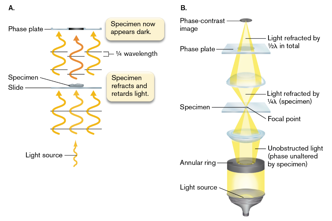
phase contrast microscopy
exploits differences in refractive index between the cytoplasm and the surrounding medium or between different organelles
depends on the principle of interference where two wavefronts interact with each other by addition or subtraction which results in alternating zones of constructive and destructive interference
addition - amplitudes in phase | subtraction - amplitudes out of phase
due to the fact that living cells have relatively high contrast because of their concentration of solutes, after light passes through the cell, about one-quarter of a wavelength behind the phase of light is transmitted directly through the medium
optical system is designed so that by the light refracted through the cell is slowed by a total of half a wavelength and when out of phase, it produces destructive interference (a region of darkness in the image of the specimen)
the light transmitted through the medium needs to be separated from the light interacting with the object
the transmitted light is separated by a ring-shaped slit (annular ring) which generates a hollow cone of light
fixation & staining for bright-field microscopy
fixation - a process which cells are adhered to a slide in a fixed position
cells can be fixed by methanol or heat treatments in order to denature the cell’s proteins so the side chains are exposed and bind to the glass
staining - the use of molecule that absorbs the incident light, usually over a wavelength that results in a distinctive color
how stains work:
stain molecules contain conjugated double bonds or aromatic rings that absorb visible light & have positively charged groups that bind cell-surface components with negative charge
different stains will depend on the strength of their binding and the degree of binding to different parts of a cell
simple stains
adds a dark color to the specimen but not to the external medium or immediate surrounding tissue
most common simple stain used is methylene blue


differential stains
this is a type of stain that differentiates among objects by staining only particular types of cells or specific subcellular structures
Gram stain - distinguishes cells that have a thick cell wall & retain a positively charged stain from cells that have a thin cell wall and outer membrane & fail to retain the stain
gram-negative cells usually stain red | gram-positive cells usually stain purple
Procedure for gram-staining
stain process: crystal violet dye, mordant (binding agent), decolorizer like ethanol, and counterstain (safranin)
during the stage of using a decolorizer, gram-positive cells appear dark purple and gram-negative cells appear colorless
the timing is important for each step because it can release the retained stain
in the decolorizer stage if over time then the gram-positive cells will release their crystal violet stain
in the counterstain stage if over time then both the gram-positive and gram-negative cells appear pink because of the safranin

acid-fast stain
a stain for mycobacteria that retain the dye fuchsin because of the presence of mycolic acids in the cell wall
giemsa stain for blood film
a mixture of methylene blue, eosin, and Azure B is used to stain blood cells & associated parasites
negative stain
a stain that colors the background and leaves the specimen unstained
a suspension of opaque particles such as India ink that is added to darken the surrounding medium and reveal transparent components of pathogens & microbes
antibody stains
stains linked to antibodies which can help identify precise strains of bacteria or molecular components of cells
the antibody may be conjugated to a reactive enzyme for detection
fluorescence microscopy
helpful to identify specific kinds of microbes and reveals specific cell parts function
for this type of microscopy, the specimen absorbs light of a defined wavelength and then emits light of lower energy
some microorganisms can fluoresce on their own due to the presence of chlorophyll
specific parts of the cell are labeled with a fluorophore
two different colors of fluorescence distinguish between the cyanobacterial cells performing photosynthesis and the heterocysts performing nitrogen fixation
excitation and emission
fluorescence occurs when a molecule absorbs light of a specific wavelength to raise an electron to a higher-energy orbital (excitation wavelength)
because this higher-energy electron state is unstable the electron will decay to an orbital of slightly lower energy, some E lost as heat
it will then fall back it’s original level by emitting a photon of less energy and longer wavelength (emission wavelength)
INSERT IMAGE HERE
the optical system for fluorescence microscopy uses filters to limit the source light to the wavelength range of excitation and the specimen’s emitted light to the wavelength range of emission
the wavelengths of excitation and emission are determined by the choice of fluorophore
fluorophores for labeling
a fluorophore’s properties is determined by the molecular structure of each fluorophore determines its peak wavelength of excitation and emission & its binding properties
the specificity of a fluorophore can be determined by:
chemical affinity - especially for biological molecules
labeled antibodies - antibodies that specifically bind to a cell component are covalently attached to a fluorophore which forms a “conjugated antibody” (immunofluorescence)
DNA hybridization - a short sequence of DNA attached to a fluorophore will hybridize to a specific sequence in the genome
gene fusion reporter - cells can engineered with a gene fusion which expresses a bacterial protein combined with either GFP or one of many GFP variants
super-resolution imaging
enables scientists to pinpoint the location of a DNA-binding protein with a precision tenfold greater than the resolution of an ordinary optical microscope
when tasked with tracking a single molecule, we must consider the shape of the magnified image of a point source of light
the uncertainty of the central peak position is about a tenth wide on the intensity profile and computation based on this profile can reveal the peak positions with high precision
confocal laser scanning microscopy
a microscopic laser light source scans across the specimen in such a way that the excitation light from at laser and the emitted light from the specimen are focused together to produce a high-resolution image
this type of imaging can be used for 3D imaging of pathogens embedded within host cell or tissue
Fluorescence In Situ Hybridization (FISH)
FISH can map specific taxa of microbes within an environment & can show the spatial location of microbial taxa
the FISH technique makes use of a fluorophore-labeled oligonucleotide probe that hybridizes to a microbe’s DNA or rRNA
doing so increases sensitivity because rRNA is present in approx 100-fold to 10,000-fold excess over DNA
chemical imaging microscopy
the use of mass spectrometry to visualize the distribution of chemicals within living cells
a high res method for chemical imaging is referred to as nanoscale secondary ion mass spectrometry (NanoSIMS)
has an ionizing probe which provides a source of energy that breaks up the large organic molecules of a sample
this instrument measures the fragment masses of the secondary ions that fly off the source which generates a mass spectrum