Grade 12 Biology- Molecular Genetics
DNA – The Molecular Basis of Life
DNA: The Hereditary Material
The Molecular Basis of Inheritance
DNA Structure and Function of DNA; A History
DNA Replication, Repair and Aging
Evidence of Hereditary Material
DNA Organization in Eukaryotes (Chromosome Structure) and Prokaryotes
DNA Replication and Aging
Protein Synthesis (Chapter 7)
Genes and Protein Synthesis
From Gene to Protein - One Gene One Polypeptide Hypothesis
Protein Synthesis Overview
Transcription: DNA-Directed RNA Synthesis
Translation: Nucleic Acid to Protein
Controlling Gene Expression
Genetic Mutations
Key Differences Between Eukaryotes and Prokaryotes
Genomes and Gene Organization
DNA: The Hereditary Material
DNA molecules are carriers of genetic information
Gregor Mendel first to discover the existence of DNA in 1853
Scientists later discovered evidence of DNA 8 years after (1861) APX
Uses Of DNA
Detection of diseases and illness
Criminal cases
Ancestry testing
Prenatal genetic tests
Paternity test
DNA As Heredity Molecules
Proteins were thought to be the heredity material
1868, physician Frederick Meischer was interested in the composition of cell nucleus
Collected pus(white blood cells) from the bandages of his patients
From these cells, he extracted large amounts of an unknown substance that was acidic and had a large amount of phosphorus
He named the unknown substances “nuclein” because he found it in the nucleus of white blood cells
Discovering DNA
Miescher demonstrated that "nuclein" was present in the nucleus.
- Hammerling showed that it was very likely that the hereditary material resided in the nucleus.
- Griffith demonstrated that a molecule, which could be extracted from dead bacteria, could transform live bacteria and transfer hereditary information to the bacteria. If this material could be identified, the chemical nature of the hereditary material would be known.
- Avery, McCarty, and Macleod identified DNA as the molecule that transforms bacteria.
- Hershey and Chase confirmed that DNA was the hereditary material—this time using bacteriophage.
- Chargaff provided the first evidence indicating that bases might be paired in DNA.
- Franklin generated X-ray diffraction data that suggested that DNA was a helical molecule.
- Watson and Crick produced the double helix model with inward-pointing paired hydrogen-bonded bases, which explained Chargaff's data and conformed with the dimensions of Franklin's image.
DNA Composition
DNA (Deoxyribonucleic Acid) is a polymer of deoxyribonucleotides
Deoxyribonucleotides consists of:
5 Carbon sugars
Nitrogenous Bases: Adenine, Thymine, Guanine, and Cytosine
Phosphate group
Nitrogenous Bases
The following four (4) types of nitrogenous bases are found in DNA:
Adenine ( A) These two are double ringed purines
Guanine (G)
Cytosine (C)
Thymine (T) These two are single ringed pyrimidines
Structure Of DNA
James Watson and Francis Crick discovered that the DNA molecule is a double-helix spiral. It consists of two complementary strands that are coiled together around a vertical axis, resembling a ladder.
Phosphodiester Bond
The nucleotides in each strand are held together by a phosphodiester bond.
This bond is between a phosphate group of one nucleotide and a hydroxyl group of the sugar on carbon 3 of the adjacent nucleotide
The reaction is a condensation reaction.
Base Pairing
In double stranded DNA, adenine can only pair with thymine while guanine can only pair with cytosine.
Adenine from one strand pairs with thymine on the adjacent strand with two hydrogen bonds. While guanine pairs with cytosine with three hydrogen bonds.
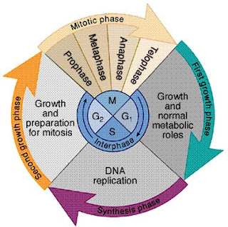
DNA Replication
A process where the DNA molecule makes a replication of itself during interphase in preparation of cell division
Before the cell can divide, DNA must be duplicated
Process in where the DNA molecule makes a replica of itself during interphase in preparation for cell division
Before the cell can divide, its DNA must be duplicated
DNA replicates during the “S” phase of the cell cycle
The cell cycle has other phases including G1, S, G2, M (mitosis)
preparation for cell divisi
How DNA Replicates
Semi-Conservative: 2 parental strands separate and each makes a copy of itself after 1 round of replication, the 2 daughter molecules each contain one old and one new strand
Conservative Model: The parental molecule separated was thought to direct the synthesize of an entire new DNA molecule
Dispersive Model: This model suggests that each strand of both daughter molecules contains a mixture of old and newly synthesized DNA, resulting in a blend of parental and daughter DNA.
The Semi-Conservative model was ultimately confirmed through experiments conducted by Meselson and Stahl, which demonstrated that the newly synthesized DNA strands indeed incorporated one parental strand and one newly formed strand in each daughter molecule.
Semi-Conservative is the correct model
proved by Meselson and Stahl in 1958
Preparing For Replication
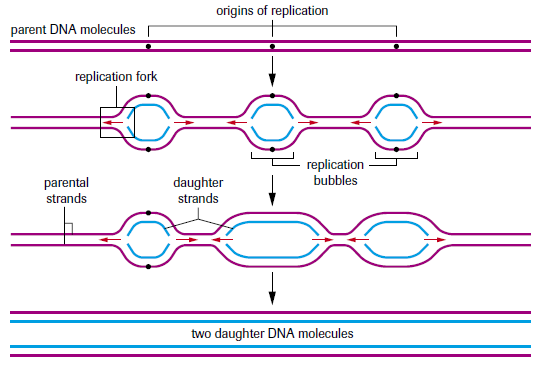
1. DNA replication begins at the origin
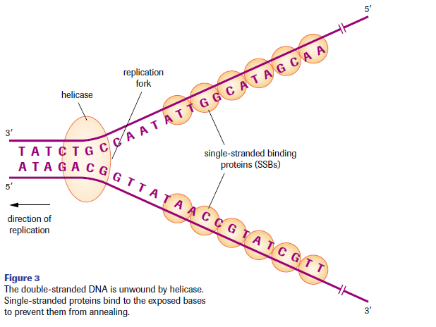
2. Replication bubbles form when DNA strands open at the origin from enzyme helicase
3. Two strands open forming replication forks (Y shape start of bubble)
4. New strands grow towards the forks (direction of replication)
5-8. Helicase separates DNA strands by breaking hydrogen bonds and single stranded binding proteins attach to keep the two DNA strands separated, topoisomerase attaches to the two forks (Y shapes) to relieve any tension and RNA primers provide new starting points
9-12. Primase enzyme synthesizes and lays down RNA primer, DNA Polymerase III adds new nucleotides and can only do so at the 3’ end (making new strand to be build in 5’ to 3’ direction)
13-17. Leading strand is synthesized from the origin towards the fork, lagging strand is synthesized away from fork making short segments called Okazaki fragments, DNA Polymerase I removes RNA primers and Ligase joins Okazaki fragments to make one strand
Proofreading:
DNA polymerase III makes about 10,000 base pairing errors
Enzymes proofread and correct errors
Damage and repair:
Chemicals and ultraviolet radiation damage the DNA in an organism’s cells
Enzymes known as exonucleases remove damaged parts of DNA
DNA polymerase and DNA ligase replace and bond new nucleotides together
Telomeres:
Repeating sequences of nucleotides at the end of chromosomes
Help prevent chromosome ends from fusing to other chromosomes
Prevent degradation form enzymes called nucleases
Play a role in determining the number of times a cell can divide (determining lifespans)
After many DNA replications, the telomere can be completely lost and can’t provide anymore protection to the chromosome which leads to essential DNA pieces being lost during following replications
If a cell loses its ability to grow and divide, its referred to as cell aging
Total number a cell can divide is called the Hayflick limit
One Gene-one polypeptide hypothesis
Hypothesis:
Each gene is responsible for directing the building of a single specific enzyme
If there was a one to one relationship between genes and specific enzymes, it should be possible to create genetic mutants that are unable to carry out specific enzymatic reactions
The results lead to the statement of each gene being responsible for directing the building of a single specific enzyme
Central Dogma of molecular genetics
According to what has been called the central dogma of molecular genetics…
The function of FNA is to store information and pass it on to RNA, while the function of RNA is to read, decode, and use the information received from DNA to make proteins
Replication: DNA replicates its information in a process involving many enzymes
Transcription: DNA codes for the production of messenger RNA
Translation: mRNA carries coded information to ribosomes and the ribosomes read the information and use it for protein synthesis
Transcription
Transfer of information from a DNA molecule into a messenger RNA molecule
During transcription, only one of the two DNA strands is read (template strand)
mRNA molecule is complementary rather than identical to the DNA template
In a prokaryotic cell, which lacks a nucleus, mRNA produced by transcription in immediately translated without additional processing
In a eukaryotic cell, two main steps protein synthesis occur in separate compartments: transcription in the nucleus and in the cytoplasm
Transcription starts with enzymes called RNA polymerase, which pry the two strands of DNA apart and hook together RNA nucleotides as they base pair along the template strand
RNA polymerase can only add nucleotides to the 3’ end of the growing polymer, thus RNA molecule can only elongate in the 5’ end to 3’ direction
RNA polymerase I is responsible for RNA synthesis and RNA polymerase III synthesis tRNA
Three key steps in transcription are polymerase binding/initiation, elongation, and termination
Three key steps in transcription are polymerase binding/initiation, elongation and termination
Initiation: RNA polymerase bind to regions of DNA called promoters (includes initiation site where transcription starts), certain regions are important for RNA polymerase to recognize (eukaryotes have TATA box which is an area rich in thymine and adenine centered at locations 25 nucleotides upstream from initiation sites) and enzymes begins to separate the two DNA strands at initiation sites
Elongation: As RNA polymerase II moves along the DNA, it untwists the double helix separating the strands and exposing 10 DNA bases for pairing with RNA nucleotides (adds to 3’ end of the growing mRNA molecule), and the enzyme peels away from the DNA template strand and the noncoding strand forms a double helix with a single gene being transcribed simultaneously by several molecules of RNA polymerase II allowing a cell to produce particular proteins in large amounts
Termination: transcription proceeds until RNA polymerase reaches a termination site on the DNA (most common sequence is AATAAA
Post transcriptional modifications are when the primary mRNA is vulnerable to conditions in the cytosol and undergo modification (only in eukaryotic cells and not prokaryotic) including a sequence of 7 “Gs” added to the start of pre-mRNA at 5’ cap and a string of adenine nucleotides added to 3’ end to protect from enzymes in the cytosol
Introns are noncoding regions while exons are coding regions (introns are removed by bully spliceosomes)
Alternative splicing is when exons may be joined in different combinations to produce different mRNAs from a single DNA gene sequence and once mRNA has been produced, it is ready to leave the nucleus and be translated by a ribosome
Translation
RNA directed synthesis of polypeptides
Requires all three RNA types
mRNA: template used for correct addition of individual amino acids to the growing polypeptide chain
tRNA: carry activated amino acids into the ribosome
rRNA: ensures correct access of activated tRNAs and contains necessary enzymes to catalyze peptide bond formation
3 codons: 3 of the 64 triplet codons are termination codons
Remaining 61 codons are recognized by individual tRNAs
Start codon: AUG, signals the start of translation
Stop codons: UAA, UAG, UGA, act as stop signals to terminate translation
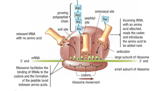
Ribosome:
Made of a small and large subunit
Large subunit has an A site (aminoacyl site), a P site (peptidyl site), and an E site (exit site)
Translation steps:
Initiation: small subunit of a ribosome forms a complex with the initiator tRNA and binds upstream of the start of the message, proceeding downstream (5’-3’) scanning mRNA until encountering start codon AUG, once found the large subunit joins the complex the initiator tRNA binds to the P site on the ribosome, and paired correctly to establish the correct reading frame
Elongation: an aminoacyl-tRNA is able to base pair with the with the next codon on the mRNA arrives at the A site (accepting site), proceeding amino acid is covalently linked to the incoming amino acid with a peptide bond (requires energy), initiation tRNA is released from the P site and opens the A site for the arrival of a new aminoacyl-tRNA, protein known as elongation factor is involved and another energy molecule is used
Termination: The end of translation occurs when the ribosome reaches one or more stop codons (UAA, UGA, UAG), there are no tRNA molecules with anticodons for stop codons, protein release factors recognize these codons when arrived at A site, binding to these proteins releases the polypeptide from the ribosome along with an energy molecule, ribosome splits into subunits which are later reassembled for another round of protein synthesis
Polysomes: a single mRNA molecule usually has many ribosomes traveling along it, in various stages of synthesizing a polypeptide (complex is called polysome)
Controlling gene expression
Not all proteins are needed by the cell all the time
Prokaryotic gene control methods:
Genes are switched off when not needed so that the cell can save energy, housekeeping genes are on all the time and regulate processes such as metabolism, growth, DNA replication and transcription, gene control in prokaryotes is in response to regulating the metabolism for prokaryotic gene control mechanisms and a control mechanism is known as an operon model of gene expression
Operon is a group of genes or a segment of DNA that functions as a single transcription unit made up of an operator, promoter and one or more structural genes that are transcribed into one mRNA which codes for more than one protein with related metabolic functions
Lac operon consists of a promoter, operator and a cluster of three genes (LacZ, LacY and LacA) when lactose is absent, the genes don’t function because a repressor protein (Lacl) is active and bound to the DNA preventing transcription and when that repressor protein is bound to the DNA, RNA polymerase cannot bind to the DNA, The protein must be removed before the genes can be transcribed, if lactose is present, E-coli needs to produce the necessary enzymes to break it down (beta-galactosidase, permease, transacetylase), and the genes LacZ, LacY and LacA respectively represent the genes that produce the 3 enzymes needed to metabolize lactose, lactose is the signal molecule or inducer due to the structural genes normally being inactive unless lactose is present
Trp Operon: tryptophan is an aminoacid needed by prokaryotes to make proteins, genes (trpE, trpD, trpC, trpB, trpA) that code for proteins that produce tryptophan are continuously transcribed, the operon that regulates the production of tryptophan is similar to the lac operon, tryptophan is a corepressor due to the repressor not binding to DNA unless its activated by binding with a tryptophan, repressible operon because the structural genes only active when tryptophan is present
Eukaryotic gene control mechanisms:
Gene expression is controlled by a number of mechanisms that range from those that prevent transcription to those that prevent expression after the protein has been produced
Four mechanisms are transcriptional (prevent transcription), post transcriptional (control/regulate mRNA after it has been produced), translational (prevent translation), post translational (act after the protein has been produced)